Augmented Innate and Adaptive Immune Responses Under Conditions of Diabetes-Filariasis Comorbidity
←
→
Page content transcription
If your browser does not render page correctly, please read the page content below
ORIGINAL RESEARCH
                                                                                                                                     published: 10 September 2021
                                                                                                                                   doi: 10.3389/fimmu.2021.716515

                                          Augmented Innate and Adaptive
                                          Immune Responses Under
                                          Conditions of Diabetes–Filariasis
                                          Comorbidity
                                          Joy Manohar Sibi 1, Viswanathan Mohan 2, Saravanan Munisankar 3, Subash Babu 3
                                          and Vivekanandhan Aravindhan 1*
                                          1 Department of Genetics, Dr A. L. Mudaliar Post Graduate Institute of Basic Medical Sciences (ALM PG IBMS), University of
                                          Madras, Chennai, India, 2 Madras Diabetes Research Foundation and Dr. Mohan’s Diabetes Specialties Centre, Chennai,
                                          India, 3 National Institute of Health–International Centre for Excellence in Research, National Institute for Research in
                                          Tuberculosis, Chennai, India

                                          Metainflammation, as seen in chronic diabetes subjects, impairs immunity and increases
                                          the susceptibility to infections. In the present study, the effect of diabetes on immune
                                          response against filariasis was studied. Both toll-like receptor (TLR)-mediated and crude
                                          antigen-induced immune responses were quantified, in whole blood cultures from
                          Edited by:      filariasis-infected subjects (LF+), with and without diabetes. Blood cultures were
                        Manuel Ritter,
                                          stimulated with TLR ligands (TLR2 and TLR4) or filarial antigen or were left unstimulated
   University Hospital Bonn, Germany
                                          (control) for 18 h. Cytokine, chemokine, and defensin secretion was quantified by ELISA.
                       Reviewed by:
               Suprabhat Mukherjee,       Expression of HLA-DR, B7-1, B7-2, activation marker (CD69), and Th (Th1, Th2, Th17,
          Kazi Nazrul University, India   and Th9) phenotypes was quantified by flow cytometry. Expression of immunomodulatory
                     Dominik Rückerl,
        The University of Manchester,
                                          effectors (Cox-2, HO-1, IDO-1, and p47Phox) and Th-polarizing transcription factors (T-
                      United Kingdom      bet, GATA3, and ROR-gt) was quantified by quantitative PCR. Secretion of IL-27, IL-1Ra,
                  *Correspondence:        IL-12, IL-33, IL-9, and SDF-1 was increased under diabetes conditions with increased
           Vivekanandhan Aravindhan
                                          Th9 polarization and increased expression of Cox-2 and IDO. Overall, diabetes was found
             cvaravindhan@gmail.com
                                          to augment both TLR-mediated and antigen-induced inflammation, which can promote
                   Specialty section:     chronic pathology in LF+ subjects.
         This article was submitted to
                 Parasite Immunology,     Keywords: diabetes, filariasis, TLR, immunomodulation, Th cell, inflammation
               a section of the journal
              Frontiers in Immunology

            Received: 28 May 2021         INTRODUCTION
        Accepted: 23 August 2021
     Published: 10 September 2021         Filarial infections, unlike viral and bacterial infections, induce immunomodulation, rather than
                            Citation:     inflammation (1). Murine studies have shown significant protection against both forms of diabetes
    Sibi JM, Mohan V, Munisankar S,       (type 1 and type 2) by filarial pre-infections (2–4). Previously, we have shown a decreased
    Babu S and Aravindhan V (2021)
                                          prevalence of filariasis among both T1DM (5) and T2DM (6) subjects in the South Indian
     Augmented Innate and Adaptive
Immune Responses Under Conditions
                                          population. Serum cytokine profiling in these subjects showed significant downregulation of pro-
   of Diabetes–Filariasis Comorbidity.    inflammatory cytokines [interleukin-6 (IL-6), tumor necrosis factor-alpha (TNF-a), and
          Front. Immunol. 12:716515.      granulocyte–macrophage-colony stimulating factor (GM-CSF)] and upregulation of anti-
   doi: 10.3389/fimmu.2021.716515         inflammatory cytokine [tumor growth factor-beta (TGF-b)], in filarial-positive diabetic

Frontiers in Immunology | www.frontiersin.org                                    1                                   September 2021 | Volume 12 | Article 716515
Sibi et al.                                                                                                    Diabetes and Filariasis Co-Morbidity

subjects (6). Interestingly, this effect was specific only to T1DM        IL-17F) sub-types (26). T cell-mediated immune responses during
and T2DM and was not seen in those with coronary artery                  filarial infection largely depend on the phase of the infection:
disease (CAD) (7). These observations were later replicated in           (1) acute phase—skewed towards the Th2 response; (2) chronic
other Asian countries like China (8) and Indonesia (9).                  phase—skewed towards “modified Th2 response”, with Tregs
    The relationship between filariasis and diabetes is bi-               playing a more prominent role compared to Th2 cells;
directional (10). While childhood filarial infection can dampen           (3) chronic pathology phase—a drastic shift from “modified
inflammation and can confer protection against diabetes, the              Th2” response to pro-inflammatory “Th1/Th17” response takes
effect of diabetes on filariasis-induced immune response is               place and happens only in those who develop lymphatic pathology
largely unknown. This is relevant in endemic zones wherein at            (22). Thus, in the present study, we looked at both TLR-mediated
least some of the subjects with lymphatic filariasis (LF) would           innate and filarial antigen-induced adaptive immune responses in
develop diabetes, during the course of infection. T2DM in                LF+ subjects, both with and without diabetes. Cytokine/
general, weakens the immune system and makes the patients                chemokine secretion, expression of immunomodulatory
more susceptible to infections (11). Immune response against             enzymes, upregulation of MHC and co-stimulatory molecules,
filariasis, in general, can be broadly classified into innate and          T-cell activation, Th polarization, and expression of T-cell
adaptive immune responses. Toll-like receptors (TLRs) serve as           polarizing transcription factors were quantified.
the first line of defense mechanism, bridging the innate and
adaptive arms of the immune responses (12). They are
abundantly present in cells of the innate immune system,                 MATERIALS AND METHODS
which include macrophages, dendritic cells, neutrophils, and
eosinophils (13). Previously, several filarial antigens were              Study Subjects
shown to bind directly to TLRs and activate them (14, 15).               This study is a follow-up of our previous publication wherein we
Upon TLR ligation, the professional antigen-presenting cells             reported decreased prevalence of LF among diabetic subjects
(APCs) that include macrophages, dendritic cells, and B cells            compared to control subjects (6). As a continuation of this study,
undergo activation followed by the secretion of cytokines and            1,001 outpatients visiting Dr. Mohan’s Diabetes Specialties
chemokines (16). The cytokines secreted by the innate immune             Centre, Chennai, India, were screened for LF. Out of 1,001,
cells can be broadly classified into (1) type 1 interferons               only 12 were found to be positive for LF antigen (1.2%). None of
(interferon-a and b), (2) pro-inflammatory cytokines (TNF-a,              them had any clinical symptoms of LF (DM-LF+). As controls,
IL-1b, IL-6, and GM-CSF), and (3) anti-inflammatory cytokines             eight normal glucose-tolerant (non-diabetic) LF+ subjects were
(IL-10, TGF-b, IL-1Ra, IL-35, and IL-27) (17). TLR ligation also         included (NGT-LF+). The control subjects were recruited from
upregulates the antigen processing and presentation machinery,           the healthy volunteers who accompanied the diabetes patients
which includes MHC (HLA) and co-stimulatory (B7-1/CD80                   and underwent OGTT testing, as part of this study. Institutional
and B7-2/CD86) molecules (18). Furthermore, certain cell-type-           ethical committee approval from the Madras Diabetes Research
specific effector functions like secretion of antibodies by B cells       Foundation Ethics Committee was obtained (Ref No-MDRF-EC/
(19), defensin by neutrophils (20), and matrix-metalloproteinases        SOC/2009//05) and written informed consent was obtained from
(MMPs) by macrophages (21) are also augmented. TLR-mediated              all the study subjects. The study was conducted as per the
expression of Cyclooxygenase-2 (Cox-2), Heme Oxygenase-1                 Declaration of Helsinki, following STROBE guidelines.
(HO-1), and Indoleamine-2,3-Dioxygenase (IDO) constitute a
immunoregulatory circuit, which controls filariasis-mediated              Inclusion and Exclusion Criteria
immunomodulation (22). The net result is the recruitment and             Only subjects who were LF+ (as determined by TropBio) were
activation of T cells, which marks the transition from innate to         included in the study. None of the subjects showed any symptoms
adaptive immune response (12).                                           of lymphatic filariasis at the time of recruitment. The exclusion
    In contrast to the cells of innate immune system, that detect        criteria were patients with type 1 diabetes and those with a previous
pathogens through TLRs, T cells are the workhorses of the                diagnosis of urolithiasis, liver cirrhosis, congestive heart failure,
adaptive immune system, largely depending on surface T-cell              chronic lung diseases, chronic infections, or viral hepatitis.
receptors (TCRs), for detecting the pathogens (23). While the
major function of CD4+ helper T (Th) cells is to regulate other          Diagnosis of T2DM
cells, CD8+ cytotoxic T (Tc) cells are mainly involved in the            The diagnosis was done following WHO guidelines. Subjects were
elimination of virally infected cells and tumor cells (24). In           grouped as Control (NGT) based on the Oral Glucose Tolerance
contrast to the innate immune cytokines, the adaptive immune             Test (OGTT) and as diabetes based on previous history (https://
cytokines are secreted by T cells (effector cytokines) and APCs          www.who.int/diabetes/publications/Definition%20and%20diagnosis
(polarizing cytokines) (25). Upon engagement of the TCRs and             %20of%20diabetes_new.pdf) (ISBN 978-92-4-159493-6).
antigen co-receptor (CD28), the Th cells undergo differentiation
into various sub-types depending on the polarizing cytokines             Detection of Bancroftian LF
secreted by the APCs (25). Depending on cytokine secretion, Th           To quantify the filarial antigen levels, sera were analyzed using
cells are classified into Th1 (IL-12, IFN-g, and IL-2), Th2 (IL-33,       the W. bancrofti Og4C3 antigen-capture ELISA (Tropbio,
IL-4, IL-5, and IL-13), Th9 (IL-9), and Th17 (IL-23, IL-17, and          Australia) according to the manufacturer’s instructions.

Frontiers in Immunology | www.frontiersin.org                        2                               September 2021 | Volume 12 | Article 716515
Sibi et al. Diabetes and Filariasis Co-Morbidity
Preparation of Filarial Antigen T-helper cells were gated based on CD3 and CD4 expression.
Crude antigen extract from Brugia malayi antigen (BmA) was The expression of cytokines and other effector molecules within
prepared by extracting somatic antigens from the live L3 larvae the gated population was analyzed as illustrated in Figures S1
in 1× PBS, as described previously (27). The extract was and S2. Both the percentage and mean fluorescence intensity
concentrated, quantified, and stored at −80°C. The endotoxin (MFI) of the gated cell population were quantified using FlowJo
levels as determined by the limulus amoebocyte lysate assay software, version v10 (BD Biosciences, USA). The list of various
(QCL-1000 kit; BioWhittaker) was below the detection limit in monoclonal antibodies used in flowcytometry is provided in
these preparations. It is important to note that both W. bancrofti Table S2.
and Brugia malayi share several overlapping antigens, and
immune cells from patients infected with W. bancrofti mount a Real-Time PCR Analysis
strong immune response against Brugia malayi antigens during RNA extraction from the stored samples was carried out using
rechallenge experiments (28). RNeasy Mini Kit (Qiagen). The quality and quantity of the
extracted RNA was quantified using nanodrop. One microgram
of RNA was converted to cDNA using reverse transcription and
Peripheral Blood Leukocyte Cultures real-time PCR was performed using TaqMan probes (Applied
Whole blood cultures from the study subjects were done as Biosystems, USA) specific for Cox-2, IDO, Phox P47, HO-1, T-bet,
described previously (29). Whole blood was collected in EDTA- GATA-3, and ROR-gt. 18S rRNA was used as a house-keeping
coated tubes. After centrifugation, the packed cell volume was control. Gene expression levels (normalized to 18S rRNA) were
diluted with RPMI medium (1:1 ratio) containing 10% FCS and analyzed using the StepOnePlus RT-PCR system (Applied
was used for in vitro culture. Cells were stimulated with TLR2 Biosystems, Foster City, CA, USA) and 2–DDCt was calculated for
ligand-PAM3CSK4 (100 ng/ml) (Invivogen, USA) or TLR4 all samples with unstimulated sample values as reference. All the
ligand-LPS (100 ng/ml) (Invivogen, USA) or were left probes used in these studies were purchased from Applied
unstimulated for 24 h in parallel cultures. For antigen Biosystems, and the list of various probes used is provided in
stimulation, cultures were stimulated with crude extract of Table S3.
BmA (10 µg/ml) for 24 h. The supernatants were harvested for
cytokine/chemokine estimation. The cell pellets were solubilized
in RNAzol and were stored at −80°C. Cell pellets from parallel Statistical Analysis
cultures were treated with golgi stop for 6 h, washed, and stained Mann–Whitney U test was used for comparing NGT versus DM
for flow cytometry. groups. Multiple comparisons were corrected using Holm’s
correction. All the analyses were done using GraphPad Prism
version 5.0 (GraphPad Software, USA). p-value less than 0.05
Measurement of Cytokines and was considered significant.
Chemokines by ELISA
The levels of cytokines (TNF-a, IL-6, IL-1b, GM-CSF, IL-10,
TGF-b, IL-27, IL-1Ra, IFN b, IL-12p70, IFN-g, IL-2, IL-23, IL-33,
IL-4, IL-17, and IL-9), chemokines (SDF-1, IL-8, MCP-1, RESULTS
RANTES, and IP-10) and a-defensin-1 in the cell supernatant
were quantified using ELISA (R&D, USA) following the Clinical Characteristics of the
manufacturer’s protocol. IL-35 was estimated using pre-coated Study Subjects
ELISA plates (Immunoconcept). The lower detection limits were Table S4 shows the clinical characteristics of the study groups.
as follows: TNF-a = 1.95 pg/ml, IL-6 = 0.59 pg/ml, IL-1b = As can be seen in the table, DM-LF+ subjects had significantly
0.24 pg/ml, GM-CSF = 0.67 pg/ml, IL-10 = 9.76 pg/ml, TGF- increased BMI, glycemic parameters (FPG, PPPG, and HbA1c),
b = 1.95 pg/ml, IL-35 = 6.25 pg/ml, IL-27 = 19.53 pg/ml, IL-1Ra = total cholesterol, and total triglyceride lipids, compared to NGT-
0.144 pg/ml, IL-12 = 1.95 pg/ml, IFN-g = 1.17 pg/ml, IL-33 = 0.016 LF+ subjects.
pg/ml, IL-4 = 1.9 pg/ml, IL-17, IL-9, SDF-1 = 107.2 pg/ml, IL-8 =
1.98 pg/ml, IP-10 = 0.97 pg/ml, and a-defensin = 62.50 pg/ml. The Effect of Diabetes on the TLR-Induced
CV was found to beSibi et al. Diabetes and Filariasis Co-Morbidity
A B C
D E F
G H I
J K
FIGURE 1 | Effect of diabetes on the TLR-induced secretion of pro- and anti-inflammatory cytokine type 1 interferons and defensins in LF+ subjects. Bar graph
showing Unstimulated (UNS), TLR2-, and TLR4-induced secretion of TNF-a (A), IL-6 (B), IL-1b (C), GM-CSF (D), IL-10 (E), IL-1Ra (F), TGF-b (G), IL-35 (H), IL-27
(I), IFN-b (J), and a-defensin (K) in the supernatants of blood cultures in NGT-LF+ and DM-LF+ subjects. Statistical significance was determined by non-parametric
Mann–Whitney U test and p < 0.05 was considered significant. *p < 0.05; **p < 0.01; ***p < 0.001.
Frontiers in Immunology | www.frontiersin.org 4 September 2021 | Volume 12 | Article 716515Sibi et al. Diabetes and Filariasis Co-Morbidity
secretion of IL-10 and IL-1Ra between the NGT-LF+ and DM- and SDF-1) in the study groups. BmA stimulation resulted in the
LF+ groups. TLR2 and TLR4 induced a-defensin-1 secretion secretion of IL-8. BmA-induced secretion of IL-12 and IL-33 was
only in the DM-LF+ group. seen only in the DM-LF+ group.
Effect of Diabetes on the TLR-Induced Effect of Diabetes on T-Cell Activation, Th
Secretion of Adaptive Immune Cytokines Polarization, Expression of Th Master
and Chemokines in LF+ Subjects Regulators, and Immunomodulatory
Figure 2 shows the TLR-induced secretion of adaptive immune
cytokines (IL-12, IFN-g, IL-2, IL-23, IL-17, IL-9, IL-33, and IL-4)
Enzymes in LF+ Subjects
Figure 6 shows the antigen-induced T-cell activation (expression
and chemokines (IL-8, IP-10, RANTES, MCP-1, and SDF-1) in
of CD69) and Th polarization (Th1, Th2, Th9, and Th17) in the
the study groups. TLR2 stimulation resulted in the secretion of
study groups. There was no significant difference in T-cell
IL-12 and IL-8 and decreased the secretion of IL-23. TLR4
activation (as determined by CD69 expression) between the
stimulation resulted in the secretion of IL-12, IL-23, IL-8, and
groups. However, in accordance to antigen-induced cytokine
IP-10. The basal secretion of IL-12 and IL-8 was decreased while
secretion, the percentage of Th9 cells was significantly higher in
that of IL-17, IL-9, IL-33, and SDF-1 was augmented in the DM-
the DM-LF+ compared to the NGT-LF+ group. Figure S4 shows
LF+ compared to the NGT-LF+ group. TLR2 and TLR4 induced
the expression of Th polarizing transcriptional regulators (T-bet,
MCP-1 and downregulated IL-33 secretion only in the
GATA3, and ROR-gt) and immunomodulatory enzymes
DM-LF+ group.
(Phox47 and HO-1) between diabetic and non-diabetic LF+
subjects. No significant difference was seen in the expression of
Effect of Diabetes on the TLR-Induced
T-bet, GATA-3, and ROR-gT between the groups. The
Expression of MHC and Costimulatory expression of Phox47 and HO-1 was not significantly different
Molecules in LF+ Subjects between the groups.
Figure S3 shows the expression of HLA-DR, CD80, and CD86 in
B cells, monocytes, and granulocytes in the study groups. No
significant difference was seen in the expression of these
molecules between the groups. DISCUSSION
Effect of Diabetes on the TLR-Induced In the present study, we elucidated the effect of diabetes on anti-
Expression of Immunomodulatory filarial response, in LF+ subjects. Anti-filarial response is complex
and multifaceted and involves both the innate and adaptive arms
Effectors in LF+ Subjects of the immune system (30). For innate immunity, we studied TLR
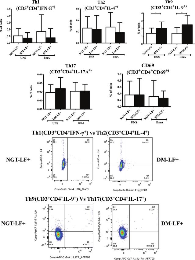
Figure 3 shows the TLR-induced expression of immunomodulatory
signaling, and for adaptive immunity, we studied filarial antigen-
effectors (Cox-2, HO-1, Phox47, and IDO), in the study groups.
induced responses. Under TLR signaling, we studied (1) secretion
TLR-induced expression of Cox-2 and IDO was significantly
of cytokines (IFN-b, TNF-a, IL-6, IL-1b, GM-CSF, IL-10, TGF-b,
augmented in the diabetic group (DM-LF+).
IL-1Ra, IL-35, IL-27, IL-1Ra, IL-12, IFN-g, IL-2, IL-33, IL-4, IL-9,
IL-23, and IL-17) and chemokines (RANTES, MCP-1, IL-8, IP-10,
Effect of Diabetes on the Antigen-Induced and SDF-1); (2) upregulation of MHC (HLA-DR) and co-
Secretion of Pro- and Anti-Inflammatory stimulatory molecules (B7-1/CD80 and B7-2/CD86); and
Cytokines, Type 1 Interferons, and (3) effector functions, which include secretion of defensins (a-
Defensins in LF+ Subjects Defensin-1) and upregulation of immunomodulatory enzymes
Figure 4 shows the antigen-induced secretion of pro- (TNF-a, (Cox-1, HO-1, IDO-1, and p47Phox). Under antigen-induced
IL-6, IL-1b, and GM-CSF) and anti- (IL-10, TGF-b, IL-1Ra, IL- responses, we studied (1) secretion of cytokines (IFN-b, TNF-a,
35, and IL-27) inflammatory cytokines, and type 1 interferon IL-6, IL-1b, GM-CSF, IL-10, TGF-b, IL-1Ra, IL-35, IL-27, IL-1Ra,
(IFN-b) and defensin (a-defensin-1) cytokines in the study IL-12, IFN-g, IL-2, IL-33, IL-4, IL-9, IL-23, and IL-17) and
groups. BmA stimulation resulted in the secretion of TNF-a, chemokines (RANTES, MCP-1, IL-8, IP-10, and SDF-1); (2) Th
IL-6, IL-1b, IL-10, and IL-27. No significant difference was seen immunophenotype (Th1, Th2, Th9, and Th17); (3) T-cell
in the secretion of these cytokines between the groups. Thus, activation (upregulation of CD69); and (4) expression of master
with respect to pro- and anti-inflammatory cytokines, antigen- controllers of Th polarization (T-bet, GATA3, and RORgt). Our
induced secretion of these cytokines remains largely unaffected major findings were as follows: (1) Even in the absence of TLR- or
by diabetes. antigen-induced activation, the basal level secretion of few
cytokines was significantly altered in the DM-LF+ compared to
Effect of Diabetes on the Antigen-Induced the NGT-LF+ group. (2) TLR2 stimulation induced the secretion
Secretion of Adaptive Immune Cytokines of TNF-a, IL-6, IL-1b, IL-10, IL-12, and IL-8; TLR4 stimulation
and Chemokines in LF+ Subjects induced the secretion of TNF-a, IL-6, IL-1b, IL-10, IL-27, IL-12,
Figure 5 shows the antigen-induced secretion of adaptive IL-23, IL-8, and IP-10; both TLR2 and 4 induced the secretion of
immune cytokines (IL-12, IFN-g, IL-2, IL-23, IL-17, IL-9, IL- a-defensin-1 and MCP-1 only in the DM-LF+ group. A significant
33, and IL-4) and chemokines (IL-8, IP-10, RANTES, MCP-1, difference was seen in the TLR2-induced secretion of IL-10,
Frontiers in Immunology | www.frontiersin.org 5 September 2021 | Volume 12 | Article 716515Sibi et al. Diabetes and Filariasis Co-Morbidity
A B C
D E F
G H I
J K L
M
FIGURE 2 | Effect of diabetes on the TLR-induced secretion of adaptive immune cytokines and chemokines in LF+ subjects. Bar graph showing Unstimulated
(UNS), TLR2-, and TLR4-induced secretion of IL-12 (A), IFN-g (B), IL-2 (C), IL-23 (D), IL-17 (E), IL-9 (F), IL-33 (G), IL-4 (H), IL-8 (I), IP-10 (J), RANTES (K), MCP-1
(L), and SDF-1 (M) in the supernatants of blood cultures in NGT-LF+ and DM-LF+ subjects. Statistical significance was determined by non-parametric Mann–
Whitney U test and p < 0.05 was considered significant. *p < 0.05; **p < 0.01; ***p < 0.001.
Frontiers in Immunology | www.frontiersin.org 6 September 2021 | Volume 12 | Article 716515Sibi et al. Diabetes and Filariasis Co-Morbidity
A B
C D
FIGURE 3 | Effect of diabetes on the TLR-induced expression of immunomodulatory effectors in LF+ subjects. Bar graph showing TLR2- and TLR4-induced
expression of Cyclooxygenase (Cox)-2 (A), p47Phox (B), Heme Oxygenase (HO)-1 (C), and Indoleamine 2,3-dioxygenase (IDO) (D) in the immune cells in NGT-LF+
and DM-LF+ subjects. Statistical significance was determined by non-parametric Mann–Whitney U test and p < 0.05 was considered significant. *p < 0.05. NGT,
normal glucose tolerance; DM, diabetes mellitus.
IL-1Ra, IL-17, and SDF-1 and TLR4-induced secretion of IL-9, IL- we studied TLR-induced secretion of (1) type 1 interferon (IFN-b),
33, MCP-1, and SDF-1, between the NGT-LF+ and DM-LF+ (2) pro-inflammatory cytokines (TNF-a, IL-6, IL-1b, and GM-
groups. (3) TLR-induced expression of Cox-2 and IDO was CSF), (3) anti-inflammatory cytokines (IL-10, TGF-b, IL-1Ra, IL-
significantly augmented by diabetes. (4) BmA stimulation 35, IL-27, and IL-1Ra), (4) adaptive immune cytokines (Th1
induced the secretion of TNF-a, IL-6, IL-1b, IL-10, IL-27, IL-8, cytokines—IL-12, IFN-g, and IL-2; Th2 cytokines—IL-33 and
and SDF-1. In the presence of diabetes, the basal secretion of IL- IL-4; Th9 cytokines—IL-9; Th17 cytokines—IL-23 and IL-17),
1b, IL-27, IL-33, and SDF-1 is augmented while that of IL-10, IL- and (5) chemokines (RANTES, MCP-1, IL-8, IP-10, and SDF-1).
12, and IL-8 was downregulated. The augmented secretion of IL-9 Out of all the cytokines, chemokines, and defensins, diabetes
in the diabetic group (DM-LF+) was associated with increased specifically altered (1) TLR2-induced secretion of IL-10, IL-1Ra,
Th9 polarization. While we expected downregulation of many of IL-17, and SDF-1; and (2) TLR4-induced secretion of IL-12, IL-9,
these responses, most of these effectors were upregulated under IL-33, MCP-1, and SDF-1. The expression of MHC (HLA-DR)
diabetes conditions. and co-stimulatory (B7-1 and B7-2) molecules on monocytes, B
The basal level secretion of IL-12 and IL-8 was decreased while cells, and granulocytes was largely unaffected. Finally, TLR2- and
that of IL-1b, IL-27, IL-17, IL-9, IL-33, and SDF-1 was increased in TLR4-induced expression of immunomodulatory effectors Cox-2
the DM-LF+ compared to the NGT-LF+ group. This could be due and IDO was significantly augmented by diabetes. Previously, it
to epigenetic modifications induced due to chronic hyperglycemia, was shown that baseline expression of TLRs was significantly
onto the immune cells (31). It is well known that chronic lower in B cells (but not in monocytes) of the LF+ subjects,
hyperglycemia, as seen in diabetes, can induce epigenetic compared to uninfected individuals (35). TLR stimulation of B
modifications onto immune cells, which can permanently alter cells from these subjects showed diminished activation and
their function, which is referred to as metabolic memory (32). a n t i b o d y s e c r e t io n , in d i c a t i n g a s t a t e o f i m m u n e
TLRs serve as a first line of defense mechanism against pathogen unresponsiveness (35). Filarial parasite was shown to actively
invasion, linking innate and adaptive arms of the immune inhibit the expression and function of TLRs in human dendritic
response (33). TLR2 polymorphisms were shown to be cells (36). In contrast, diabetes is largely a chronic inflammatory
associated with bancroftian filariasis (34). In the present study, state that augments TLR expression and increases the activation in
Frontiers in Immunology | www.frontiersin.org 7 September 2021 | Volume 12 | Article 716515Sibi et al. Diabetes and Filariasis Co-Morbidity
A B C
D E F
G H I
J K
FIGURE 4 | Effect of diabetes on the antigen-induced secretion of pro- and anti-inflammatory cytokines, type 1 interferons, and defensins in LF+ subjects. Bar
graph showing Unstimulated (UNS) and filarial antigen (BmA)-induced secretion of TNF-a (A), IL-6 (B), IL-1b (C), GM-CSF (D), IL-10 (E), IL-1Ra (F), TGF-b (G),
IL-35 (H), IL-27 (I), IFN-b (J), and a-defensin (K) in the supernatants of blood cultures in NGT-LF+ and DM-LF+ subjects. Statistical significance was determined by
non-parametric Mann–Whitney U test and p < 0.05 was considered significant. *p < 0.05; **p < 0.01; ***p < 0.001.
Frontiers in Immunology | www.frontiersin.org 8 September 2021 | Volume 12 | Article 716515Sibi et al. Diabetes and Filariasis Co-Morbidity
A B C
D E F
G H I
J K L
M
FIGURE 5 | Effect of diabetes on the antigen-induced secretion of adaptive immune cytokines, and chemokines in LF+ subjects. Bar graph showing Unstimulated
(UNS) and filarial antigen (BmA)-induced secretion of IL-12 (A), IFN-g (B), IL-2 (C), IL-23 (D), IL-17 (E), IL-9 (F), IL-33 (G), IL-4 (H), IL-8 (I), IP-10 (J), RANTES (K),
MCP-1 (L), and SDF-1 (M) in the supernatants of blood cultures in NGT-LF+ and DM-LF+ subjects. Statistical significance was determined by non-parametric
Mann–Whitney U test and p < 0.05 was considered significant. *p < 0.05; **p < 0.01; ***p < 0.001.
Frontiers in Immunology | www.frontiersin.org 9 September 2021 | Volume 12 | Article 716515Sibi et al. Diabetes and Filariasis Co-Morbidity
A B C
D E
FIGURE 6 | Effect of diabetes on T-cell activation and Th polarization in LF+ subjects. Bar graph showing Unstimulated (UNS) and filarial antigen (BmA)-induced Th1
(A), Th2 (B), Th9 (C), Th17 (D), and activated T cells (E) in NGT-LF+ and DM-LF+ subjects. Statistical significance was determined by non-parametric Mann–
Whitney U test and p < 0.05 was considered significant. *p < 0.05.
various cell types (37). Recently, fetuin-A was found to act as an all filarial antigens have an anti-inflammatory effect (14, 15). Thus,
endogenous ligand for TLR4 to promote free fatty acid-induced under conditions of diabetes–filariasis comorbidity, one can
insulin resistance, in the adipose tissue (38). Several filarial expect either augmentation or inhibition of TLR responses,
antigens are known to directly bind to TLR4 and down- depending on the expression pattern of the filarial antigens,
modulate their effect (39, 40). However, few filarial antigens under the given set of conditions. In the present study, at least
were also found to augment TLR-induced inflammation, some of the TLR-induced responses were found to be augmented
especially in macrophages and dendritic cells, indicating that not under conditions of diabetes–filariasis comorbidity (22).
Frontiers in Immunology | www.frontiersin.org 10 September 2021 | Volume 12 | Article 716515Sibi et al. Diabetes and Filariasis Co-Morbidity
Interestingly, the TLR downregulation gets reversed when filarial characterized and were found to be involved in allergies,
patients develop chronic pathology, with augmented secretion of autoimmunity, and tumor evasion (47, 48). Their role in both
pro-inflammatory cytokines (41). They also show enhanced TLR- diabetes and filariasis is largely unknown. Recently, expansion of
induced secretion of the pro-angiogenic factor, which promotes filarial antigen-specific Th9 cells in whole blood culture was found
neo lymphangiogenesis leading to lymphedema and elephantiasis, to be associated with chronic pathology (49). In the present study,
a hallmark feature of lymphatic filariasis (42). As per the above diabetes augmented IL-9 secretion and increased Th9 population.
model, diabetes can at least partially augment chronic pathology in Like IL-9, the basal level of SDF-1a was found to be significantly
LF+ subjects by enhancing TLR-mediated immune responses. high in LF+ subjects with diabetes. Previously, SDF-1/CXCR4 was
Compared to TLR-induced cytokine/chemokine secretion, the found to promote chronic pathology in LF+ subjects (50). Thus,
expression of immunomodulatory effectors like Cox-2, HO-1, and diabetes, by augmenting IL-9 and SDF-1, can promote chronic
IDO-1 is poorly studied in filariasis and diabetes. These effectors pathology in LF+ subjects.
produce second messengers like prostaglandin H, CO, and In conclusion, in the present report, the effect of diabetes on
kynurenine, respectively, which have an immunomodulatory TLR pathway and antigen-induced immune response in LF+
effect. In the present study, diabetes specifically augmented TLR- subjects was studied. Several parameters including cytokines and
induced expression of Cox-2 and IDO in LF+ subjects. While Cox-2 chemokines, antigen processing and presentation molecules, T-
expression induces a pro-inflammatory milieu, IDO expression cell activation marker, Th polarization, expression of master
induces an anti-inflammatory environment, as a counter regulators of Th polarization, and immunomodulatory effectors
mechanism (43). At least in a tumor microenvironment, this were studied. The most interesting and unexpected finding of
positive feedback loop augments inflammation and inhibits T-cell this study is that, while we expected downregulation of TLR and
response, augmenting tumor growth and immune evasion, antigenic responses, diabetes had a modest but significant
respectively (44). Whether the same strategy has been hijacked by augmenting effect on specific TLR-mediated inflammatory
filariasis and is augmented by diabetes, aiding pathology, is an markers. Most of the inflammatory markers that were found to
interesting possibility that needs further exploration. be augmented by diabetes have previously been shown to
Next to TLRs, T cells play a vital role in orchestrating the promote chronic pathology in LF+ subjects (51, 52). The major
immune response against filariasis. Antigen stimulation induced limitations of this study are the limited sample size and the cross-
the secretion of pro-inflammatory cytokines (TNF-a, IL-6, and sectional study design, which makes it impossible to decipher the
IL-1b), anti-inflammatory cytokines (IL-10 and IL-27), and cause–effect phenomenon. However, the major strength of the
chemokines (IL-8 and SDF-1). Out of all the cytokines, study is that it was conducted in a filarial endemic population
chemokines, and defensins secreted, diabetes specifically wherein the prevalence of diabetes is rapidly increasing (53).
altered antigen-induced secretion of IL-12, IL-9, and SDF-1. T- Overall, diabetes was found to augment certain inflammatory
cell-mediated immune responses during filariasis depend on the components that can promote chronic pathology in LF+
phase of the infection: (1) the acute phase is characterized by subjects, and this needs further validation in a large cohort study.
skewed Th2 response (with enhanced secretion of IL-4, IL-5, and
IL-13); (2) the chronic phase is associated with “modified Th2
response” with Tregs (with enhanced IL-10 and TGF-b
secretion) playing a major role compared to Th2 cells; and
DATA AVAILABILITY STATEMENT
(3) the chronic pathology phase is characterized by a shift The raw data supporting the conclusions of this article will be
towards “Th1/Th17” (enhanced secretion of IFN-g, IL-2, and made available by the authors, without undue reservation.
IL-17) response, which takes place in individuals who develop
pathology (45). Stimulation of T cells from LF+ subjects with B.
malayi antigen showed impaired Th1 response with decreased ETHICS STATEMENT
expression of T-bet, Suppressor of cytokine signaling-1 (SOCS-
1), SOCS-5, and SOCS-7 (27). Furthermore, in vitro culture of The studies involving human participants were reviewed and
PBMCs from LF+ subjects with live larval stage 3 (L3) and approved by the MDRF Institutional Ethical Committee. The
microfilarial (mf) worms showed impaired expression of T-bet patients/participants provided their written informed consent to
and GATA-3 and augmented expression of FoxP3 (46). In participate in this study.
accordance with these reports, no significant Th1 or Th17
polarization was seen in LF+ subjects. While TLR-induced IL-
12 and basal IL-33 secretion was augmented under diabetes AUTHOR CONTRIBUTIONS
condition, this was not seen at the level of Th1 and Th2
polarization. This indicates that, at least under in vivo VM, SB, and VA conceived and designed the experiment. JS and
condition, apart from IL-12 and IL-33, other factors might SM performed the experiment. JS and VA analyzed the data. JS
play a significant role in Th1/Th2 polarization. and VA drafted the manuscript. All the authors contributed to
Compared to TLR stimulation, antigen stimulation induced the the discussion and reviewed the manuscript. VA is the guarantor
secretion of only pro-inflammatory cytokines and chemokines. of the data. All authors contributed to the article and approved
Compared to Th1, Th2, and Th17 cell types, Th9 cells were recently the submitted version.
Frontiers in Immunology | www.frontiersin.org 11 September 2021 | Volume 12 | Article 716515Sibi et al. Diabetes and Filariasis Co-Morbidity
showing monocyte, lymphocyte and granulocyte gates (A). FSe-A vs FSe-H plot
FUNDING showing gated single cells oflymphocytes (B), monocytes (C), Granulocytes (D).
CD19 vs FSC-H plot showing the gated CD19+ B cells (E). CD14 vs FSC-H plot
The Department of Genetics, University of Madras, has received
showing the gated CD14+ Monocytes (F). Expression of HLA DR on lymphocytes
funds for infrastructural support from DST-FIST and UGC-SAP (G), monocytes (H), Granulocytes (I). Expression of B7-1 (CD80) on lymphocytes
programs. The funders had no role in study design, data (J), monocytes (K), Granulocytes (L). Expression ofB7-2 (CD86) on lymphocytes
collection and analysis, decision to publish, or preparation of (M), monocytes (N), Granulocytes (O).
the manuscript.
Supplementary Figure 2 | Gating strategy used for analysis of T helper cell
subtype analysis. FSC vs SSC plot showing the gated lymphocyte population (A).
FSC-A vs FSC-H plot showing gated single cells of lymphocytes (B). CD3 vs CD4
plot showing the gated CD3+CD4+ T helper cells (C). IFN-y Vs IL-4 plot showing
SUPPLEMENTARY MATERIAL the gated Thl and Th2 population (E). IL-9 Vs IL-17 plot showing the gated Th 17
and Th9 population (F).
The Supplementary Material for this article can be found online
at: https://www.frontiersin.org/articles/10.3389/fimmu.2021. Supplementary Figure 3 | Effect of diabetes on the TLR induced expression of
716515/full#supplementary-material MHC and costimulatory molecules in LF+ subjects.
Supplementary Figure 1 | Gating strategy used for the analysis ofHLA-DR, B7-l Supplementary Figure 4 | Effect of diabetes on expression of Th master
(eD80), B7-2 (eD86) on Monocytes and Granulocytes and B cells. FSe vs SSe plot regulators and immunomodulatory enzymes in LF+ subjects.
13. Tartey S, Takeuchi O. Pathogen Recognition and Toll-Like Receptor Targeted
REFERENCES Therapeutics in Innate Immune Cells. Int Rev Immunol (2017) 36:57–73. doi:
1. Evans H, Mitre E. Worms as Therapeutic Agents for Allergy and Asthma: 10.1080/08830185.2016.1261318
Understanding Why Benefits in Animal Studies Have Not Translated Into 14. Mukherjee S, Mukherjee S, Maiti TK, Bhattacharya S, Sinha Babu SP. A Novel
Clinical Success. J Allergy Clin Immunol (2015) 135:343–53. doi: 10.1016/ Ligand of Toll-Like Receptor 4 From the Sheath of Wuchereria Bancrofti
j.jaci.2014.07.007 Microfilaria Induces Proinflammatory Response in Macrophages. J Infect Dis
2. Hubner MP, Stocker JT, Mitre E. Inhibition of Type 1 Diabetes in Filaria- (2017) 215:954–65. doi: 10.1093/infdis/jix067
Infected Non-Obese Diabetic Mice is Associated With a T Helper Type 2 Shift 15. Mukherjee S, Karnam A, Das M, Babu SPS, Bayry J. Wuchereria Bancrofti
and Induction of FoxP3+ Regulatory T Cells. Immunology (2009) 127:512–22. Filaria Activates Human Dendritic Cells and Polarizes T Helper 1 and
doi: 10.1111/j.1365-2567.2008.02958.x Regulatory T Cells via Toll-Like Receptor 4. Commun Biol (2019) 2:169.
3. Hubner MP, Shi Y, Torrero MN, Mueller E, Larson D, Soloviova K, et al. doi: 10.1038/s42003-019-0392-8
Helminth Protection Against Autoimmune Diabetes in Nonobese Diabetic 16. Zeytun A, Chaudhary A, Pardington P, Cary R, Gupta G. Induction of
Mice Is Independent of a Type 2 Immune Shift and Requires TGF-Beta. Cytokines and Chemokines by Toll-Like Receptor Signaling: Strategies for
J Immunol (2012) 188:559–68. doi: 10.4049/jimmunol.1100335 Control of Inflammation. Crit Rev Immunol (2010) 30:53–67. doi: 10.1615/
4. Berbudi A, Surendar J, Ajendra J, Gondorf F, Schmidt D, Neumann AL, et al. CritRevImmunol.v30.i1.40
Filarial Infection or Antigen Administration Improves Glucose Tolerance in Diet- 17. Lata S, Raghava GP. CytoPred: A Server for Prediction and Classification
Induced Obese Mice. J Innate Immun (2016) 8:601–16. doi: 10.1159/000448401 of Cytokines. Protein Eng Des Sel (2008) 21:279–82. doi: 10.1093/protein/
5. Aravindhan V, Mohan V, Surendar J, Rao MM, Ranjani H, Kumaraswami V, gzn006
et al. Decreased Prevalence of Lymphatic Filariasis Among Subjects With 18. Guo X, Wu N, Shang Y, Liu X, Wu T, Zhou Y, et al. The Novel Toll-Like
Type-1 Diabetes. Am J Trop Med Hyg (2010) 83:1336–9. doi: 10.4269/ Receptor 2 Agonist SUP3 Enhances Antigen Presentation and T Cell Activation
ajtmh.2010.10-0410 by Dendritic Cells. Front Immunol (2017) 8:158. doi: 10.3389/fimmu.2017.00158
6. Aravindhan V, Mohan V, Surendar J, Muralidhara Rao M, Pavankumar N, Deepa 19. Buchta CM, Bishop GA. Toll-Like Receptors and B Cells: Functions and
M, et al. Decreased Prevalence of Lymphatic Filariasis Among Diabetic Subjects Mechanisms. Immunol Res (2014) 59:12–22. doi: 10.1007/s12026-014-8523-2
Associated With a Diminished Pro-Inflammatory Cytokine Response (CURES 20. Sabroe I, Dower SK, Whyte MK. The Role of Toll-Like Receptors in the
83). PloS Negl Trop Dis (2010) 4:e707. doi: 10.1371/journal.pntd.0000707 Regulation of Neutrophil Migration, Activation, and Apoptosis. Clin Infect
7. Aravindhan V, Mohan V, Surendar J, Rao MM, Anuradha R, Deepa M, et al. Dis (2005) 41(Suppl 7):S421–6. doi: 10.1086/431992
Effect of Filarial Infection on Serum Inflammatory and Atherogenic 21. McCoy CE, O’Neill LA. The Role of Toll-Like Receptors in Macrophages.
Biomarkers in Coronary Artery Disease (CURES-121). Am J Trop Med Hyg Front Biosci (2008) 13:62–70. doi: 10.2741/2660
(2012) 86:828–33. doi: 10.4269/ajtmh.2012.11-0773 22. Aravindhan V, Anand G. Cell Type-Specific Immunomodulation Induced by
8. Chen Y, Lu J, Huang Y, Wang T, Xu Y, Xu M, et al. Association of Previous Helminthes: Effect on Metainflammation, Insulin Resistance and Type-2
Schistosome Infection With Diabetes and Metabolic Syndrome: A Cross- Diabetes. Am J Trop Med Hyg (2017) 97:1650–61. doi: 10.4269/ajtmh.17-0236
Sectional Study in Rural China. J Clin Endocrinol Metab (2013) 98:E283–7. 23. Milam AV, Allen PM. Functional Heterogeneity in CD4(+) T Cell Responses
doi: 10.1210/jc.2012-2517 Against a Bacterial Pathogen. Front Immunol (2015) 6:621. doi: 10.3389/
9. Wiria AE, Hamid F, Wammes LJ, Prasetyani MA, Dekkers OM, May L, et al. fimmu.2015.00621
Infection With Soil-Transmitted Helminths Is Associated With Increased Insulin 24. Taniuchi I. CD4 Helper and CD8 Cytotoxic T Cell Differentiation. Annu Rev
Sensitivity. PloS One (2015) 10:e0127746. doi: 10.1371/journal.pone.0127746 Immunol (2018) 36:579–601. doi: 10.1146/annurev-immunol-042617-053411
10. Raizada N, Feingold KR, Anawalt B, Boyce A, Chrousos G, de Herder WW, 25. Beadling C, Slifka MK. Regulation of Innate and Adaptive Immune Responses
et al. Endotext (Internet). N Raizada, KR Feingold, B Anawalt, A Boyce, G by the Related Cytokines IL-12, IL-23, and IL-27. Arch Immunol Ther Exp
Chrousos, WW de Herder, K Dhatariya, et al. eds. South Dartmouth, MA: (Warsz) (2006) 54:15–24. doi: 10.1007/s00005-006-0002-6
Endotext (2000). 26. Wang W, Sung N, Gilman-Sachs A, Kwak-Kim J. T Helper (Th) Cell Profiles
11. Unnikrishnan R, Misra A. Infections and Diabetes: Risks and Mitigation With in Pregnancy and Recurrent Pregnancy Losses: Th1/Th2/Th9/Th17/Th22/Tfh
Reference to India. Diabetes Metab Syndr (2020) 14:1889–94. doi: 10.1016/ Cells. Front Immunol (2020) 11:2025. doi: 10.3389/fimmu.2020.02025
j.dsx.2020.09.022 27. Babu S, Kumaraswami V, Nutman TB. Transcriptional Control of Impaired
12. Mukherjee S, Huda S, Sinha Babu SP. Toll-Like Receptor Polymorphism in Th1 Responses in Patent Lymphatic Filariasis by T-Box Expressed in T Cells
Host Immune Response to Infectious Diseases: A Review. Scand J Immunol and Suppressor of Cytokine Signaling Genes. Infect Immun (2005) 73:3394–
(2019) 90:e12771. doi: 10.1111/sji.12771 401. doi: 10.1128/IAI.73.6.3394-3401.2005
Frontiers in Immunology | www.frontiersin.org 12 September 2021 | Volume 12 | Article 716515Sibi et al. Diabetes and Filariasis Co-Morbidity
28. Babu S, Bhat SQ, Pavan Kumar N, Lipira AB, Kumar S, Karthik C, et al. Inhibitors Through Regulatory T Cells. J Immunother (2009) 32:22–8. doi:
Filarial Lymphedema is Characterized by Antigen-Specific Th1 and Th17 10.1097/CJI.0b013e31818ac2f7
Proinflammatory Responses and a Lack of Regulatory T Cells. PloS Negl Trop 44. Chen JY, Li CF, Kuo CC, Tsai KK, Hou MF, Hung WC. Cancer/stroma
Dis (2009) 3:e420. doi: 10.1371/journal.pntd.0000420 Interplay via Cyclooxygenase-2 and Indoleamine 2,3-Dioxygenase Promotes
29. Madhumitha H, Mohan V, Babu S, Aravindhan V. TLR-Induced Secretion of Breast Cancer Progression. Breast Cancer Res (2014) 16:410. doi: 10.1186/
Novel Cytokine IL-27 Is Defective in Newly Diagnosed Type-2 Diabetic s13058-014-0410-1
Subjects. Cytokine (2018) 104:65–71. doi: 10.1016/j.cyto.2017.09.032 45. Bancroft AJ, Grencis RK, Else KJ, Devaney E. The Role of CD4 Cells in
30. Kwarteng A, Ahuno ST. Immunity in Filarial Infections: Lessons From Protective Immunity to Brugia Pahangi. Parasite Immunol (1994) 16:385–7.
Animal Models and Human Studies. Scand J Immunol (2017) 85:251–7. doi: 10.1111/j.1365-3024.1994.tb00364.x
doi: 10.1111/sji.12533 46. Babu S, Blauvelt CP, Kumaraswami V, Nutman TB. Regulatory Networks
31. Villeneuve LM, Reddy MA, Lanting LL, Wang M, Meng L, Natarajan R. Induced by Live Parasites Impair Both Th1 and Th2 Pathways in Patent
Epigenetic Histone H3 Lysine 9 Methylation in Metabolic Memory and Lymphatic Filariasis: Implications for Parasite Persistence. J Immunol (2006)
Inflammatory Phenotype of Vascular Smooth Muscle Cells in Diabetes. 176:3248–56. doi: 10.4049/jimmunol.176.5.3248
Proc Natl Acad Sci USA (2008) 105:9047–52. doi: 10.1073/pnas.0803623105 47. He Y, Dong L, Cao Y, Bi Y, Liu G. IL-9 and Th9 Cells in Tumor Immunity.
32. Mossel DM, Moganti K, Riabov V, Weiss C, Kopf S, Cordero J, et al. Adv Exp Med Biol (2020) 1240:35–46. doi: 10.1007/978-3-030-38315-2_3
Epigenetic Regulation of S100A9 and S100A12 Expression in Monocyte- 48. Badolati I, Sverremark-Ekstrom E, van der Heiden M. Th9 Cells in Allergic
Macrophage System in Hyperglycemic Conditions. Front Immunol (2020) Diseases: A Role for the Microbiota? Scand J Immunol (2020) 91:e12857. doi:
11:1071. doi: 10.3389/fimmu.2020.01071 10.1111/sji.12857
33. Venugopal PG, Nutman TB, Semnani RT. Activation and Regulation of Toll- 49. Anuradha R, George PJ, Hanna LE, Chandrasekaran V, Kumaran P, Nutman
Like Receptors (TLRs) by Helminth Parasites. Immunol Res (2009) 43:252–63. TB, et al. IL-4-, TGF-Beta-, and IL-1-Dependent Expansion of Parasite
doi: 10.1007/s12026-008-8079-0 Antigen-Specific Th9 Cells Is Associated With Clinical Pathology in
34. Junpee A, Tencomnao T, Sanprasert V, Nuchprayoon S. Association Between Human Lymphatic Filariasis. J Immunol (2013) 191:2466–73. doi: 10.4049/
Toll-Like Receptor 2 (TLR2) Polymorphisms and Asymptomatic Bancroftian jimmunol.1300911
Filariasis. Parasitol Res (2010) 107:807–16. doi: 10.1007/s00436-010-1932-9 50. Nathan AA, Dixit M, Babu S, Balakrishnan AS. Comparison and Functional
35. Babu S, Blauvelt CP, Kumaraswami V, Nutman TB. Diminished Expression and Characterisation of Peripheral Blood Mononuclear Cells Isolated From
Function of TLR in Lymphatic Filariasis: A Novel Mechanism of Immune Filarial Lymphoedema and Endemic Normals of a South Indian Population.
Dysregulation. J Immunol (2005) 175:1170–6. doi: 10.4049/jimmunol.175.2.1170 Trop Med Int Health (2017) 22:1414–27. doi: 10.1111/tmi.12969
36. Semnani RT, Venugopal PG, Leifer CA, Mostbock S, Sabzevari H, Nutman 51. Satapathy AK, Sartono E, Sahoo PK, Dentener MA, Michael E, Yazdanbakhsh M,
TB. Inhibition of TLR3 and TLR4 Function and Expression in Human et al. Human Bancroftian Filariasis: Immunological Markers of Morbidity and
Dendritic Cells by Helminth Parasites. Blood (2008) 112:1290–8. doi: Infection. Microbes Infect (2006) 8:2414–23. doi: 10.1016/j.micinf.2006.05.003
10.1182/blood-2008-04-149856 52. Anuradha R, George PJ, Pavan Kumar N, Fay MP, Kumaraswami V, Nutman
37. Aravindhan V, Madhumitha H. Metainflammation in Diabetic Coronary TB, et al. Circulating Microbial Products and Acute Phase Proteins as Markers
Artery Disease: Emerging Role of Innate and Adaptive Immune Responses. of Pathogenesis in Lymphatic Filarial Disease. PloS Pathog (2012) 8:e1002749.
J Diabetes Res (2016) 2016:6264149. doi: 10.1155/2016/6264149 doi: 10.1371/journal.ppat.1002749
38. Pal D, Dasgupta S, Kundu R, Maitra S, Das G, Mukhopadhyay S, et al. Fetuin- 53. Anjana RM, Deepa M, Pradeepa R, Mahanta J, Narain K, Das HK, et al.
A Acts as an Endogenous Ligand of TLR4 to Promote Lipid-Induced Insulin Prevalence of Diabetes and Prediabetes in 15 States of India: Results From the
Resistance. Nat Med (2012) 18:1279–85. doi: 10.1038/nm.2851 ICMR-INDIAB Population-Based Cross-Sectional Study. Lancet Diabetes
39. Panda SK, Kumar S, Tupperwar NC, Vaidya T, George A, Rath S, et al. Endocrinol (2017) 5:585–96. doi: 10.1016/S2213-8587(17)30174-2
Chitohexaose Activates Macrophages by Alternate Pathway Through TLR4
and Blocks Endotoxemia. PloS Pathog (2012) 8:e1002717. doi: 10.1371/
journal.ppat.1002717 Conflict of Interest: The authors declare that the research was conducted in the
40. Goodridge HS, Marshall FA, Else KJ, Houston KM, Egan C, Al-Riyami L, et al. absence of any commercial or financial relationships that could be construed as a
Immunomodulation via Novel Use of TLR4 by the Filarial Nematode potential conflict of interest.
Phosphorylcholine-Containing Secreted Product, ES-62. J Immunol (2005)
174:284–93. doi: 10.4049/jimmunol.174.1.284 Publisher’s Note: All claims expressed in this article are solely those of the authors
41. Babu S, Anuradha R, Kumar NP, George PJ, Kumaraswami V, Nutman TB. and do not necessarily represent those of their affiliated organizations, or those of
Filarial Lymphatic Pathology Reflects Augmented Toll-Like Receptor- the publisher, the editors and the reviewers. Any product that may be evaluated in
Mediated, Mitogen-Activated Protein Kinase-Mediated Proinflammatory this article, or claim that may be made by its manufacturer, is not guaranteed or
Cytokine Production. Infect Immun (2011) 79:4600–8. doi: 10.1128/ endorsed by the publisher.
IAI.05419-11
42. Babu S, Anuradha R, Kumar NP, George PJ, Kumaraswami V, Nutman TB. Copyright © 2021 Sibi, Mohan, Munisankar, Babu and Aravindhan. This is an open-
Toll-Like Receptor- and Filarial Antigen-Mediated, Mitogen-Activated access article distributed under the terms of the Creative Commons Attribution
Protein Kinase- and NF-kappaB-Dependent Regulation of Angiogenic License (CC BY). The use, distribution or reproduction in other forums is permitted,
Growth Factors in Filarial Lymphatic Pathology. Infect Immun (2012) provided the original author(s) and the copyright owner(s) are credited and that the
80:2509–18. doi: 10.1128/IAI.06179-11 original publication in this journal is cited, in accordance with accepted academic
43. Lee SY, Choi HK, Lee KJ, Jung JY, Hur GY, Jung KH, et al. The Immune practice. No use, distribution or reproduction is permitted which does not comply with
Tolerance of Cancer Is Mediated by IDO That is Inhibited by COX-2 these terms.
Frontiers in Immunology | www.frontiersin.org 13 September 2021 | Volume 12 | Article 716515You can also read