Chemical Science - Royal ...
←
→
Page content transcription
If your browser does not render page correctly, please read the page content below
Volume 10 Number 13 7 April 2019 Pages 3691–3894

Chemical
Science
rsc.li/chemical-science

                          ISSN 2041-6539

                           EDGE ARTICLE
                           Wei Wei, Songqin Liu et al.
                           Telomerase and poly(ADP-ribose) polymerase-1 activity
                           sensing based on the high fluorescence selectivity and
                           sensitivity of TOTO-1 towards G bases in single-stranded
                           DNA and poly(ADP-ribose)
Chemical
Science
This article is licensed under a Creative Commons Attribution-NonCommercial 3.0 Unported Licence.
View Article Online
EDGE ARTICLE View Journal | View Issue
Telomerase and poly(ADP-ribose) polymerase-1
Cite this: Chem. Sci., 2019, 10, 3706
activity sensing based on the high fluorescence
All publication charges for this article
selectivity and sensitivity of TOTO-1 towards G
bases in single-stranded DNA and poly(ADP-
Open Access Article. Published on 19 2019. Downloaded on 13.01.2021 16:08:20.
have been paid for by the Royal Society
of Chemistry
ribose)†
Haitang Yang,a Fangjia Fu,b Wei Li, b
Wei Wei, *a Yuanjian Zhang a
and Songqin Liu *a
Telomerase and poly(ADP-ribose) polymerase-1 (PARP-1) are two potential cancer biomarkers and are
closely related to tumor initiation and malignant progression. TOTO-1 is well-known for differentiating
ss-DNA from ds-DNA because it is virtually non-fluorescent without DNA and exhibits very low
fluorescence with ss-DNA, while it emits strong fluorescence with ds-DNA. In this paper, for the first
time, it was found that TOTO-1 has high fluorescence selectivity and sensitivity towards the G bases in
single-stranded DNA and poly(ADP-ribose) (PAR). Poly(dG) was used as the model target to explore its
possible mechanism. Molecular dynamics (MD) simulation proved that intramolecular p–p stacking
existed in TOTO-1 (in an aqueous solution), while intermolecular p–p stacking formed between TOTO-1
and poly(dG) in a similar way as that observed for dsDNA. Interestingly, telomerase and PARP-1 catalyzed
the formation of G-rich DNA and PAR in vivo, respectively. Therefore, TOTO-1 was explored in detecting
both of them, obtaining satisfactory results. To the best of our knowledge, no probe has been reported
to recognize PAR. It is also the first time where telomerase is detected based on the specific recognition
Received 26th December 2018
Accepted 18th February 2019
of G bases. Importantly, integrating multiple functions into one probe that can detect not only
telomerase but also PARP-1 will significantly raise the specificity of screening cancer and decrease false
DOI: 10.1039/c8sc05770b
positive proportion, which make TOTO-1 a promising candidate probe for clinical diagnosis and
rsc.li/chemical-science pharmaceutical screening.
(TTAGGG)n-30 . First, the extended primer is a powerful
Introduction displacement strand that can be used to initiate the signal
Telomerase and poly(ADP-ribose) polymerase-1 (PARP-1) are “turn-on” or “turn-off” based on the hybridization of DNA.3–5
two potential cancer biomarkers and are closely related to Second, the extended G-rich primer forms a G-quadruplex,
tumor initiation and malignant progression.1,2 Integrating which has high peroxidase-like activity and can initiate
multiple functions into one probe that can detect not only signals closely related to the catalyzed reaction.6–9 Third, the
telomerase but also PARP-1 will signicantly raise the specicity extended DNA sequence has ample negative charges that have
of screening cancer and decrease false positive proportion. To great inuence on the signal depending on the electrostatic
the best of our knowledge, few probes have been reported to interactions.10–12 Only few telomerase detection methods have
detect not only telomerase but also PARP-1. been reported based on the specic recognition of bases.
Most telomerase detection methods depend on the unique Most detection methods for PARP-1 are based on its cata-
property of its extended primer, 50 -AAT CCG TCG AGC AGA GTT lyzed production of PAR that has a large number of negative
charges, which can adsorb positively charged probes by elec-
a
Jiangsu Engineering Laboratory of Smart Carbon-Rich Materials and Device, Jiangsu
trostatic attraction.13–15 These methods do not have good
Province Hi-Tech Key Laboratory for Bio-medical Research, School of Chemistry and selectivity because of the high background signals. Because PAR
Chemical Engineering, Southeast University, Nanjing, 211189, China. E-mail: lacks unique properties as the extended telomerase primer, very
weiw@seu.edu.cn; liusq@seu.edu.cn; Fax: +86-25-52090618; Tel: +86-25-52090613 few methods have been developed for its detection even though
b
Institution of Theoretical and Computational Chemistry, School of Chemistry and its activity is very important for assessing cancer
Chemical Engineering, Nanjing University, Nanjing 210023, People's Republic of
development.16,17
China
† Electronic supplementary information (ESI) available. See DOI:
Fluorescence analysis has been widely applied in detecting
10.1039/c8sc05770b biomolecules because of its inherent advantages of being
3706 | Chem. Sci., 2019, 10, 3706–3714 This journal is © The Royal Society of Chemistry 2019
View Article Online
Edge Article Chemical Science
simple, convenient and sensitive. The uorescence properties constructed using AutoDock 4.0 with the Lamarckian genetic
of dyes differ widely when they are in different environments, algorithm.40 The ground-state geometry of free TOTO-1 mole-
and they are strongly dependent on the local environment of the cule was rst optimized at the M06-2X/6-311++G(d,p) level with
macromolecules and their circumstances.18 DNA carries the the polarizable continuum model (PCM), and the RESP charge
This article is licensed under a Creative Commons Attribution-NonCommercial 3.0 Unported Licence.
genetic information of all known living organisms. The ability of the TOTO-1 molecule was calculated using the GAUSSIAN09
to specically discriminate DNA sequences is signicant for program.41 The congurations of TOTO-1 without and with
disease screening and oncology studies.19 Some uorescence poly(dG) at the center with 8000 water molecules were simu-
probes that specically recognize DNA sequences have been lated at 298 K in cubic boxes of 56.61 56.61 56.61 Å3 and
developed.20–22 Fluorescence nanoparticles such as DNA-silver 62.46 62.46 62.46 Å3, respectively. The generalized amber
nanoclusters23–26 and small organic dyes such as thioavin force eld (GAFF) and FF99BSC0 force eld were used for the
T,27–29 ethidium bromide (EtBr),30 thiazole orange (TO),31 TOTO-1 molecule and poly(dG), respectively. All the water
TOTO,32 and iridium complex33 can bind to DNA chains and molecules were described by the TIP4PFB force eld, which was
Open Access Article. Published on 19 2019. Downloaded on 13.01.2021 16:08:20.
show various uorescence properties depending on their found to be able to reproduce the experimental properties of
manner of binding and binding ability. Some of them can liquid water.42 Cl and Na+ counterions were added to
distinguish ss-DNA from ds-DNA or G-quadruplex DNA. neutralize the system of TOTO-1 without and with poly(dG) in
However, only a few uorescence probes are known to effec- aqueous solutions, respectively. The simulations of the two
tively distinguish deoxynucleotides34 even though they are systems were run on the NPT ensemble at 1.0 bar with
signicant for DNA sequence discrimination, bio-analysis and a Berendsen barostat. Periodic boundary conditions (PBC) were
clinical diagnosis. carried out and the long-range electrostatic interactions were
TOTO-1, an unsymmetrical cyanine dye dimer, is well-known taken into account using the particle mesh Ewald (PME)
for differentiating ss-DNA from ds-DNA (Fig. S1†).35,36 It is method. The velocity Verlet algorithm was employed to solve
virtually non-uorescent without DNA and exhibits strong Newton's motion equations with the bonds of hydrogen atoms
uorescence when it binds to ds-DNA (Fig. S2†).37,38 In this constrained. The van der Waals cutoff was set as 10 Å for the two
paper, for the rst time, we found that TOTO-1 has unique systems. For TOTO-1 without and with poly(dG) in aqueous
uorescence selectivity and high sensitivity towards the G bases solutions, two simulation trajectories of 20 ns aer 2 ns of
in single-stranded DNA and PAR. Its uorescence intensity was equilibrium were carried out and snapshots were collected at
much stronger with poly(dG) than those with poly(dA), poly(dC) the interval of 1 ps with every time step of 1 fs.
and poly(dT) (Fig. S2A†). More interestingly, the uorescence of
TOTO-1 with poly(dG) was far stronger than that with dGTP
even when the concentration of dGTP was 4 orders of magni- Hybrid quantum mechanical and molecular mechanical (QM/
tude higher than that of the G bases in poly(dG). Furthermore, MM) calculation
the uorescence of TOTO-1 with PAR increased sharply
The electronic absorption spectra of TOTO-1 without and with
compared to those with higher concentrations of ADP or
poly(dG) in aqueous solutions were calculated using the QM/
poly(dA).
MM method at the level of time-dependent density functional
These ndings urged us to develop a simple and sensitive
theory (TDDFT) with the M06-2X functional and the 6-31G(d)
method to assay the activities of both telomerase and PARP-1;
basis set. TOTO-1 molecule and TOTO-1 with poly(dG) were
they catalyzed the formation of a G-rich elongated TS primer
treated in the QM part for the two systems, and the remaining
and PAR in vivo, respectively, which were highly sensitive to
molecules including the counterions and water molecules were
TOTO-1. The high background signals produced by the telo-
treated as background point charges in the MM region.
merase primer and the activated dsDNA were reduced by 9
times and 38 times, respectively, under the inuence of diges-
tion by the Exo III enzyme. As a result, the detection sensitivity
improved greatly. A linear relationship between the uores- Procedures of telomerase activity and inhibition efficiency
cence intensities and logarithmic concentrations of telomerase evaluation
was observed between 13 and 4000 cells per mL, and the limit of Cell cultivation, telomerase extraction and primer extension
detection (LOD) was determined to be 13 cells per mL. Also, the reaction were performed according to previously described
uorescence intensities showed a linear response between 0.02 methods (ESI†). The extended G-rich sequence was hybridized
U and 1.5 U for PARP-1 with LOD of 0.02 U. This simple and with cDNA (complementary to the TS primer) at 95 C for 5 min,
sensitive approach is promising for clinical diagnosis and is followed by cooling down slowly to room temperature. Then,
a powerful tool for pharmaceutical screening. Exo III was used to digest the duplex part formed by cDNA and
TS primer at 37 C. The resulting solution (30 mL) was diluted to
Experimental section 200 mL with PBS buffer containing 250 nM TOTO-1. Then, the
uorescence spectra were recorded. Various concentrations of
Molecular dynamics (MD) simulation BIBR 1532 or curcumin were incubated with 750 cells per mL
The initial single-stranded poly(dG) structure was constructed A549 cells to evaluate their telomerase inhibition efficiency.
using the NAB module in the AmberTools16 package.39 Then, Heated A549 cells (95 C, 5 min) were used for control
the conguration of TOTO-1 binding to poly(dG) was experiments.
This journal is © The Royal Society of Chemistry 2019 Chem. Sci., 2019, 10, 3706–3714 | 3707
View Article Online
Chemical Science Edge Article
Procedures of PARP-1 activity and inhibition efficiency telomerase. The absorbance and uorescence spectra of TOTO-
evaluation 1 are shown in Fig. 1. From curves (a and b) in Fig. 1A–D, it can
be seen that their absorption spectra vary signicantly in the
First, active dsDNA was prepared by hybridizing two comple-
presence of different oligodeoxynucleotides, indicating that the
mentary sequences ssDNA1 and ssDNA2 in a hybridization
This article is licensed under a Creative Commons Attribution-NonCommercial 3.0 Unported Licence.
molecular orbitals of TOTO-1 were changed by these oligo-
buffer at 95 C for 5 min, followed by cooling to room temper-
deoxynucleotides. The orbital distribution of the excited state
ature slowly. Then, dsDNA (150 nM) was incubated with PARP-1,
transitions for TOTO-1/poly(dG) corresponding to the absor-
NAD+ (250 mM) and reaction buffer (R-buffer) for 1 h at room
bance at 454 nm and 542 nm were simulated (Fig. S4†). The
temperature. Aer PARP-1 catalyzed the polymerization of PAR,
uorescence intensity of TOTO-1 in the presence of different
dsDNA was digested by Exo III at 37 C to reduce the back-
types of oligonucleotides varied greatly (curve c, Fig. 1A–D).
ground signal. The resulting solution was mixed with PBS buffer
Specically, TOTO-1 in the presence of poly(dG) yielded much
containing 250 nM TOTO-1. Fluorescence spectra were recor-
higher signals than those with the three other types of oligo-
ded. For PARP-1 inhibitor assay, the same procedure was
Open Access Article. Published on 19 2019. Downloaded on 13.01.2021 16:08:20.
nucleotides. The quantum yields in the presence of poly(dG),
carried out except for the treatment of PARP-1 with different
poly(dA), poly(dC), and poly(dT) were determined to be 0.25,
concentrations of AG014699.
0.11, 0.05 and 0.02, respectively. These data indicated that
TOTO-1 showed strong uorescence selectivity towards four
Result and discussion types of bases: poly(dG) [ poly(dA) > poly(dC) > poly(dT).
High uorescence selectivity and sensitivity of TOTO-1 Fig. 1E shows the comparison of uorescence intensities of
towards the G bases in single-stranded DNA and PAR TOTO-1 in the presence of different types of oligonucleotides.
The uorescence selectivities of TOTO-1 towards 15 mM dGTP,
dATP, dCTP, dTTP and ADP were detected (Fig. S3†). The results MD simulation of the interaction between TOTO-1 and
showed that TOTO-1 has much stronger uorescence with dGTP poly(dG)
or ADP than that with dATP, dCTP or dTTP. Considering that MD simulation indicated that intramolecular p–p stacking
TOTO-1 was used to respond to G-rich DNA sequences catalyzed existed in free TOTO-1, while intermolecular p–p stacking
by telomerase, poly(dG) was used as the model target in this formed between TOTO-1 and poly(dG) (Fig. 2A). As a result,
study. Compared with the result for 15 mM of dGTP, the uo- a folding structure similar to that of duplex DNA was formed.
rescence intensity of TOTO-1 increased from 1.5 to 25 when only The absorption spectrum calculated by M06-2X/6-31G(D) was
86 nM poly(dG) (690 nM of G bases) was present. These data highly consistent with the experimental spectrum (Fig. 2B),
indicated that TOTO-1 is more sensitive to the G bases existing which demonstrated the intermolecular p–p stacking in
in single-stranded DNA than single dGTP, which is signicant poly(dG)/TOTO-1. Other uorescent dyes such as uorescein
for the detection of extended DNA sequences catalyzed by sodium, thioavin T and acriavine did not have uorescence
Fig. 1 Spectra of TOTO-1 (250 nM) and TOTO-1 in the presence of 86 nM of poly(dG) (A), poly(dA) (B), poly(dC) (C) and poly(dT) (D). Absorption
spectra of free TOTO-1 (a); absorption (b) and fluorescence (c) spectra of TOTO-1 in the presence of oligodeoxynucleotides. (E) Fluorescence
intensity comparison with four types of oligodeoxynucleotides.
3708 | Chem. Sci., 2019, 10, 3706–3714 This journal is © The Royal Society of Chemistry 2019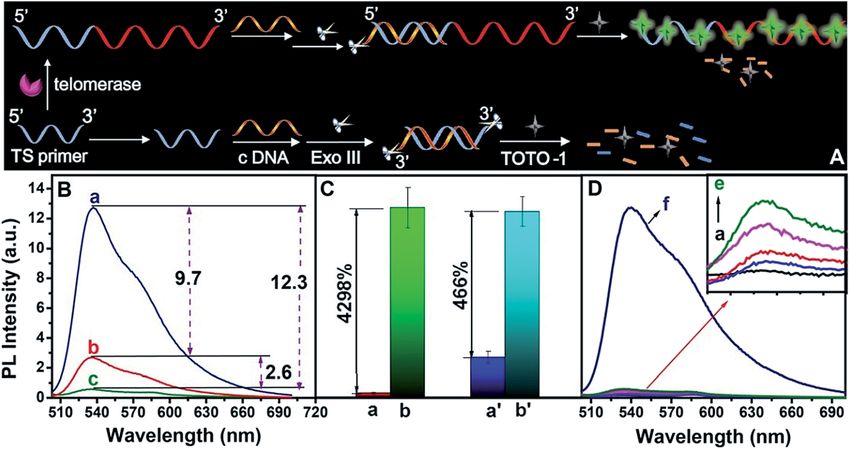
View Article Online
Edge Article Chemical Science
This article is licensed under a Creative Commons Attribution-NonCommercial 3.0 Unported Licence.
Fig. 2(A) Intramolecular p–p stacking of free TOTO-1 and intermolecular p–p stacking of TOTO-1/poly(dG). (B) Simulated and experimental
absorption spectra of TOTO-1 in the presence of poly(dG).
Open Access Article. Published on 19 2019. Downloaded on 13.01.2021 16:08:20.
selectivity towards the bases (Fig. S5†). Although TO showed maximum uorescence emission wavelength changed
selectivity similar to that of TOTO-1, its PL intensity was much slightly, its uorescence intensity increased by 2.5 times. The
lower (30%) than that of TOTO-1 in the presence of poly(dG) and corresponding quantum yield also increased from 0.11 to
poly(dA) (Fig. S5C†). 0.32, which denitely contributed to improving PARP-1
activity detection. Furthermore, compared with the result for
15 mM of ADP, the uorescence intensity of TOTO-1 increased
Fluorescence of TOTO-1 in the presence of G-rich elongated from 0.7 to 7 in the presence of PAR produced by 0.8 U PARP-1.
TS primer and PAR This also indicates that TOTO-1 is more sensitive to poly-
The absorbance and uorescence spectra of TOTO-1 in the merized ADP than single ADP. Thus, it is possible to construct
presence of G-rich elongated TS primer (Fig. 3A) and PAR a sensitive and simple method to detect telomerase and PARP-
(Fig. 3B) were also studied. In the presence of G-rich elongated 1 activities based on the unique uorescence selectivity and
TS primer, the absorbance and uorescence spectra of TOTO-1 sensitivity of TOTO-1 towards the G-rich elongated primer and
changed obviously compared with those in the presence of PAR.
poly(dG) and poly(dA). Compared with the observation for pol-
y(dG), the uorescence intensity for the elongated TS primer
decreased from 25 to 12.6, indicating that thymine and adenine The strategy for telomerase activity evaluation
certainly decreased the uorescence emission of TOTO-1. The The scheme for telomerase activity detection is illustrated in
corresponding quantum yield decreased to 0.16. Under this Fig. 4A. The telomerase primer was incubated with dNTPs and
circumstance, its uorescence was still sensitive enough to telomerase was extracted from tumor A549 cells. The TTAGGG
distinguish the G-rich elongated TS primer, making it possible repeat units were continuously synthesized on the 30 end of the
to detect telomerase activity. TS primer to form a G-rich extended primer. In the presence of
PAR, synthesized by PARP-1, is composed of adenosine telomerase, the G-rich elongated primer led to a sharp increase
diphosphate ribose (ADP-ribose) monomers (Fig. S6†). In the in the uorescence intensity of TOTO-1 (a, Fig. 4B). In the
presence of PAR, the absorbance spectrum of TOTO-1 showed presence of TS primer without telomerase, the background
signicant changes with a new absorbance peak at 425 nm signal was 2.9 (b, Fig. 4B). A primer was designed to form ds-
and increased absorbance values at 480 nm and 508 nm, DNA by incubation with complementary DNA (c-DNA), fol-
indicating that the molecular orbitals of TOTO-1 were also lowed by digesting with Exo III; thus, the background signal
different from those in the presence of poly(dA). Although its decreased to 0.30 (c, Fig. 4B). The signal-to-noise ratio (S/N)
Fig. 3Absorption spectra of free TOTO-1 (a in A and B) and TOTO-1 in the presence of G-rich extended primer (b, A) and PAR (b, B). Fluo-
rescence spectra of TOTO-1 in the presence of G-rich extended primer (c, A) and PAR (c, B).
This journal is © The Royal Society of Chemistry 2019 Chem. Sci., 2019, 10, 3706–3714 | 3709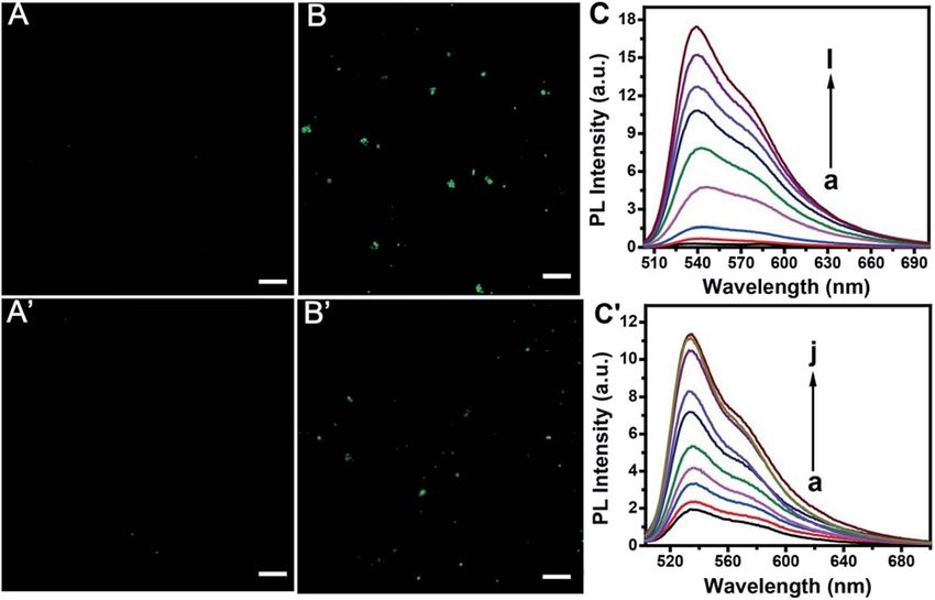
View Article Online
Chemical Science Edge Article
This article is licensed under a Creative Commons Attribution-NonCommercial 3.0 Unported Licence.
Open Access Article. Published on 19 2019. Downloaded on 13.01.2021 16:08:20.
Fig. 4 (A) Scheme of sensing telomerase activity by TOTO-1. (B) PL spectra of TOTO-1/extended-primer/c-DNA/Exo III (a) primer/TOTO-1 (b)
and TOTO-1/primer/c-DNA/Exo III (c). (C) PL intensity of TOTO-1 in the presence of primer (a) and elongated primer (b) digested with Exo III; (a0 )
and (b0 ) correspond to (a) and (b) without Exo III. (D) PL spectra of TOTO-1 (a), TOTO-1/primer/Exo III/c-DNA (b), TOTO-1/primer/Exo III/c-DNA
in the presence of dNTPs (c), A549 cells (d), dNTPs/Heated A549 cells (e), dNTPs/A549 cells (f); 750 A549 cells per mL and 250 nM TOTO-1 were
used.
increased by nearly 9 times (Fig. 4C). The control experiment The strategy for PARP-1 activity evaluation
results are shown in Fig. 4D. The uorescence intensities of
The scheme for PARP-1 activity detection is illustrated in
TOTO-1 in the presence of dNTPs, heated A549 cells and Exo III
Fig. 5A. PARP-1 was activated by specic dsDNA. Then, PARP-1
were negligible. Its uorescence increased sharply when TOTO-
transferred the rst ADP-ribose unit from nicotinamide adenine
1 was incubated with the primer, dNTPs and A549 cells, indi-
dinucleotide (NAD+) to an acceptor protein and sequentially
cating that telomerase triggered the uorescence of the system
added multiple ADP-ribose units to the preceding ones to form
(f, Fig. 4D). hyper-branched PAR. However, the background uorescence
Fig. 5 (A) Scheme of fluorescence assay of PARP-1 based on TOTO-1. (B) PL spectra of TOTO-1 with ds-DNA (b) and after ds-DNA digestion by
Exo III (a). (C) PL spectra of TOTO-1 (a), TOTO-1/ds-DNA/Exo III (b) TOTO-1/ds-DNA/Exo III with PARP-1 (c), NAD+ (d), NAD+/heated PARP-1 (e),
NAD+/PARP-1 (f); 0.8 U PARP-1 was used.
3710 | Chem. Sci., 2019, 10, 3706–3714 This journal is © The Royal Society of Chemistry 2019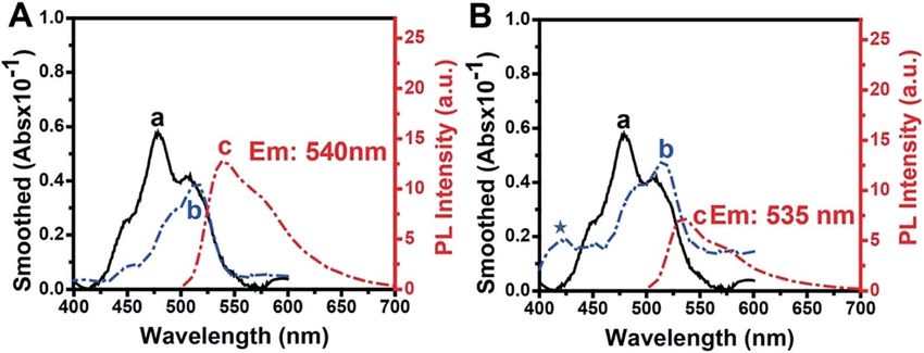
View Article Online
Edge Article Chemical Science
produced by the activated dsDNA was very high because TOTO-1 for telomerase detection listed in Table 1, it can be concluded
showed strong uorescence in the presence of dsDNA (curve b, that this method is more sensitive than the UV methods.6,43 It
Fig. 5B). It was also necessary to use Exo III to reduce the is also comparable to other sensitive methods such as
background signal. From curve a in Fig. 5B, we can see that the ECL,40–47 CL,9 SERS,48 and uorescence methods that use other
This article is licensed under a Creative Commons Attribution-NonCommercial 3.0 Unported Licence.
background signal is reduced by 38 times, which makes it probes.49–53 It can also be seen that the detection sensitivity
possible to detect the PARP-1 activity sensitively. The control for PARP-1 is comparable to those of other reported methods
experiments for PARP-1 detection are shown in Fig. 5C. Only (Table S2†).
activated PARP-1 could trigger the uorescence of the system, The practicality and selectivity for telomerase activity detec-
while other substances such as NAD+, heated PARP-1, and Exo tion were veried by using other kinds of cell lines, proteins and
III exhibited negligible effects on the uorescence intensity. enzymes (Fig. 7A). The PL intensity increased signicantly for
A549, HeLa, and MCF-7 cells and was negligible for heated A549
Performances for telomerase and PARP-1 analysis cells, PARP-1, HRP, BSA, and HSA, indicating that high PL
Open Access Article. Published on 19 2019. Downloaded on 13.01.2021 16:08:20.
resulted from telomerase in these cancer cell lines. As shown in
Confocal imaging gave further evidence for the feasibility of the
Fig. 7B, we also studied the selectivity of the biosensor for PARP-
proposed method. As is shown in Fig. 6, TOTO-1 exhibits very
1. The experimental results indicate no obvious change in PL
weak uorescence in the presence of the telomerase and PARP-1
when PARP-1 was replaced by BSA, heated PARP-1, and telo-
detection system without telomerase (A) or PARP-1 (A0 ). The
merase, suggesting satisfactory selectivity for PARP-1 detection.
presence of telomerase (B) or PARP-1 (B0 ) triggered the uo-
rescence of TOTO-1 sharply due to the formation of G-rich
elongated primer and PAR, respectively.
The uorescence spectra for telomerase and PARP-1 detec- Application of the strategy to detect telomerase in urine and
tions are shown in Fig. 6C and C0 , respectively. The optimization PARP-1 in cells
of the detection conditions is shown in Fig. S7 and S8.† PL Telomerase was detected by the proposed method in 11 early
intensity showed a linear correlation with increasing concen- morning urine samples provided by the Nanjing General
tration of telomerase from 13 to 4000 A549 cells per mL. The Hospital of Chinese People's Liberation Army. A threshold value
regression equation is Y ¼ 0.75 + 4.79 lg C, (R2 ¼ 0.997). A of 0.59 was calculated from the mean uorescence intensity of
linear relationship between the PL intensity and PARP-1 100 blank samples plus 3 times the relative standard deviation.
ranging from 0.02 U to 1.5 U (0.04 nM to 3 nM) was obtained The detection results are shown in Table 2. Sample 1 is the blank
Y ¼ 2.66 + 5.23C (R2 ¼ 0.992), where Y is the PL intensity and C is solution and samples 2–8 were obtained from normal, vesical
the content of A549 cells or PARP-1. The detection limit for calculus, kidney stone and inammation patients. All PL inten-
telomerase and PARP-1 was 13 cells per mL and 0.02 U, sities were lower than the threshold value. Samples 9–12 were
respectively. Compared with the previously reported methods obtained from bladder cancer patients. All their results were far
Fig. 6 Fluorescence images of TOTO-1 in the absence and presence of telomerase (7500 cells per mL) (A and B) and PARP-1 (0.8 U/20 mL) (A0
and B0 ). The scale bar is 5 mm. Fluorescence spectra of the detection system with increasing concentration of telomerase from 2 to 4000 A549
cells per mL (C) and PARP-1 from 0.02 U to 1.5 U (C0 ).
This journal is © The Royal Society of Chemistry 2019 Chem. Sci., 2019, 10, 3706–3714 | 3711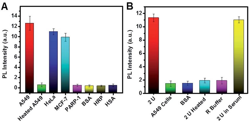
View Article Online
Chemical Science Edge Article
Table 1 Comparison of analytical performances of various methods for the determination of telomerase activity
Method System Detection range Detection limit Reference
UV-vis Hemin-graphene conjugate-based biosensor 100–2300 cells per mL 60 cells per mL 10
This article is licensed under a Creative Commons Attribution-NonCommercial 3.0 Unported Licence.
UV-vis Primer-modied GNPs 0–200 cells per mL 29 cells per mL 43
ECL DNA tetrahedral scaffold-based platform 0–32 000 cells per mL 70 cells per mL 44
ECL Porphyrin-functionalized graphene 10–750 cells per mL 10 cells per mL 45
ECL Ruthenium polyethylenimine complex-doped ZIF-8 50–106 cells 11 cells 46
ECL Polyluminol-PtNPs composite 15–9000 cells 15 cells 47
CL Hemin-G-quadruplex 1–1000 cells 1 cell 9
SERS ABT modied AuNPs 5–100 cells 1 cell 48
Fluorescence TS primer-modied AuNPs 10–1000 cells 1 cell 49
Fluorescence Cy3-labeled DNA 10–2000 cells 10 cells 50
Fluorescence AIEgens 5–10 000 cells 5 cells 51
Open Access Article. Published on 19 2019. Downloaded on 13.01.2021 16:08:20.
Fluorescence GNRs-based FRET 1–500 cells 1 cell 52
Fluorescence FRET-based TS-FAM/CCP platform 5–2000 cells 5 cells 53
Fluorescence TOTO-1 2–4000 cells per mL 13 cells per mL This work
higher than the threshold value. Therefore, this painless method Table 2 Detection results of telomerase in urine samples and PARP-1
has great potential to be used for bladder cancer diagnosis. in cellsa
The PARP-1 levels in normal cells of IOSE80 and breast
Telomerase Patient ID Clinical outcome TRAP PL intensity
cancer cells of MCF-7 and SK-BR-3 were evaluated. The detection
results for samples 13–15 showed that PARP-1 was present at a low 1 Water 0.25
level in normal human cells, while it was signicantly up-regulated 2 Normal Normal 0.3
in the malignant SK-BR-3 cells and MCF-7 cells. PARP-1 was higher 3 1003295831 Vesical calculus 0.3
in the nucleus than that in the cytoplasm of both SK-BR-3 cells and 4 1008455348 Bladder cancer 10.5
5 1002693440 Kidney stone 0.35
MCF-7 cells, which was consistent with previous results.54,55
6 1008455719 Kidney stone 0.31
Recovery experiments were conducted in human serum, normal 7 1008461668 Kidney stone 0.29
cells IOSE80 and human ovarian cancer cells A2780 to prove the 8 1007144483 Inammation 0.25
accuracy of the sensor in complex biological matrices. Recoveries 9 1007677148 Bladder cancer + 5
were from 94% to 109% and the relative standard derivations were 10 1007922178 Bladder cancer + 4
11 1007045669 Bladder cancer + 12
in the range of 2.48–8.21%, indicating that the method has good
12 1007924814 Bladder cancer + 7
accuracy and high precision (Table S4†). PARP-1 Cells Nucleus (U) Plasma (U)
13 IOSE80 0.05 0.02
Detection of inhibition efficiency on telomerase and PARP-1 14 MCF-7 0.67 0.53
15 SK-BR-3 0.74 0.5
To test the inhibitor screening function of the proposed sensor, a
“” or “+” means negative or positive for telomerase, respectively.
BIBR1532 and curcumin were chosen as the model inhibitors
for telomerase and AG014699 was selected as the model
Fig. 7 (A) Selectivity of the biosensor for telomerase activity detection; 750 cells per mL was used for each cell line. (B) Selectivity of TOTO-1 as
a PARP-1 biosensor; 2.0 U PARP-1 was used. Error bars show the standard deviation of three experiments.
3712 | Chem. Sci., 2019, 10, 3706–3714 This journal is © The Royal Society of Chemistry 2019
View Article Online
Edge Article Chemical Science
inhibitor for PARP-1. The PL intensities decreased continually References
to a stable value when the amounts of BIBR 1532 and curcumin
increased (Fig. S9A and C†). Decreased percentages of the PL 1 L. J. Wang, F. Ma, B. Tang and C. Y. Zhang, Chem. Sci., 2017,
intensity in the presence of various concentrations of BIBR 1532 8, 2495–2502.
This article is licensed under a Creative Commons Attribution-NonCommercial 3.0 Unported Licence.
and curcumin are shown in Fig. S9B and D.† A549 without an 2 M. F. Langelier, J. L. Planck, S. Roy and J. M. Pascal, Science,
inhibitor was used as the control (750 cells per mL). The IC50 2012, 336, 728.
(concentration of inhibitor producing 50% inhibition) values 3 R. C. Qian, L. Ding, L. Yan, M. Lin and H. X. Ju, J. Am. Chem.
for BIBR1532 and curcumin against telomerase were 251 nM Soc., 2014, 136, 8205–8208.
and 8.64 mM, respectively. These results were in accordance with 4 M. Hong, L. D. Xu, Q. W. Xue, L. Li and B. Tang, Anal. Chem.,
previously reported values.56,57 The uorescence spectra in the 2016, 88, 12177–12182.
presence of PARP-1 inhibited by different concentrations of 5 X. W. Xu, L. Wang, K. Li, Q. H. Huang and W. Jiang, Anal.
AG014699 are shown in Fig. S9E.† The decreased percentages of Chem., 2018, 90, 3521–3530.
Open Access Article. Published on 19 2019. Downloaded on 13.01.2021 16:08:20.
the PL intensity are shown in Fig. S9F.† The IC50 value of 6 H. T. Yang, A. R. Liu, M. Wei, Y. J. Liu, B. J. Lv, W. Wei,
AG014699 was determined to be 8.2 nM; it was also in good Y. J. Zhang and S. Q. Liu, Anal. Chem., 2017, 89, 12094–12100.
agreement with the reported IC50 value in literature.58 7 R. Freeman, E. Sharon, C. Teller, A. Henning, Y. Tzfati and
I. Willner, ChemBioChem, 2010, 11, 2362–2367.
Conclusions 8 C. L. Wang, H. T. Yang, S. S. Wu, Y. J. Liu, W. Wei, Y. J. Zhang,
M. Wei and S. Q. Liu, TrAC, Trends Anal. Chem., 2018, 105,
In summary, the unique uorescence selectivity and sensitivity 404–412.
of TOTO-1 towards the G bases in single-stranded DNA and PAR 9 L. J. Wang, Y. Zhang and C. Y. Zhang, Anal. Chem., 2013, 85,
were found for the rst time and studied elaborately. MD 11509–11517.
simulation proved that intermolecular p–p stacking formed 10 X. L. Xu, M. Wei, Y. J. Liu, X. Liu, W. Wei, Y. J. Zhang and
between TOTO-1 and poly(dG) and TOTO-1/poly(dG) folded in S. Q. Liu, Biosens. Bioelectron., 2017, 87, 600–606.
a way analogous to that of duplex DNA, which explained why 11 X. Liu, M. Wei, E. S. Xu, H. T. Yang, W. Wei, Y. J. Zhang and
TOTO-1 has high selectivity towards poly(dG). TOTO-1 is also S. Q. Liu, Biosens. Bioelectron., 2017, 91, 347–353.
the rst probe that has been reported to have high selectivity 12 X. Liu, M. Wei, Y. J. Liu, B. J. Lv, W. Wei, Y. J. Zhang and
and sensitivity towards PAR. A label-free, simple and sensitive S. Q. Liu, Anal. Chem., 2016, 88, 8107–8114.
uorescence biosensor was constructed for telomerase and 13 S. Y. Tang, Z. Nie, W. Li, D. Q. Li, Y. Huang and S. Z. Yao,
PARP-1 sensing based on TOTO-1 and Exo III-assisted back- Chem. Commun., 2015, 51, 14389–14392.
ground noise reduction. This strategy shows high sensitivity 14 Y. Y. Xu, L. Liu, Z. Y. Wang and Z. H. Dai, ACS Appl. Mater.
and selectivity towards telomerase and PARP-1. Integrating Interfaces, 2016, 8, 18669–18674.
multiple functions into one simple probe, i.e., detecting not 15 S. S. Wu, M. Wei, H. T. Yang, J. H. Fan, W. Wei, Y. J. Zhang
only telomerase but also PARP-1 can signicantly raise the and S. Q. Liu, Sens. Actuators, B, 2018, 259, 565–572.
specicity of screening cancer and decrease false positive 16 X. J. Yang, K. Zhang, T. T. Zhang, J. J. Xu and H. Y. Chen,
proportion. This makes TOTO-1 a promising candidate probe Anal. Chem., 2017, 89, 4216–4222.
for clinical diagnosis. However, the probe cannot be used to 17 R. Krishnakumar and W. L. Kraus, Mol. Cell, 2010, 39, 8–24.
image telomerase or PARP-1 in vivo because Exo III is necessary 18 N. Dai and E. T. Kool, Chem. Soc. Rev., 2011, 40, 5756–5770.
to decrease the background signals and it is difficult to be 19 J. Liang, B. Z. Tang and B. Liu, Chem. Soc. Rev., 2015, 44,
transferred into cells. 2798–2811.
20 H. Zhou, J. Liu, J. J. Xu, S. S. Zhang and H. Y. Chen, Chem.
Conflicts of interest Soc. Rev., 2018, 47, 1996–2019.
21 H. Zhang, F. Li, B. Dever, X. F. Li and X. C. Le, Chem. Rev.,
There are no conicts to declare. 2013, 113, 2812–2841.
22 H. Peng, A. M. Newbigging, Z. Wang, J. Tao, W. Deng,
Acknowledgements X. C. Le and H. Zhang, Anal. Chem., 2018, 90, 190–207.
23 H. C. Yeh, J. Sharma, J. J. Han, J. S. Martinez and
We genuinely express our gratitude to Prof. Jing Ma, Institution J. H. Werner, Nano Lett., 2010, 10, 3106–3110.
of Theoretical and Computational Chemistry, Nanjing Univer- 24 H. C. Yeh, J. Sharma, M. Shih Ie, D. M. Vu, J. S. Martinez and
sity, for her MD simulation of the interaction between TOTO-1 J. H. Werner, J. Am. Chem. Soc., 2012, 134, 11550–11558.
and poly (dG). This work was supported by the National 25 W. Zhou, J. Zhu, D. Fan, Y. Teng, X. Zhu and S. Dong, Adv.
Natural Science Foundation of China (No. 21775019, 21475020, Funct. Mater., 2017, 27, 1704092.
21635004, 81730087). Fundamental Research Funds for the 26 Y. Zhang, C. Zhu, L. Zhang, C. Tan, J. Yang, B. Chen, L. Wang
Central Universities and A Project Funded by the Priority and H. Zhang, Small, 2015, 11, 1385–1389.
Academic Program Development of Jiangsu Higher Education 27 J. Mohanty, N. Barooah, V. Dhamodharan, S. Harikrishna,
Institutions (No. 2242018K3DN04), The Open Project of The Key P. I. Pradeepkumar and A. C. Bhasikuttan, J. Am. Chem.
Laboratory of Modern Toxicology of Ministry of Education, Soc., 2013, 135, 367–376.
Nanjing Medical University (NMUMT201804).
This journal is © The Royal Society of Chemistry 2019 Chem. Sci., 2019, 10, 3706–3714 | 3713View Article Online
Chemical Science Edge Article
28 A. Renaud de la Faverie, A. Guedin, A. Bedrat, L. A. Yatsunyk 44 Q. M. Feng, Z. Zhou, M. X. Li, W. Zhao, J. J. Xu and
and J. L. Mergny, Nucleic Acids Res., 2014, 42, e65. H. Y. Chen, Biosens. Bioelectron., 2017, 90, 251–257.
29 S. Liu, P. Peng, H. Wang, L. Shi and T. Li, Nucleic Acids Res., 45 L. Wu, J. S. Wang, L. Y. Feng, J. S. Ren, W. L. Wei and
2017, 45, 12080–12089. X. G. Qu, Adv. Mater., 2012, 24, 2447–2452.
This article is licensed under a Creative Commons Attribution-NonCommercial 3.0 Unported Licence.
30 S. Ding, X. Qiao, J. Suryadi, G. S. Marrs, G. L. Kucera and 46 C. Y. Xiong, W. B. Liang, Y. N. Zheng, Y. Zhuo, Y. Q. Chai and
U. Bierbach, Angew. Chem., Int. Ed., 2013, 52, 3350–3354. R. Yuan, Anal. Chem., 2017, 89, 3222–3227.
31 H. S. Rye, M. A. Quesada, K. Peck, R. A. Mathies and 47 H. R. Zhang, B. X. Li, Z. M. Sun, H. Zhou and S. S. Zhang,
A. N. Giazer, Nucleic Acids Res., 1991, 19, 327–333. Chem. Sci., 2017, 8, 8025–8029.
32 Y. Liu, M. Wei, Y. Li, A. Liu, W. Wei, Y. Zhang and S. Liu, 48 M. L. Shi, J. Zheng, C. H. Liu, G. X. Tan, Z. H. Qing, S. Yang,
Anal. Chem., 2017, 89, 3430–3436. J. F. Yang, Y. J. Tan and R. H. Yang, Biosens. Bioelectron.,
33 D. L. Ma, S. Lin, K. H. Leung, H. J. Zhong, L. J. Liu, 2016, 77, 673–680.
D. S. H. Chan, A. Bourdoncle, J. L. Mergny, H.-M. D. Wang 49 Y. F. Gao, J. Xu, B. X. Li and Y. Jin, ACS Appl. Mater. Interfaces,
Open Access Article. Published on 19 2019. Downloaded on 13.01.2021 16:08:20.
and C.-H. Leung, Nanoscale, 2014, 6, 8489–8494. 2016, 8, 13707–13713.
34 N. Jan, S. Nicke and K. Mikael, Biopolymers, 1998, 46, 39–51. 50 X. Su, Z. H. Li, X. Z. Yan, L. Wang, X. Zhou, L. Wei, L. H. Xiao
35 H. S. Rye and A. N. Glazer, Nucleic Acids Res., 1995, 23, 1215– and C. Y. Yu, Anal. Chem., 2017, 89, 3576–3582.
1222. 51 Y. Zhuang, M. Zhang, B. Chen, R. Duan, X. Min, Z. Zhang,
36 V. L. Singer, L. J. Jones, S. T. Yue and R. P. Haugland, Anal. F. Zheng, H. Liang, Z. Zhao, X. Lou and F. Xia, Anal.
Biochem., 1997, 249, 228–238. Chem., 2015, 87, 9487–9493.
37 H. S. Rye, S. Yue, D. E. Wemmer, M. A. Quesada, 52 Y. Wang, L. Yang, B. Li and Y. Jin, Analyst, 2016, 141, 6133–
R. P. Haugland, R. A. Mathies and A. N. Glazer, Nucleic 6139.
Acids Res., 1992, 20, 2803–2812. 53 C. Chen, M. Wei, Y. Liu, E. Xu, W. Wei, Y. Zhang and S. Liu,
38 A. Castro, F. R. Faireld and E. B. Shera, Anal. Chem., 1993, Microchim. Acta, 2017, 184, 3453–3460.
65, 849–852. 54 P. Domagala, T. Huzarski, J. Lubinski, K. Gugala and
39 D. A. Case, R. M. Betz, D. S. Cerutti, T. E. Cheatham III, W. Domagala, Breast Cancer Res. Treat., 2011, 127, 861–869.
T. A. Darden, R. E. Duke, T. J. Giese, H. Gohlke, 55 F. Rojo, J. Garcı́a-Parra, S. Zazo, I. Tusquets, J. Ferrer-Lozano,
A. W. Goetz, N. Homeyer, et al., AMBER, University of S. Menendez, P. Eroles, C. Chamizo, S. Servitja, N. Ramı́rez-
California, San Francisco, 2016. Merino, F. Lobo, B. Bellosillo, J. M. Corominas, J. Yelamos,
40 G. M. Morris, D. S. Goodsell, R. S. Halliday, R. Huey, S. Serrano, A. Lluch, A. Rovira and J. Albanell, Ann. Oncol.,
W. E. Hart, R. K. Belew and A. J. Olson, J. Comput. Chem., 2012, 23, 1156–1164.
1998, 19, 1639–1662. 56 E. Pascolo, C. Wenz, J. Lingner, N. Hauel, H. Priepke,
41 M. J. Frisch, G. W. Trucks, H. B. Schlegel, G. E. Scuseria, I. Kauffmann, P. Garin-Chesa, W. J. Rettig, K. Damm and
M. A. Robb, J. R. Cheeseman, G. Scalmani, V. Barone, A. Schnapp, J. Biol. Chem., 2002, 277, 15566–15572.
G. A. Petersson, H. Nakatsuji, X. Li, M. Caricato, 57 S. Chakraborty, U. Ghosh, N. P. Bhattacharyya,
A. V. Marenich, et al., Gaussian 09, revision D.01, Gaussian, R. K. Bhattacharya and M. Roy, Mutat. Res., Fundam. Mol.
Inc., Wallingford, CT, 2016. Mech. Mutagen., 2006, 596, 81–90.
42 L. P. Wang, T. J. Martinez and V. S. Pande, J. Phys. Chem. 58 R. Plummer, C. Jones, M. Middleton, R. Wilson, J. Evans,
Lett., 2014, 5, 1885–1891. A. Olsen, N. Curtin, A. Boddy, P. McHugh, D. Newell,
43 L. Zhang, S. Zhang, W. Pan, Q. Liang and X. Song, Biosens. A. Harris, P. Johnson, H. Steinfeldt, R. Dewji, D. Wang,
Bioelectron., 2016, 77, 144–148. L. Robson and H. Calvert, Clin. Cancer Res., 2008, 14,
7917–7923.
3714 | Chem. Sci., 2019, 10, 3706–3714 This journal is © The Royal Society of Chemistry 2019You can also read