Expression and inhibitory effects of p53 upregulated modulator of apoptosis in gallbladder carcinoma
←
→
Page content transcription
If your browser does not render page correctly, please read the page content below
ONCOLOGY LETTERS 21: 234, 2021

                 Expression and inhibitory effects of p53‑upregulated
                  modulator of apoptosis in gallbladder carcinoma
                            ZHIDE LIU1*, CHENG YAN1*, YANGYAN XIAO2, WEICHANG ZHANG1,
                                       LI WANG3, QINGLONG LI1 and WENWU CAI1

                          Departments of 1General Surgery and 2Ophthalmology, The Second Xiangya Hospital,
                             Central South University, Changsha, Hunan 410011; 3Department of Oncology,
                            Peking University Shenzhen Hospital, Shenzhen, Guangdong 518036, P.R. China

                                         Received January 8, 2020; Accepted January 6, 2021

                                                     DOI: 10.3892/ol.2021.12495

Abstract. The p53‑upregulated modulator of apoptosis                 those in the control group. Low PUMA expression levels were
(PUMA) has been reported to be involved in various types             associated with a short overall survival time in patients with
of cancer. However, its potential biological role in gallbladder     GBC. In conclusions, PUMA may act as a tumor suppressor in
carcinoma (GBC) has not been fully elucidated. The present           GBC and may serve as a potential novel treatment target for
study aimed to determine the expression levels of PUMA               human GBC.
and its biological effects on GBC. The mRNA and protein
expression levels of PUMA in GBC tissues and cell lines              Introduction
were measured using reverse transcription‑quantitative PCR
and western blotting, respectively. The effects of PUMA              Gallbladder carcinoma (GBC) was the fifth most common
overexpression on cell viability, proliferation and invasive         malignant tumor of the digestive system and the most common
ability were determined in vitro using the MTT, colony               malignant tumor of the biliary tract worldwide in 2015 (1,2).
formation and Transwell invasion assays, respectively. The           Early diagnosis and radical surgical resection are the most
apoptotic rates were detected using the Annexin V‑FITC               effective and preferred treatment approaches for patients with
apoptosis detection kit. Furthermore, follow‑up of patients          GBC, resulting in improved long‑term survival (3,4). However,
with GBC was performed to identify the association between           due to the atypical early symptoms, and frequently rapid and
PUMA expression levels and GBC prognosis. The results of             asymptomatic progression, a large proportion of patients with
the present study demonstrated that the expression levels of         GBC are likely to be diagnosed with late stage cancer; thus,
PUMA were significantly lower in the GBC tissues and cell            resectable GBC accounts for only 15‑47% of all cases (5,6).
lines compared with those in adjacent normal gallbladder             The aforementioned features of GBC may account for the
tissues and normal gallbladder cells, respectively. Further          unsatisfactory prognosis of patients for GBC. The median
experiments indicated that overexpression of PUMA inhibited          survival time of patients with GBC is only ~6 months, with a
the viability, proliferation and invasive ability of GBC cells       5‑year survival rate of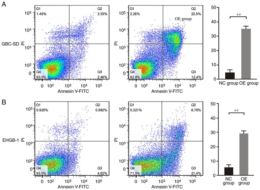
2 LIU et al: PUMA IN GALLBLADDER CARCINOMA
Materials and methods GCTGTAGCCAAA‑3'. The expression levels of GAPDH were
used to normalize the results and the 2‑ΔΔCq method was used
Clinical specimen collection. A total of 18 paired GBC to calculate the relative expression levels of target gene (16).
and adjacent normal gallbladder tissues were collected
from patients who underwent surgical treatment between Western blotting. Total protein was extracted from clinical
December 2016 and December 2018 in the Second Xiangya tissues and cell lines using RIPA lysis buffer (50 mM Tris
Hospital (Changsha, China), including 6 men and 12 women, pH 8.0, 120 mM NaCl, 0.5% sodium deoxycholate, 0.5%
with the mean age of 52 years (range, 42‑68 years) in Nevin's NP‑40, 0.1% SDS, 1 mM EDTA, 50 mM NaF, 1 mM Na2VO4,
stage I‑III (15). All patients enrolled in the present study had 1 mM PMSF and 2 µg/ml aprotinin) on ice for 30 min.
not undergone any treatment prior surgery. The fresh GBC The protein concentration was determined by BCA assay,
and adjacent normal gallbladder tissues were stored at ‑80˚C and 50 µg protein/lane was separated by 10% SDS‑PAGE
in liquid nitrogen until RNA extraction. Written informed at 120 V for 60 min and transferred to PVDF membranes.
consent for participation and publication were obtained from The membranes were blocked with 5% non‑fat milk in TBS
all patients. Follow‑up was performed after surgery until (10 mM Tris pH 7.4 and 150 mM NaCl) and 0.1% Tween‑20
December 2019, and no cases were lost to the follow‑up. This (TBST) at room temperature for 60 min, followed by
study was approved by the Research Ethics Committee of the incubation with primary antibodies against PUMA (1:1,000;
Second Xiangya Hospital (approval no. 179 in 2017). cat. no. ab33906), E‑cadherin (1:10,000; cat. no. ab40772),
vimentin (1:1,000; cat. no. ab92547), Bax (1:1,000;
Cell culture. The normal gallbladder cells were obtained by cat. no. ab32503), Bcl‑2 (1:2,000; cat. no. ab182858) (all from
primary culture of cells isolated from healthy gallbladder Abcam) and β‑actin (1:1,000; cat. no. A2228; Sigma‑Aldrich;
wall tissue, which was obtained from six patients with Merck KGaA) at 4˚C for 12 h. Subsequently, the membranes
gallbladder polyps undergoing cholecystectomy, including were washed three times with TBST for 5 min and incu‑
three men and three women, with the mean age of 50 years bated with HRP‑conjugated goat anti‑rabbit IgG (1:2,000;
(range, 44‑65 years). Written informed consent for participa‑ cat. no. sc‑2004) or anti‑mouse IgG (1:2,000; cat. no. sc‑2005)
tion and publication were obtained from all patients. The (both from Santa Cruz Biotechnology, Inc.) secondary anti‑
gallbladder wall tissue was transferred to ice‑cold Ca2+‑free body at 37˚C for 60 min. The protein bands were visualized
Hanks solution with 0.5 mM EGTA and cut into 1‑cm 2 frag‑ with an enhanced chemiluminescence system (Thermo Fisher
ments under sterile conditions. Subsequently, the tissue was Scientific, Inc.) according to the manufacturer's instructions.
placed on a 60‑mm culture dish and treated for 45 min at room The Gel Doc 2000 imaging system (Bio‑Rad Laboratories,
temperature with 0.025% trypsin (Thermo Fisher Scientific, Inc.) was used for densitometric analysis of the protein bands
Inc.) in PBS containing 0.02% EDTA‑2Na. The tissue was with ImageJ software (version 1.8.0.112; National Institutes of
agitated every 15 min using a pipette to promote the release of Health) according to the manufacturer's instructions. β‑actin
the cells. The gallbladder cells were collected by centrifuga‑ was used as the internal control.
tion at 1,200 x g for 5 min and counted in a CC‑108 microcell
counter (TOA Corporation). Plasmid generation and cell transfection. The PUMA sequence
Human GBC cell lines GBC‑SD, SGC‑996, NOZ and was synthesized and subcloned into the LV‑BBC3 vector
EHGB‑1 obtained from the Institute of Biochemistry and (22944‑1) (Shanghai GeneChem Co., Ltd.). Ectopic expression
Cell Biology of the Chinese Academy of Sciences. The GBC of PUMA was achieved by the PUMA lentivirus transfection,
cells were cultured in Dulbecco's modified Eagle's medium and an empty vector (KL8781‑1) (Shanghai GeneChem Co.,
(Gibco; Thermo Fisher Scientific, Inc.) containing 10% fetal Ltd.) used as a negative control. Prior to transfection, human
bovine serum supplemented with 100 U/ml penicillin and GBC cell lines were cultured in complete medium without
100 U/ml streptomycin (Invitrogen; Thermo Fisher Scientific, antibiotics for at least 24 h. When the GBC‑SD and EHGB‑1
Inc.) at 37˚C with 5% CO2. cell confluence reached 30‑50%, the cells were washed with
1X PBS once and transfected with the LV‑BBC3 (22944‑1)
RNA extraction and reverse transcription‑quantitative (RT‑q) (5x10 8 TU/ml; 40 µl; MOI, 20) and empty (KL8781‑1)
PCR. The TRIzol® Reagent (Invitrogen; Thermo Fisher (5x10⁸ TU/ml; 40 µl; MOI, 20) vector at 37˚C for 72 h, in the
Scientific, Inc.) was used to extract RNA from clinical tissues presence of Lipofectamine® 2000 (Invitrogen, Thermo Fisher
and cell lines, which was subsequently treated with DNase I Scientific, Inc.) according to the manufacturer's instructions.
(Invitrogen; Thermo Fisher Scientific, Inc.) for purification At 48 h post‑transfection, the cells were harvested, and the
according to the manufacturer's instructions. The miRcute expression levels of PUMA were determined by RT‑qPCR.
miRNA First‑Strand cDNA Synthesis kit (Takara Bio, Inc.)
was used to perform reverse transcription according to the MTT assay. At 48 h post‑transfection, the cells were seeded
manufacturer's instructions. qPCR was performed using the into 96‑well plates (2x10⁴ cells/well). Following culture for 4 h
SYBR® Green PCR detection kit (Takara, Bio, Inc.) in a 7500 at 37˚C, the supernatant was discarded, and 150 µl DMSO was
Real‑Time PCR system (Applied Biosystems; Thermo Fisher added to each well. After shaking for 10 min, 100 µl cell suspen‑
Scientific, Inc.) according to the manufacturer's instructions sion was placed into another 96‑well plate with 100 µl DMSO
with the following primers: PUMA forward, 5'‑TGAAGA solution as a control. The Cell Proliferation Reagent Kit I (MTT;
GCAAATGAGCCAAACG‑3' and reverse, 5'‑CAGAGCACA Roche Applied Science) was used at 37˚C for 4 h to determine
GGAT TCACAGTCT‑3'; and GAPDH forward, 5'‑TGACTT the cell viability. The absorbance values were determined
CAACAGCGACACCCA‑3' and reverse, 5'‑CACCCTGTT at 450 nm using a spectrophotometer (Omega Bio‑Tek, Inc.).
ONCOLOGY LETTERS 21: 234, 2021                                                             3

Figure 1. PUMA mRNA and protein expression levels in GBC tissues and cell lines. (A) PUMA mRNA expression levels were significantly lower in GBC
tissues compared with those in adjacent tissues, and in GBC cell lines compared with those in the normal group. (B) PUMA protein expression levels
in GBC tissues were significantly decreased compared with those in the adjacent tissues. (C) PUMA protein expression levels in GBC cell lines and
PUMA‑overexpressing EHGB‑1 and GBC‑SD cell lines. *P
4                                               LIU et al: PUMA IN GALLBLADDER CARCINOMA

Figure 2. PUMA overexpression inhibits gallbladder carcinoma cell proliferation. (A) Fluorescence of GBC‑SD cells following transfection with the LV‑BBC3
vector. (B) PUMA mRNA expression were levels significantly upregulated in the GBC‑SD and EHGB‑1 OE groups compared with those in the corresponding
NC groups. (C and D) Cell proliferation was significantly inhibited in the (C) GBC‑SD and (D) EHGB‑1 OE groups compared with that in the corresponding
NC groups. *P
ONCOLOGY LETTERS 21: 234, 2021                                                                 5

Figure 3. PUMA overexpression inhibits gallbladder carcinoma cell colony formation and invasion. (A) The colony formation and (B) invasive abilities were
significantly inhibited in the GBC‑SD and EHGB‑1 OE groups compared with those in the corresponding NC groups. **P
6                                               LIU et al: PUMA IN GALLBLADDER CARCINOMA

Figure 4. PUMA overexpression regulates the expression of proteins associated with the epithelial‑mesenchymal transition and apoptosis. (A) The protein
levels of E‑cadherin were upregulated, whereas the levels of vimentin were downregulated in the GBC‑SD and EHGB‑1 OE groups compared with those in
the corresponding NC groups. (B) The protein expression levels of Bax were upregulated, whereas the expression levels of Bcl‑2 were downregulated in the
GBC‑SD and EHGB‑1 OE groups compared with those in the NC groups. *P
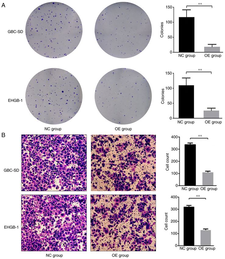
ONCOLOGY LETTERS 21: 234, 2021                                                           7

Figure 5. PUMA inhibits apoptosis in gallbladder carcinoma cells. The apoptotic rates were significantly higher in the (A) GBC‑SD and (B) EHGB‑1 OE
groups compared with those in the NC groups. **P
8                                           LIU et al: PUMA IN GALLBLADDER CARCINOMA

further studies are needed to elucidate the precise underlying            The follow‑up results in the present study demonstrated
mechanism of PUMA‑induced apoptosis.                                  that patients with GBC in the PUMA high group exhibited a
    Our previous study has reported that PUMA is a marker             longer survival time compared with those in the PUMA low
that may be associated with the clinical behavior of GBC and          group, which suggested that PUMA may be a new clinical
may reflect the occurrence, development and prognosis of              marker for the prognosis of GBC and may serve as a potential
GBC in patient tissues (23). In the present study, the expression     target for the diagnosis and treatment of patients with GBC.
levels of PUMA were detected in GBC tissues and cell lines,           However, a larger sample and long‑term studies are required
and its functional roles on the biological behaviors of GBC           to verify this hypothesis.
cell lines were further investigated. The results demonstrated            In the present study, the expression levels of PUMA and its
that the expression levels of PUMA were significantly lower           inhibitory effects on GBC were only investigated by a limited
in GBC tissues compared with those in the adjacent tissues,           number of clinical specimen and cell experiments. Further
and were low in GBC cell lines. Further experiments were              studies are required to confirm these results in a large sample
performed in vitro to demonstrate the functions of PUMA               and in vivo animal experiments.
in the biological behavior of GBC; the results demonstrated               In conclusion, the results of the present study demonstrated
that following overexpression, PUMA significantly inhibited           that PUMA inhibited the proliferative and invasive capabilities
the proliferative and invasive abilities of GBC cells compared        of GBC cells through the Bax/Bcl‑2 signaling pathway and
with those of the control groups, which suggested that PUMA           partially by regulating the EMT. These results suggested that
may serve as a tumor suppressor in GBC.                               PUMA may serve as a potential tumor maker for the diagnosis
    The potential mechanisms of PUMA‑inhibited invasion               and treatment of GBC in clinical practice. In the last decade,
were further investigated in the present study. The EMT is a          studies on circulating tumor cells (CTCs) have enriched the
process during which epithelial cells trans‑differentiate into        methods of detecting tumor cells (35,36); in our future work,
mesenchymal cells (26,27). The EMT endows tumor cells                 the potential functions of PUMA in CTCs will be investigated.
with the traits of migration and invasion and induces cancer
stem cell properties (27,28). However, the effects of the EMT         Acknowledgements
in PUMA‑induced GBC cell apoptosis remain unknown. The
results of the present study demonstrated that PUMA overex‑           Not applicable.
pression in GBC cell lines increased the protein expression
levels of E‑cadherin and decreased the levels of vimentin             Funding
compared with those in the negative control group. These
results suggested that PUMA‑induced EMT reversal may                  This work was supported by the National Natural Science
account for the inhibitory effects of PUMA on GBC cell                Foundation of China, Beijing (grant no. 81703767 to WC) and
invasion. However, further studies are needed to unravel the          the Hunan Natural Science Foundation of China, Changsha
precise signaling pathways involved in the PUMA‑mediated              (grant no. 2019JJ50891 to WC).
inhibition of the EMT.
    The results of the present study also demonstrated that the       Availability of data and materials
protein expression levels of Bax were upregulated, whereas
the levels of Bcl‑2 were downregulated following PUMA                 The datasets used and/or analyzed during the current study
overexpression compared with those in the negative control            are available from the corresponding author on reasonable
groups. The Bcl‑2 family serves a vital role in apoptosis (29).       request.
As a member of the Bcl‑2 family, Bax promotes apoptosis;
the protein expression levels of Bax are associated with the          Authors' contributions
regulation of apoptosis, since high levels of the Bax protein
induce apoptosis (30,31). As a suppressor of apoptosis, Bcl‑2         QL and WC conceived and designed the study. ZL and CY
negatively regulates apoptosis by regulating mitochondrial            obtained the clinical samples and were the major contributors
cytochrome c release (32). In addition, Bcl‑2 binds to Bax to         in acquiring the data. WZ and LW performed the cell experi‑
form a Bax/Bcl‑2 polymer to inhibit the apoptotic process (33).       ments. WC and YX analyzed and interpreted the data, and
High expression levels of Bcl‑2 also maintain cell stability by       drafted the manuscript. QL and WC revised the manuscript.
inducing glutathione aggregation in the cell nucleus to reduce        ZL and WC confirm the authenticity of all the raw data. All
caspase activity (34). The results of the present study suggested     authors read and approved the final manuscript.
that PUMA may inhibit GBC invasion at least partially by
regulating the expression of Bax and Bcl‑2. However, further          Ethics approval and consent to participate
studies are needed to clarify the exact mechanism of this
regulation process.                                                   This study was approved by the Research Ethics Committee of
    In the present study, a normal gallbladder epithelial cell line   Second Xiangya Hospital (approval no. 179 in 2017; Changsha,
was intended to be used as a control for the in vitro experi‑         China). Informed consent was obtained from all included
ments; however, careful review of the available literature did not    patients.
identify any normal gallbladder epithelial cell lines. Primary
culture of cells isolated from normal gallbladder wall tissues        Patient consent for publication
were subsequently considered. However, these cells could not
be used as a control cell line due to their short survival time.      Not applicable.
ONCOLOGY LETTERS 21: 234, 2021 9
Competing interests 20. Sun YL, Jiang WQ, Luo QY, Yang DJ, Cai YC, Huang HQ and
Sun J: A novel Bcl‑2 inhibitor, BM‑1197, induces apoptosis in
malignant lymphoma cells through the endogenous apoptotic
The authors declare that they have no competing interests. pathway. BMC Cancer 20: 1, 2019.
21. Letai A: Puma strikes Bax. J Cell Biol 185: 189‑191, 2009.
22. Erlacher M, Labi V, Manzl C, Böck G, Tzankov A, Häcker G,
References Michalak E, Strasser A and Villunger A: Puma cooperates with
Bim, the rate‑limiting BH3‑only protein in cell death during
1. Kanthan R, Senger JL, Ahmed S and Kanthan SC: Gallbladder lymphocyte development, in apoptosis induction. J Exp Med 203:
Cancer in the 21st Century. J Oncol 2015: 967472, 2015. 2939‑2951, 2006.
2. Jiahong D, Jianming W and Jianping Z: Guidelines for diagnosis 23. Cai W, Li Q, Yang Z, Miao X, Wen Y, Huang S and Ouyang J:
and treatment of gallbladder cancer (2015 edition). J Clin Expression of p53 upregulated modulator of apoptosis (PUMA)
Hepatol 32: 411‑419, 2015. and C‑myb in gallbladder adenocarcinoma and their pathological
3. Kakaei F, Beheshtirouy S, Nejatollahi SM, Zarrintan S and significance. Clin Transl Oncol 15: 818‑824, 2013.
Mafi MR: Surgical treatment of gallbladder carcinoma: A critical 24. Shu GS, Lv F, Yang ZL and Miao XY: Immunohistochemical
review. Updates Surg 67: 339‑351, 2015. study of PUMA, c‑Myb and p53 expression in the benign and
4. Cziupka K, Partecke LI, Mirow L, Heidecke CD, Emde C, malignant lesions of gallbladder and their clinicopathological
Hoffmann W, Siewert U, van den Berg N, von Bernstorff W significances. Int J Clin Oncol 18: 641‑650, 2013.
and Stier A: Outcomes and prognostic factors in gallbladder 25. Wei D, Zhang X, Zou H, Wang L, Fu B, Wu X, Luo Z, Li X,
cancer: A single‑centre experience. Langenbecks Arch Surg 397: Ge J, Li Y, et al: WW domain containing oxidoreductase
899‑907, 2012. induces apoptosis in gallbladder‑derived malignant cell by
5. Lazcano‑Ponce EC, Miquel JF, Muñoz N, Herrero R, Ferrecio C, upregulating expression of P73 and PUMA. Tumour Biol 35:
Wistuba II, Alonso de Ruiz P, Aristi Urista G and Nervi F: 1539‑1550, 2014.
Epidemiology and molecular pathology of gallbladder cancer. 26. T h ier y J P, Acloque H, Hua ng RY a nd Nieto M A:
CA Cancer J Clin 51: 349‑364, 2001. Epithelial‑mesenchymal transitions in development and desease.
6. Mekeel KL and Hemming AW: Surgical management of Cell 139: 871‑890, 2009.
gallbladder carcinoma: A review. J Gastrointest Surg 11: 27. Fu XT, Dai Z, Song K, Zhang ZJ, Zhou ZJ, Zhou SL, Zhao YM,
1188‑1193, 2007. Xiao YS, Sun QM, Ding ZB and Fan J: Macrophage‑secreted
7. Zhang LF, Hou CS, Xu Z, Guo LM, Ling XF, Wang LX and IL‑8 induces epithelial‑mesenchymal transition in hepatocellular
Xiu DR: Laparoscopic treatment for incidental gallbladder carcinoma cells by activating the JAK2/STAT3/Snail pathway.
cancer: A retrospective 10 years study from a single institution. Int J Oncol 46: 587‑596, 2015.
Zhonghua Wai Ke Za Zhi 57: 277‑281, 2019 (In Chinese). 28. Mani SA, Guo W, Liao MJ, Eaton EN, Ayyanan A, Zhou AY,
8. Butte JM, Matsuo K, Gönen M, D'Angelica MI, Waugh E, Brooks M, Reinhard F, Zhang CC, Shipitsin M, et al: The
Allen PJ, Fong Y, DeMatteo RP, Blumgart L, Endo I, et al: epithelial‑mesenchymal transition generates cells with properties
Gallbladder cancer: Differences in presentation, surgical of stem cells. Cell 133: 704‑715, 2008.
treatment, and survival in patients treated at centers in three 29. Babu PP, Suzuki G, Ono Y and Yoshida Y: Attenuation of
countries. J Am Coll Surg 212: 50‑61, 2011. ischemia and/or reperfusion injury during myocardial infarc‑
9. Yu J: PUMA kills stem cells to stall cancer? Mol Cell tion using mild hypothermia in rats: An immunohistochemical
Pharmacol 1: 112‑118, 2009. study of bcl‑2, bax, Bak and TUNEL. Pathol Int 54: 896‑903,
10. Yu J, Wang Z, Kinzler KW, Vogelstein B and Zhang L: PUMA 2004.
mediates the apoptotic response to p53 in colorectal cancer cells. 30. Duan XX, Ou JS, Li Y, Su JJ, Ou C, Yang C, Yue HF and
Proc Natl Acad Sci USA 100: 1931‑1936, 2003. Ban KC: Dynamic expression of apoptosis related genes during
11. He S, Ma X, Ye Y, Zhang M, Zhuang J, Song Y and Xia W: development of laboratory hepatocellular carcinoma and its
HEATR1 modulates cell survival in non‑small cell lung cancer relation to apoptosis. World J Gastroenterol 11: 4740‑4744,
via activation of the p53/PUMA signaling pathway. Onco Targets 2005.
Ther 12: 4001‑4011, 2019. 31. Martin LJ: Neuronal cell death in nervous system development,
12. Yee KS, Wilkinson S, James J, Ryan KM and Vousden KH: disease, and injury (Review). Int J Mol Med 7: 455‑478, 2001.
PUMA‑ and Bax‑induced autophagy contributes to apoptosis. 32. Cecconi F, Alvarez‑Bolado G, Meyer BI, Roth KA and Gruss P:
Cell Death Differ 16: 1135‑1145, 2009. Apaf1 (CED‑4 homolog) regulates programmed cell death in
13. Sinicrope FA, Rego RL, Okumura K, Foster NR, O'Connell MJ, mammalian development. Cell 94: 727‑737, 1998.
Sargent DJ and Windschitl HE: Prognostic impact of bim, puma, 33. McDonnell TJ, Troncoso P, Brisbay SM, Logothetis C,
and noxa expression in human colon carcinomas. Clin Cancer Chung LW, Hsieh JT, Tu SM and Campbell ML: Expression of
Res 14: 5810‑5818, 2008. the protooncogene Bcl‑2 in the prostate and its association with
14. Diallo JS, Aldejmah A, Mouhim AF, Péant B, Fahmy MA, emergence of androgen‑independent prostate cancer. Cancer
Koumakpayi IH, Sircar K, Bégin LR, Mes‑Masson AM and Res 52: 6940‑6944, 1992.
Saad F: NOXA and PUMA expression add to clinical markers 34. Roos A, Sato T, Maier H, van Kooten C and Daha MR: Induction
in predicting biochemical recurrence of prostate cancer patients of renal cell apoptosis by antibodies and compiement. Exp
in a survival tree model. Clin Cancer Res 13: 7044‑7052, 2007. Nephrol 9: 65‑70, 2001.
15. Nevin JE, Moran TJ, Kay S and King R: Carcinoma of the gall‑ 35. Fan ZC, Yan J, Liu GD, Tan XY, Weng XF, Wu WZ, Zhou J and
bladder: Staging, treatment, and prognosis. Cancer 37: 141‑148, Wei XB: Real‑time monitoring of rare circulating hepatocelluar
1976. carcinoma cells in an orthotopic model by in vivo flow cytom‑
16. Livak KJ and Schmittgen TD: Analysis of relative gene expres‑ etry assesses resection on metastasis. Cancer Res 72: 2683‑2691,
sion data using real‑time quantitative PCR and the 2(‑Delta Delta 2012.
C(T)) method. Methods 25: 402‑408, 2001. 36. Yan J, Fan Z, Wu X, Xu M, Jiang J, Tan C, Wu W, Wei X and
17. Sharma A, Kumar A, Kumari N, Krishnani N and Rastogi N: Zhou J: Circulating tumor cells are correlated with disease
Mutational frequency of KRAS, NRAS, IDH2, PIK3CA, progression and treatment response in an orthotopic hepatocel‑
and EGFR in North Indian gallbladder cancer patients. lular carcinoma model. Cytometry A 87: 1020‑1028, 2015.
Ecancermedicalscience 11: 757, 2017.
18. Kim YW, Huh SH, Park YK, Yoon TY, Lee SM and Hong SM: This work is licensed under a Creative Commons
Expression of the c‑erb‑B2 and p53 protein in gallbladder Attribution-NonCommercial-NoDerivatives 4.0
carcinomas. Oncol Rep 8: 1127‑1132, 2001. International (CC BY-NC-ND 4.0) License.
19. Niizuma K, Endo H, Nito C, Myer DJ and Chan PH: Potential
role of PUMA in delayed death of hippocampal CA1 neurons
after transient global cerebral ischemia. Stroke 40: 618‑625,
2009.You can also read