Smaller bladder capacity and stronger bladder contractility in patients with ketamine cystitis are associated with elevated TRPV1 and TRPV4 - Nature
←
→
Page content transcription
If your browser does not render page correctly, please read the page content below
www.nature.com/scientificreports
OPEN Smaller bladder capacity
and stronger bladder contractility
in patients with ketamine cystitis
are associated with elevated TRPV1
and TRPV4
Hsueh‑Hui Yang1, Jia‑Fong Jhang2, Yung‑Hsiang Hsu3, Yuan‑Hong Jiang2, Wei‑Jun Zhai1 &
Hann‑Chorng Kuo2*
Stronger contractility and smaller bladder capacity are common symptoms in ketamine cystitis (KC).
This study investigates the association between expression levels of transient receptor potential
cation channel subfamily V (TRPV) proteins and the clinical characteristics of KC. Bladder tissues
were obtained from 24 patients with KC and four asymptomatic control subjects. Video urodynamic
parameters were obtained before surgical procedures. The TRPV proteins were investigated by
immunoblotting, immunofluorescence staining, and immunohistochemistry. The Pearson test
was used to associate the expression levels of TRPV proteins with clinical characteristics of KC. The
expression level of TRPV1 and TRPV4 was significantly higher in the severe KC bladders than in mild
KC or control bladders. The TRPV1 proteins were localized in all urothelial cell layers, and TRPV4 was
located in the basal cells and lamina propria. The expression of TRPV1 was negatively associated with
maximal bladder capacity (r = − 0.66, P = 0.01). The expression of TRPV4 was positively associated with
the velocity of detrusor pressure rise to the maximum flow rate (r = 0.53, P = 0.01). These observations
suggest smaller bladder capacity and stronger contractility in KC are associated with an elevated
expression of TRPV1 and TRPV4, respectively.
Ketamine can inhibit the function of the N-methyl-d-aspartate receptor and is a commonly used anesthetic agent
in human and veterinary procedures. In recent years, its easy accessibility, low price, and relatively short-acting
effect have led to an increase in its illegal use by teenagers as a recreational drug. It is categorized as a schedule 3
controlled drug in Taiwan and has emerged as the most common illegal drug1. In 2007, Shahani et al. reported
a new clinical entity of ulcerative cystitis related to chronic ketamine use2. It is a florid nonbacterial cystitis
condition called ketamine cystitis (KC) 2,3. These patients usually experience urgency, severe bladder pain, and
small bladder capacity.
The precise cause of KC is still not clear, although several possible pathologic mechanisms have been pro-
posed. One of the mechanisms suggests that direct toxicity by ketamine or its metabolites on the urothelial cells
causes an inflammatory response, inducing interstitial fibrosis and disrupting the proliferation, or activating the
intrinsic apoptotic pathway3–6. Enhanced oxidative stress is another potential mechanism that may contribute to
urothelial barrier defects and bladder cell a poptosis7. Although downregulated urothelial structural proteins have
been observed in KC patients8,9, intact urothelial barrier function and morphology have been reported in KC
mice, which indicates that disrupted urothelial barrier function may not be the direct cause of KC10. A recently
demonstrated novel pathway that results in ketamine-induced smooth muscle dysfunction happens through the
inhibition of the L-type voltage-gated calcium channel11.
Urothelium is more than a barrier protecting the bladder stroma and signaling the bladder’s voiding
function12. It acts as a sensory structure to control the bladder’s contractile activity, which is one of the parameters
used to evaluate the voiding function of the bladder. Recently, receptors in the urothelium such as the P2X, M2,
1
Department of Medical Research, Hualien Tzu Chi Hospital, Buddhist Tzu Chi Medical Foundation, Hualien 970,
Taiwan. 2Department of Urology, Hualien Tzu Chi Hospital, Buddhist Tzu Chi Medical Foundation and Tzu Chi
University, 707, Sec. 3, Chung Yang Rd., Hualien 970, Taiwan. 3Department of Pathology, Hualien Tzu Chi Hospital,
Buddhist Tzu Chi Medical Foundation and Tzu Chi University, Hualien 970, Taiwan. *email: hck@tzuchi.com.tw
Scientific Reports | (2021) 11:5200 | https://doi.org/10.1038/s41598-021-84734-4 1
Vol.:(0123456789)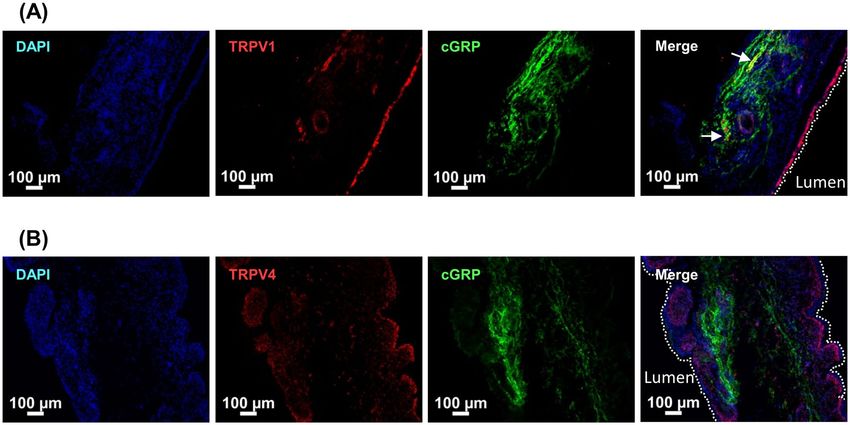
www.nature.com/scientificreports/
KC
Control Total Mild KC Severe KC P valuea P valueb
Number 4 24 12 12
Gender M2, F2 M17, F7 M10, F2 M7, F5
Age (years) 55.0 ± 4.2 27.8 ± 5.1 28.2 ± 4.9 27.4 ± 5.2 < 0.001 0.718
VUDS
CBC (ml) NA 71.3 ± 54.9 138.8 ± 50.5 48.9 ± 12.2 0.001
MBC (ml) NA 215.7 ± 128.9 333.3 ± 68.3 127.5 ± 82.2 0.003
ΔBC (ml) NA 137.0 ± 86.1 194.5 ± 60.6 87.7 ± 75.0 0.022
Pdet (cmH2O) NA 34.3 ± 26.6 30.6 ± 13.1 38.3 ± 36.9 0.743
Qmax (ml/s) NA 8.8 ± 5.5 10.7 ± 6.1 6.3 ± 3.7 0.125
VPdet.max (cmH2O/s) NA 4.2 ± 2.4 3.1 ± 1.1 5.3 ± 2.9 0.076
VAS NA 6.4 ± 2.8 2.2 ± 1.5 7.5 ± 2.3 0.002
Western blot analysis
TRPV1/GAPDH 0.08 ± 0.04 0.58 ± 0.54 0.25 ± 0.27 0.91 ± 0.55 0.017 0.002
TRPV4/GAPDH 0.02 ± 0.01 0.27 ± 0.28 0.08 ± 0.04 0.47 ± 0.29 0.002 < 0.001
Table 1. Clinical characteristics and Western blot analysis data of KC patients and control subjects. The
Western blot analysis was normalized against GAPDH. CBC cystometric bladder capacity, MBC maximal
bladder capacity, ΔBC the difference between MBC and CBC, Pdet detrusor pressure, VPdet.max velocity to reach
maximal Pdet (cm H2O/s, defined as the slope of pressure to time from the start of detrusor contraction to reach
Pdet.max), Qmax maximum flow rate, VAS visual analog score. a P values between all KC patients and controls, bP
values between mild KC and severe KC patients.
M3, p75 low-affinity nerve growth factor receptor, and β-3 adrenergic receptors have been shown to participate
in the pathophysiology of K C9,13,14.
The other possible sensor molecules in the urothelium are ion channels. Transient receptor potential (TRP)
channels expressed in the lower urinary tract are thought to be involved in the bladder’s pathologic f unction15.
However, to our knowledge, little research has been done to study what roles these proteins have in KC. Only
one research group used TRP cation channel subfamily V member 1 (TRPV1) as a marker to evaluate the effect
of lipotoxin and Ba‐Wei‐Die‐Huang‐Wan treatment on the KC rat model16,17. Instead of using an animal model,
the present study used human patients’ bladder samples to investigate the associations between the expression
levels of TRPV proteins and the clinical characteristics of KC. The results of this study may reveal the possible
roles of these proteins in KC.
Results
Clinical data. A total of 24 patients with KC (17 men and seven women, mean age 27.8 ± 5.1 years) and
four control subjects (two men and two women, mean age 55.0 ± 4.2 years) were recruited. Table 1 lists the
profile, urodynamic parameters, and Western blot analysis results of TRPV1 and TRPV4 levels for the different
patient groups. Among the patients with KC, 12 were classified as mild KC (10 men and two women, mean age
28.2 ± 4.9 years), and 12 were classified as severe KC (seven men and five women, mean age 27.4 ± 5.2 years).
Both the maximal bladder capacity (MBC) and cystometric bladder capacity (CBC) were significantly smaller
in patients with severe KC than in those with mild KC. On the other hand, the visual analogue scale (VAS) of
pain and velocity to reach maximal detrusor pressure ( VPdet.max) were significantly greater in bladders with
severe KC than in those with mild KC. Figure 1 shows the associations between these parameters. There was a
significantly positive association between MBC and CBC (r = 0.82, P < 0.001). The MBC, CBC, and the difference
between MBC and CBC (ΔBC) were significantly negatively associated with VAS (r = − 0.75, P = 0.002; r = − 0.68,
P = 0.006; and r = − 0.67, P = 0.01, respectively). Only CBC was significantly negatively associated with V
Pdet.max
(r = − 0.53, P = 0.04).
Elevated TRPV1 and TRPV4 in patients with KC. Western blot analysis was performed to assay the
protein expression level in bladder specimens of all 24 patients with KC and of the four control subjects. Fig-
ure 2A and Table 1 show the results of Western blot analysis. Patients with KC had a significantly higher expres-
sion of TRPV1 and TRPV4 than control subjects. The expressions of TRPV1 and TRPV4 in the bladders of
patients with severe KC were significantly higher than in the bladders of those with mild KC.
Immunostaining was subsequently used to validate the results of Western blot analysis. However, because
the urothelium is prone to nonspecific adsorption of antibodies, a competitive binding assay using a blocking
peptide was first performed to determine the antibody’s binding specificity. Only the TRPV4 antibody was tested
because the blocking peptide (101 amino acids) of TRPV1 was not available. The TRPV4 staining disappeared
when the antibody was preincubated with its blocking peptide, indicating the stain was specific (Supplementary
Fig. S2). The immunofluorescence staining from similar bladder regions shown in Fig. 2B indicated that the pro-
tein expressions of TRPV1 and TRPV4 in the bladders of control subjects were lower than those of KC patients
(semi-quantification was in the Supplementary Table S1), which is consistent with the Western blot analysis.
Scientific Reports | (2021) 11:5200 | https://doi.org/10.1038/s41598-021-84734-4 2
Vol:.(1234567890)
www.nature.com/scientificreports/
Figure 1. Significant associations between urodynamic parameters from KC patients: (A) between MBC and
CBC, (B) between CBC and VPdet.max, and (C) between MBC (black circle), ΔBC (red triangle), or CBC (blue
square) and VAS.
Figure 2. Higher expression of TRPV1 and TRPV4 in KC bladders than in control bladders. (A) Representative
results of Western blot analysis (from different experiments) and scatter plot of the quantification of TRPV1
and TRPV4 to GAPDH in control, mild KC, and severe KC bladder specimens. The bar graph was quantified
using ImageMaster TotalLab. *P < 0.05 and **P < 0.01. Images for GAPDH and from all samples are available in
Supplementary Fig. S1. (B) Representative results of immunofluorescence staining with TRPV1 and TRPV4 in
control (arrows), mild KC, and severe KC bladder specimens. Stronger TRPV1 and TRPV4 fluorescence (red)
was detected in severe KC specimens than in mild KC specimens. Nuclei were labeled with DAPI (blue).
Scientific Reports | (2021) 11:5200 | https://doi.org/10.1038/s41598-021-84734-4 3
Vol.:(0123456789)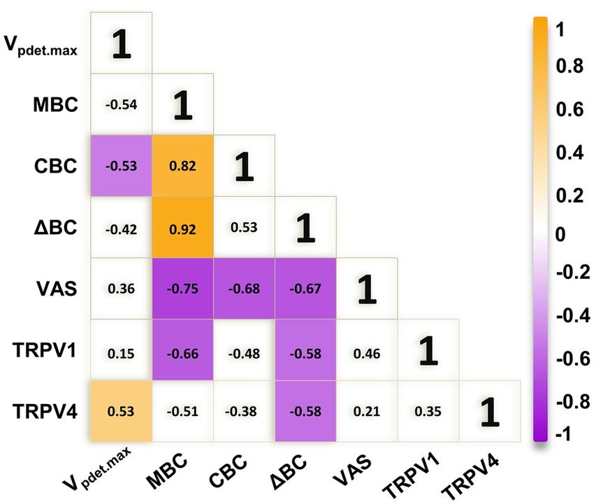
www.nature.com/scientificreports/
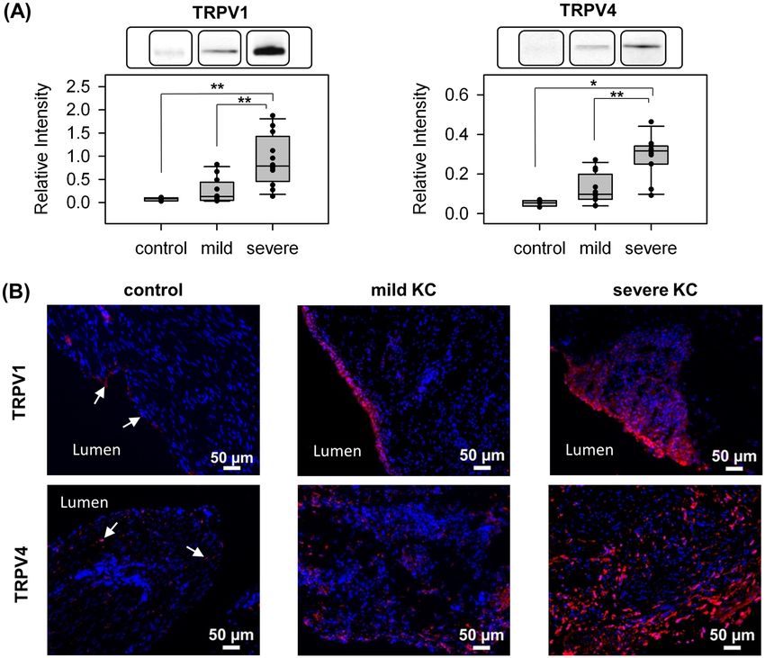
Figure 3. Representative results of immunofluorescence localization of TRPV1 (A) and TRPV4 (B) in mild KC
bladder specimens. Double immunofluorescence of TRPV1 or TRPV4 (red) with CGRP (green) was performed.
Nuclei were labeled with DAPI (blue). TRPV1-positive nerve fibers are indicated by arrows. Dashed white lines
indicate the apical surface of the umbrella cells. The double immunochemical staining revealed that the lack of
co-expression with CGRP for TRPV4, and the expression of TRPV1 in only a few nerve fibers.
Figure 4. Representative results of IHC of TRPV1 (A) and TRPV4 (B) in severe KC bladder specimens. Arrows
indicate TRPV1 or TRPV4 positive cells. The expression of TRPV1 is mainly distributed in all urothelial cell
layers. In contrast, TRPV4 was found in the basal cells and lamina propria. U urothelium, LP lamina propria.
Localization of TRPV1 and TRPV4 in the bladder. Calcitonin gene-related peptide (CGRP), a neuron-
specific marker, typically presents in the nerves distributed within the suburothelium region18. Both TRPV1
and TRPV4 were subsequently costained with CGRP to study the localization of these proteins. The results
of immunofluorescence double-staining for TRPV4 and TRPV1 with CGRP are shown in Fig. 3. Only a few
TRPV1-positive nerve fibers (yellow) were found. No TRPV4-positive nerve fibers were found. To further illus-
trate the localization of TRPV1 and TRPV4, Immunohistochemistry (IHC) was then carried out. Figure 4 shows
that TRPV1 was distributed in all urothelial cell layers. On the other hand, TRPV4 was found in the basal cells
and lamina propria.
Association between urodynamic parameters and expressions of TRPV1 and TRPV4. To
study the possible roles of TPRV1 and TRPV4 in KC, the association between TRPV1 and TRPV4 and between
these two proteins and urodynamic parameters were evaluated. Figure 5 shows the scatter plots with significant
association and all the association results were summarized in Fig. 6. Increased expressions of both TRPV1
and TRPV4 were observed in the bladders of KC patients, but they were not significantly associated (r = 0.35,
Scientific Reports | (2021) 11:5200 | https://doi.org/10.1038/s41598-021-84734-4 4
Vol:.(1234567890)www.nature.com/scientificreports/
Figure 5. Significant associations between urodynamic parameters and the expression of TRPV1 or TRPV4
form KC patients: (A) between MBC (black circle) or ΔBC (red triangle) and TRPV1, (B) between ΔBC and
TRPV4, and (C) between VPdet.max and TRPV4.
Figure 6. Summary of association between urodynamic parameters and TRPVs. Colored boxes represent a
significant association coefficient between the variables (P < 0.05). Chrome yellow and purple indicate positive
and negative associations, respectively.
P = 0.09). In addition, the expression of TRPV1 was significantly negatively associated with MBC and ΔBC
(r = − 0.66, P = 0.01 and r = − 0.58, P = 0.04, respectively). However, the expression of TRPV4 was significantly
positively associated with VPdet.max (r = 0.53, P = 0.01) and significantly negatively associated with ΔBC (r = − 0.58,
P = 0.04).
Discussion
Although there have been studies of TRPV1 and TRPV4 in the bladders, most were based on animal tissue or
cell culture models. Only a few studies were conducted on human bladders. The data presented in the current
study are from bladder tissue of patients with mild and severe KC. The data show, for the first time, that expres-
sion levels of TRPV1 and TRPV4 observed in patients with severe KC are significantly increased over those
in patients with mild KC and in control subjects. According to our previous report, KC patients usually show
significantly greater VPdet.max and smaller bladder capacity than control subjects19. In the present study, the asso-
ciation between different urodynamic parameters and between urodynamic data and protein expression were
studied. The results summarized in Fig. 6 reveal a significant association between (1) reduced bladder capacity
and increased VAS of pain, (2) increased TRPV1 expression and reduced bladder capacity, and (3) increased
TRPV4 expression and increased V Pdet.max.
Both TRPV1 and TRPV4 are nonselective cationic channels activated by a diversity of stimuli such as heat,
acidity, and chemicals. Because mechanical and chemical stimuli also stimulate bladder functions, these two
proteins are likely urothelial sensors for bladder distention. Thus, they are interesting targets for the study of
controlling sensory and motor activity of the bladder. However, although many investigations have been done
on the expression, distribution, and functions of TRPV1 and TRPV4 in the bladder, the conclusions of the role
of TRPV1 and TRPV4 in bladders are still unclear.
Scientific Reports | (2021) 11:5200 | https://doi.org/10.1038/s41598-021-84734-4 5
Vol.:(0123456789)www.nature.com/scientificreports/
The expression of TRPV1 was originally reported to be exclusively expressed in sensory g anglia20. Birder et al.
were the first one to describe the presence of TRPV1 in the urothelial cells from rats21. Although subsequent
studies have not been able to reproduce the expression of TRPV1 in mouse urothelial cells, the presence of nerve
terminals that extend into the urothelium has been r eported22,23.
Similarly, the localization of TRPV1 in human bladders is also controversial. The non-neuronal localiza-
tion of TRPV1 in detrusor smooth muscle cells and interstitial cells of Cajal and the localization of TRPV1 in
urothelial cells have been r eported24–27. In accordance with the previous r eports26,27, the expression of TRPV1
was observed in both the urothelium and suburothelium in the present study. The urothelium layerwas labeled
stronger than suburothelium layers. As the severity of KC increased, TRPV1 was distributed into deeper bladder
layers. However, only a few TRPV1 stains of the suburothelium were positively confirmed as neuronal processes
by co-staining with CGRP.
Several studies have shown higher TRPV1 expression in patients with overactive b ladder26–29. When patients
with neurogenic detrusor overactivity (DO) responded to intravesical resiniferatoxin (RTX) therapy, a significant
decrease in TRPV1-immunoreactive nerve fibers was observed26. This significant decrease in TRPV1-immuno-
reactivity was only noted in the basal cell layer, and the percentage of changes was comparable to the changes in
suburothelial TRPV1 nerve fiber density27. However, for idiopathic DO patients with successful intravesical RTX
treatment, TRPV1 was observed to be overexpressed in the urothelium and s uburothelium28. Despite different
localizations of TRPV1 in these studies, the involvement of TRPV1 in the pathophysiology of DO was suggested.
In this study, the expression of TRPV1 was significantly greater in both the urothelium and suburothelium of
specimens from patients with severe KC compared with those from patients with mild KC or control subjects.
This suggested that TRPV1 might also be involved in the pathophysiology of KC.
In our study, a significant negative association between the expression of TRPV1 with MBC was observed.
The possible role of TRPV1 in bladder function has been studied using TRPV1-knockout animals. Daly et al.
reported attenuated low threshold afferent responses in TRPV1-knockout mice but an unchanged high-threshold
afferent sensitivity, suggesting that neuronal TRPV1 channels in the suburothelium are needed in the excit-
ability of low-threshold bladder afferents30. Birder et al. demonstrated an increase in bladder capacity in anes-
thetized TRPV1-knockout mice but an unaffected micturition frequency in conscious TRPV1-knockout mice.
This suggests that TRPV1-mediated mechanisms are responsible for setting the micturition threshold under
anesthesia31,32. These studies revealed the negative association between bladder capacity and the expression of
TRPV1, which is consistent with our observation.
With regard to TRPV4, it is a broadly expressed ion channel in the body, such as the central and peripheral
nervous system, hair, skin, and the bladder. Within the rat and mouse urinary bladder, the localization of TRPV4
has been demonstrated mainly in the plasma membrane of the intermediate and basal cells and to a less extent
in the detrusor smooth muscle c ells22,23,32–35. Our observations agree with these reports in that we observed that
TRPV4 is localized in the basal cells and lamina propria.
Apart from the localization of TRPV4 in the bladder, our results also revealed that the expression of TRPV4
increased as the severity of KC increased, indicating that the possible involvement of TRPV4 in the pathophysi-
ology of KC could not be ruled out. Similarly, the increased expression of TRPV4 was detected in the bladder
urothelium in rats subjected to repeated variate stress (RVS)34. Studies indicated that the intravesical adminis-
tration of the TRPV4 antagonist HC067047 to block TRPV4 could ameliorate decreased bladder capacity and
increased voiding frequency in both RVS and cyclophosphamide-induced cystitis a nimals34,36. More recently,
the TRPV4 agonist, GSK1016790A, was used to treat detrusor underactivity. After the intravesical application of
GSK1016790A, increased voiding frequency and reduced bladder capacity, voided volume, and post-void residu-
als were observed, without an increase in nonvoiding c ontractions35,37. Based on these studies, our observation
of increased expression of TRPV4 indicates that TRPV4 might participate in the KC symptoms of decreased
bladder capacity and increased voiding frequency.
Previously, Gevaert et al. made two observations. First, the TRPV4-knockout mice exhibited a lower fre-
quency of voiding contractions and a higher frequency of nonvoiding contractions. Second, the amplitude
of spontaneous contractions in explanted bladder strips and intravesical stretch-evoked ATP release in iso-
lated whole bladders from TRPV4-knockout mice were significantly r educed32. Based on these observations,
they raised the possibility that TRPV4 plays a critical role in urothelium-mediated transduction of intravesical
mechanical pressure. To confirm this possibility, Mochizuki et al. established a cell-stretch system to investigate
stretch-evoked changes in intracellular Ca2+ concentration and ATP release, and further indicated that TRPV4
induces robust Ca2+ influx and contributes to ATP release upon extension. Thus TRPV4 is critically involved in
the sensing mechanical stretch stimuli in the b ladder33. On the other hand, Janssen et al. suggested that TRPV4
channels could be activated by urothelial stretch because TRPV4 channels are connected to adherence junctions
and the actin c ytoskeleton38,39. Taken together, the significantly positive association between the expression of
TRPV4 with V Pdet.max in the present study might be correlated with the mechanosensory properties of TRPV4.
To our knowledge, this is the first study that uses human samples to provide the association between urody-
namic parameters and the expression of TRPV1 and TRPV4. Although the present study demonstrated remark-
able changes in the expression of TRPV1 and TRPV4 and a significant association between the expression of these
proteins and the clinical characteristics of KC, there are still limitations. First, only a small number of patients
was studied and a larger sample size should be investigated in the future. Second, control subjects were not age-
matched with KC patients, and there were no urodynamic parameter data for control subjects. Therefore, the
association between urodynamic parameters and the protein expression can only be obtained between patients
with mild and severe KC and not between KC patients and control subjects. Third, the tissue collection was not
the same in all analysed groups resulting only bladder tissue form severe KC contained muscle layers. However,
this problem could be solved using immunostaining from the similar region of the bladders.
Scientific Reports | (2021) 11:5200 | https://doi.org/10.1038/s41598-021-84734-4 6
Vol:.(1234567890)www.nature.com/scientificreports/
In conclusion, this study reveals that in KC patients, a higher degree of VAS is associated with a smaller value
of MBC and CBC, and a greater magnitude of V Pdet.max is associated with a smaller value of CBC. Our data also
show that both TRPV1 and TRPV4 are upregulated in the urothelium of KC patients, and the degree of upregu-
lation increases with the degree of severity. Elevated TRPV1 and TRPV4 are associated with the smaller MBC
and the greater VPdet.max, respectively. Detailed studies are needed to prove the possible involvements of these
two channels as mechanosensors in the pathogenesis of KC bladders.
Materials and methods
Patients. Twenty-four patients with proven KC and four control subjects were enrolled in the study. The
control subjects were patients with bladder cancer or prostate cancer undergoing radical surgery without urinary
tract infection or irritative bladder symptoms. Of the patients with proven KC, 12 showed signs of mild blad-
der dysfunction (defined as MBC ≥ 300 ml under cystoscopic hydrodistention) and 12 showed signs of severe
bladder dysfunction (defined as MBC < 300 ml under cystoscopic hydrodistention). The inclusion criteria for
KC included the regular misuse of ketamine for more than six months and new onset of lower urinary tract
symptoms without bacterial urinary tract infection, stone disease, or malignancy. Computed tomography and
cystoscopic hydrodistention confirmed that the patients with KC had a contracted bladder with severe erosive
bladder mucosa and profuse bleeding after hydrodistention.
Video urodynamic study. Patient evaluations included history taking, physical examination, clinical
symptoms, VAS of pain, video urodynamic study (VUDS), cystoscopy, and renal ultrasound. During VUDS, a
6-Fr transurethral dual-channel catheter was inserted into the urinary bladder to record the intravesical pres-
sure (Pves) and post-void residual urine volume. The intra-abdominal pressure (Pabd) was measured by placing an
8-Fr catheter mounted with a water-filled balloon. Perineal surface electrodes were placed for external sphincter
electromyography. The VUDS was routinely performed at least twice to confirm bladder and bladder outlet con-
ditions during storage and voiding phases by infusing 20% urography in saline at a rate of 10–20 ml/min. The
detrusor pressure (Pdet) was calculated by subtracting the P abd from the P
ves electronically. A C-arm cinefluoro-
scope was used to visualize the bladder neck and urethra during the filling and voiding phases. The urodynamic
parameters were recorded, including P ves, Pdet, CBC, and maximum flow rate ( Qmax). During the voiding phase,
the slope of detrusor pressure rise to reach Qmax was calculated as VPdet.max. The terminology used in this study
followed the recommendations of the International Continence Society40.
Cystoscopic hydrodistention and bladder tissue retrieval. All patients underwent cystoscopic hydr-
odistention under anesthesia at an intravesical pressure of 80 cm of water, and bladder glomerulations and MBC
were recorded. The difference between MBC and CBC was defined as ΔBC, indicating the residual detrusor
distensibility under anesthesia. Random biopsies of the posterior bladder wall were obtained after cystoscopic
hydrodistention. Each specimen was 2 mm in diameter, and only bladder mucosa was obtained. The bladder
biopsy specimens were sent to the hospital pathology department for investigation of malignancy.
Patients were treated conservatively by intravesical hyaluronic acid instillations, non-steroid anti-inflam-
matory drugs, or botulinum toxin A injections. Patients who continued to have bladder pain, severe urinary
frequency, and a small MBC less than 150 ml after cystoscopic hydrodistention and conservative management
were offered augmentation enterocystoplasty with partial cystectomy for rapid relief of symptoms and early
ork41. Bladder tissue was harvested from the partial cystectomy specimen.
return to w
The study was approved by the institutional review board and Ethics Committee of the Buddhist Tzu Chi Gen-
eral Hospital (IRB number 104-163-A). The bladder tissue samples were collected after obtaining an informed
consent form. All research activities were performed in accordance with the guidelines and regulations of the
Declaration of Helsinki. Bladder specimens were retrieved from partial cystectomy in patients with severe KC
and from bladder biopsies in those with mild KC. The bladder biopsies were taken at the same sites in the control
subjects and prepared using the same methods.
Protein extraction. Bladder tissue was dissected out and homogenized in liquid nitrogen. The bladder
powder was then transferred to centrifuge tubes containing lysis buffer and centrifuged at 4 °C for 20 min at
16,000 g. The remaining insoluble pellet was discarded and the soluble bladder protein extracts were either used
immediately or stored at − 80 °C. The protein concentration was measured using a Bio-Rad protein assay kit.
Western blot analysis. Western blot analysis was performed similarly to previously described19. Tissue
lysates from 12 patients with mild KC, 12 patients with severe KC, and four control subjects were separated by
1-dimensional sodium dodecyl sulfate–polyacrylamide gel electrophoresis and transferred onto an Immobilon-
P polyvinylidene fluoride transfer membrane (Millipore, Bedford, MA) by electroblotting. The first applied anti-
bodies included anti-TRPV1 and anti-TRPV4 (Abcam, #ab111973 and #ab94868, Cambridge, MA). The second
applied antibody was IgG-conjugated horseradish peroxidase (anti-rabbit, Gene Tex, Inc., Irvine, CA). Signals
were visualized by the enhanced chemiluminescence kit (BioRad, Madrid, Spain). The scanned films were quan-
tified using ImageMaster TotalLab, Version 2.01 (GE Healthcare, Piscataway, NJ), and data were expressed as
relative fold to GAPDH.
Immunofluorescence staining. Immunofluorescence stain was performed according to the previous
ethod42. The urinary bladder specimens were first fixed with an ice-cold solution of 4% formaldehyde in phos-
m
phate-buffered saline (PBS, pH 7.4) and then rinsed with ice-cold PBS containing 15% sucrose. Four sections per
Scientific Reports | (2021) 11:5200 | https://doi.org/10.1038/s41598-021-84734-4 7
Vol.:(0123456789)www.nature.com/scientificreports/
specimen were cut using a cryostat at a thickness of 8 μm and collected on new silane III-coated glass slides. The
sections were then incubated overnight at 4 °C with primary antibodies (-CGRP, Abcam, #ab81887, Cambridge,
MA, and others were the same as used in Western blotting), rinsed with 0.1% Tween-20 in PBS, and incubated
with conjugated Alexa 594 secondary antibodies (Thermo Fisher Scientific, Waltham, MA). The sections were
counterstained with 4′,6-diamidino-2-phenylindole (Sigma Chemical Company, St. Louis, MO). Negative con-
trols included the isotype of the primary antibody.
Peptide competition assay for TRPV4 antibody. The TRPV4-blocking peptide corresponding to the
amino acid 720–769 of Human TRPV4 isoform 2 (NP_671737) was provided by Prof. Cheng-Kang Chiang
(National Dong Hwa University, Hualien, Taiwan). The TRPV4 antibody was diluted in blocking buffer (1:100
dilution) and a 5-times excess of blocking peptide by weight was added to the antibody solution. The mixture
was incubated with agitation overnight at 4 °C. The staining procedure was done with neutralized or unblocked
antibody on the two samples.
Immunohistochemistry. IHC was performed using the UltraVision Quanto Detection System HRP DAB
(ThermoScientific, Cheshire, UK). Slides were first treated with hydrogen peroxide block reagent (ThermoSci-
entific) and rinsed with PBS. Following that, Ultra V Block reagent (ThermoScientific) was used to block non-
specific binding. The slides were subsequently incubated with primary antibodies (the same as used in Western
blotting), Primary Antibody Amplifier Quanto (ThermoScientific), and HRP Polymer Quanto (ThermoScien-
tific) and DAB Quanto Chromogen and DAB Quanto Substrate (ThermoScientific) were used to visualize. The
slides were then counterstained with hematoxylin.
Statistical analysis. Data were expressed as mean ± standard deviation (SD). Because the sample distribu-
tion is not normal and the sample size is small, differences in protein expressions between mild KC, severe KC,
and controls were analyzed using the nonparametric Mann–Whitney test. The association between the bladder
protein expressions and urodynamic parameters was analyzed using the Pearson correlation. A P value of less
than 0.05 was considered statistically significant.
Received: 26 November 2020; Accepted: 18 February 2021
References
1. Wang, L. J. et al. Difference in long-term relapse rates between youths with ketamine use and those with stimulants use. Subst.
Abuse Treat. Prev. Policy 13, 50 (2018).
2. Shahani, R., Streutker, C., Dickson, B. & Stewart, R. J. Ketamine-associated ulcerative cystitis: a new clinical entity. Urology 69,
810–812 (2007).
3. Chu, P. S. K. et al. The destruction of the lower urinary tract by ketamine abuse: a new syndrome?. BJU Int. 102, 1616–1622 (2008).
4. Baker, S. C., Shabir, S., Georgopoulos, N. T. & Southgate, J. Ketamine-induced apoptosis in normal human urothelial cells: a direct,
N-methyl-d-aspartate receptor-independent pathway characterized by mitochondrial stress. Am. J. Pathol. 186, 1267–1277 (2016).
5. Gu, D. et al. Long-term ketamine abuse induces cystitis in rats by impairing the bladder epithelial barrier. Mol. Biol. Rep. 41,
7313–7322 (2014).
6. Wood, D. et al. Recreational ketamine: from pleasure to pain. BJU Int. 107, 1881–1884 (2011).
7. Liu, K. M. et al. Ketamine-induced ulcerative cystitis and bladder apoptosis involve oxidative stress mediated by mitochondria
and the endoplasmic reticulum. Am. J. Physiol. Ren. Physiol. 309, F318–F331 (2015).
8. Lee, C. L., Jiang, Y. H. & Kuo, H. C. Increased apoptosis and suburothelial inflammation in patients with ketamine-related cystitis:
a comparison with non-ulcerative interstitial cystitis and controls. BJU Int. 112, 1156–1162 (2013).
9. Tsai, Y. C., Birder, L. & Kuo, H. C. Abnormal sensory protein expression and urothelial dysfunction in ketamine-related cystitis
in humans. Int. Neurourol. J. 20, 197 (2016).
10. Rajandram, R. et al. Intact urothelial barrier function in a mouse model of ketamine-induced voiding dysfunction. Am. J. Physiol.
Ren. Physiol. 310, F885–F894 (2016).
11. Chen, H. et al. Disruption of Cav 1. 2-mediated signaling is a pathway for ketamine-induced pathology. Nat. Commun. 11, 1–13
(2020).
12. Keay, S. K., Birder, L. A. & Chai, T. C. Evidence for bladder urothelial pathophysiology in functional bladder disorders. Biomed.
Res. Int. https://doi.org/10.1155/2014/865463 (2014).
13. Baker, S. C. et al. Nerve hyperplasia: a unique feature of ketamine cystitis. Acta Neuropathol. Commun. 1, 64 (2013).
14. Meng, E. et al. Involvement of purinergic neurotransmission in ketamine induced bladder dysfunction. J. Urol. 186, 1134–1141
(2011).
15. Merrill, L., Gonzalez, E. J., Girard, B. M. & Vizzard, M. A. Receptors, channels, and signalling in the urothelial sensory system in
the bladder. Nat. Rev. Urol. 13, 193 (2016).
16. Lee, W. C. et al. Potential orphan drug therapy of intravesical liposomal onabotulinumtoxin-A for ketamine-induced cystitis by
mucosal protection and anti-inflammation in a rat model. Sci. Rep. 8, 5795 (2018).
17. Lee, W. C. et al. Ba-Wei-Die-Huang-Wan (Hachimi-jio-gan) can ameliorate ketamine-induced cystitis by modulating neurorecep-
tors, inflammatory mediators, and fibrogenesis in a rat model. Neurourol. Urodyn. 38, 2159–2169 (2019).
18. Smet, P., Moore, K. & Jonavicius, J. Distribution and colocalization of calcitonin gene-related peptide, tachykinins, and vasoactive
intestinal peptide in normal and idiopathic unstable human urinary bladder. Lab. Invest. 77, 37–49 (1997).
19. Yang, H. H., Zhai, W. J. & Kuo, H. C. The putative involvement of actin-binding proteins and cytoskeleton proteins in pathologi-
cal mechanisms of ketamine cystitis—revealed by a prospective pilot study using proteomic approaches. Proteom. Clin. Appl. 11,
1600085 (2017).
20. Caterina, M. J. et al. The capsaicin receptor: a heat-activated ion channel in the pain pathway. Nature 389, 816–824 (1997).
21. Birder, L. A. et al. Vanilloid receptor expression suggests a sensory role for urinary bladder epithelial cells. PNAS 98, 13396–13401
(2001).
Scientific Reports | (2021) 11:5200 | https://doi.org/10.1038/s41598-021-84734-4 8
Vol:.(1234567890)www.nature.com/scientificreports/
22. Yamada, T. et al. Differential localizations of the transient receptor potential channels TRPV4 and TRPV1 in the mouse urinary
bladder. J. Histochem. Cytochem. 57, 277–287 (2009).
23. Yu, W., Hill, W. G., Apodaca, G. & Zeidel, M. L. Expression and distribution of transient receptor potential (TRP) channels in
bladder epithelium. Am. J. Physiol. Ren. Physiol. 300, F49–F59 (2011).
24. Aa, F. V., Roskams, T., Blyweert, W. & Ridder, D. D. Interstitial cells in the human prostate: a new therapeutic target?. Prostate 56,
250–255 (2003).
25. Lazzeri, M. et al. Immunohistochemical evidence of vanilloid receptor 1 in normal human urinary bladder. Eur. Urol. 46, 792–798
(2004).
26. Brady, C. et al. Parallel changes in bladder suburothelial vanilloid receptor TRPV1 and pan-neuronal marker PGP9: 5 immunore-
activity in patients with neurogenic detrusor overactivity after intravesical resiniferatoxin treatment. BJU Int. 93, 770–776 (2004).
27. Apostolidis, A. et al. Capsaicin receptor TRPV1 in urothelium of neurogenic human bladders and effect of intravesical resinifera-
toxin. Urology 65, 400–405 (2005).
28. Liu, H. T. & Kuo, H. C. Increased expression of transient receptor potential vanilloid subfamily 1 in the bladder predicts the
response to intravesical instillations of resiniferatoxin in patients with refractory idiopathic detrusor overactivity. BJU Int. 100,
1086–1090 (2007).
29. Li, M., Sun, Y., Simard, J. M. & Chai, T. C. Increased transient receptor potential vanilloid type 1 (TRPV1) signaling in idiopathic
overactive bladder urothelial cells. Neurourol. Urodyn. 30, 606–611 (2011).
30. Daly, D., Rong, W., Chess-Williams, R., Chapple, C. & Grundy, D. Bladder afferent sensitivity in wild-type and TRPV1 knockout
mice. J. Physiol. 583, 663–674 (2007).
31. Birder, L. et al. Altered urinary bladder function in mice lacking the vanilloid receptor TRPV1. Nat. Neurosci. 5, 856–860 (2002).
32. Gevaert, T. et al. Deletion of the transient receptor potential cation channel TRPV4 impairs murine bladder voiding. J. Clin. Invest.
117, 3453–3462 (2007).
33. Mochizuki, T. et al. The TRPV4 cation channel mediates stretch-evoked Ca2+ influx and ATP release in primary urothelial cell
cultures. J. Biol. Chem. 284, 21257–21264 (2009).
34. Merrill, L. & Vizzard, M. A. Intravesical TRPV4 blockade reduces repeated variate stress-induced bladder dysfunction by increas-
ing bladder capacity and decreasing voiding frequency in male rats. Am. J. Physiol. Regul. Integr. Comp. Physiol. 307, R471–R480
(2014).
35. Deruyver, Y. et al. Intravesical activation of the cation channel TRPV4 improves bladder function in a rat model for detrusor
underactivity. Eur. Urol. 74, 336–345 (2018).
36. Everaerts, W. et al. Inhibition of the cation channel TRPV4 improves bladder function in mice and rats with cyclophosphamide-
induced cystitis. PNAS 107, 19084–19089 (2010).
37. Takaoka, E. I. et al. Effect of TRPV4 activation in a rat model of detrusor underactivity induced by bilateral pelvic nerve crush
injury. Neurourol. Urodyn. 37, 2527–2534 (2018).
38. Janssen, D. A. et al. The mechanoreceptor TRPV4 is localized in adherence junctions of the human bladder urothelium: a mor-
phological study. J. Urol. 186, 1121–1127 (2011).
39. Janssen, D. et al. TRPV 4 channels in the human urogenital tract play a role in cell junction formation and epithelial barrier. Acta
Physiol. 218, 38–48 (2016).
40. Abrams, P. et al. The standardisation of terminology of lower urinary tract function: report from the Standardisation Sub-committee
of the International Continence Society. Neurourol. Urodyn. 21, 167–178 (2002).
41. Chung, S. D., Wang, C. C. & Kuo, H. C. Augmentation enterocystoplasty is effective in relieving refractory ketamine-related blad-
der pain. Neurourol. Urodyn. 33, 1207–1211 (2014).
42. Jhang, J. F., Hsu, Y. H., Jiang, Y. H. & Kuo, H. C. The role of immunoglobulin E in the pathogenesis of ketamine related cystitis and
ulcerative interstitial cystitis: an immunohistochemical study. Pain Physician 19, E581–E587 (2016).
Acknowledgements
This research was supported by a Grant from the Ministry of Science and Technology of Republic of China
(Taiwan) for financial support under contract No. NSC 105-2113-M-303-001.
Author contributions
H.-H.Y.: study design, data analysis, manuscript writing. J.-F.J.: patient enrolment, urodynamic study. Y.-H.H.:
data analysis. Y.-H.J.: patient enrolment. W.-J.Z.: Data collection. H.-C.K.: study concept, manuscript writing
about the clinical part, and critical comment.
Competing interests
The authors declare no competing interests.
Additional information
Supplementary Information The online version contains supplementary material available at https://doi.
org/10.1038/s41598-021-84734-4.
Correspondence and requests for materials should be addressed to H.-C.K.
Reprints and permissions information is available at www.nature.com/reprints.
Publisher’s note Springer Nature remains neutral with regard to jurisdictional claims in published maps and
institutional affiliations.
Open Access This article is licensed under a Creative Commons Attribution 4.0 International
License, which permits use, sharing, adaptation, distribution and reproduction in any medium or
format, as long as you give appropriate credit to the original author(s) and the source, provide a link to the
Creative Commons licence, and indicate if changes were made. The images or other third party material in this
article are included in the article’s Creative Commons licence, unless indicated otherwise in a credit line to the
material. If material is not included in the article’s Creative Commons licence and your intended use is not
permitted by statutory regulation or exceeds the permitted use, you will need to obtain permission directly from
the copyright holder. To view a copy of this licence, visit http://creativecommons.org/licenses/by/4.0/.
© The Author(s) 2021
Scientific Reports | (2021) 11:5200 | https://doi.org/10.1038/s41598-021-84734-4 9
Vol.:(0123456789)You can also read