Opposing Functions of the Ets Factors NERF and ELF-1 During Chicken Blood Vessel Development
←
→
Page content transcription
If your browser does not render page correctly, please read the page content below
Opposing Functions of the Ets Factors NERF and ELF-1
          During Chicken Blood Vessel Development
               John Gaspar, Shelley Thai, Carole Voland, Antoinise Dube, Towia A. Libermann,
                                    M. Luisa Iruela-Arispe, Peter Oettgen

Objective—The purpose of this study was to evaluate the role of the Ets factor NERF in the regulation of the Tie1 and Tie2
  genes during chicken blood vessel development.
Methods and Results—We have isolated the full-length cDNA for the chicken homologue of the human Ets factor NERF2
  (cNERF2). Northern blot analysis and in situ hybridization demonstrate that cNERF2 is enriched in the developing
  blood vessels of the chicken chorioallantoic membrane. Interestingly, cNERF2 functions as a competitive inhibitor of
  a highly related Ets factor cELF-1, which we have previously shown to be enriched in chicken blood vessel
  development. Although in vitro–translated cELF-1 and cNERF2 can bind equally well to conserved Ets binding sites
  in the promoters of the Tie1 and Tie2 genes, cELF-1 preferentially binds to the Ets sites in these promoters during early
  stages of chicken blood vessel development, suggesting that cNERF may bind during later stages of blood vessel
  development and vascular remodeling.
Conclusions—cNERF2 is enriched during embryonic and extraembryonic blood vessel development in the chicken and
  facilitates tight control of Tie1 and Tie2 gene regulation. (Arterioscler Thromb Vasc Biol. 2002;22:1106-1112.)
                Key Words: angiogenesis 䡲 vasculogenesis 䡲 transcription 䡲 vascular biology 䡲 endothelium

V     asculogenesis is the development of a primary vascular
      network during embryogenesis. The Tie1 and Tie2
genes constitute a family of endothelium-specific receptor
                                                                               in the Ets binding sites of the promoter of the Flk-1 gene lead
                                                                               to a marked reduction in endothelium-specific LacZ-directed
                                                                               gene expression.9 Likewise, in transgenic animals in which
tyrosine kinases that are critical for vascular development.1                  LacZ expression is directed throughout the vasculature by the
They are expressed predominantly on endothelial cells of the                   Tie2 promoter and enhancer, a mutation in an Ets binding site
developing vasculature. Targeted disruption of the Tie genes                   in the core enhancer leads to a marked reduction in vascularly
leads to abnormalities in vascular development, including the                  directed LacZ gene expression. We have recently identified
formation of leaky dilated blood vessels, with associated                      conserved Ets binding sites in the Tie1 and Tie2 promoters,
edema and hemorrhage that result in early embryonic death.2                    which are necessary for the vascular-specific expression of
Mutations in the Tie2 gene have been identified in humans,                     these genes.10,11
resulting in venous malformations.3 Tie1 and Tie2 are also                        The Ets genes are a family of transcription factors that play
upregulated during tumor angiogenesis.4,5                                      a central role in regulating the genes involved in develop-
   Transcription factors have been shown to serve as master                    ment, cellular differentiation, and proliferation.12 All Ets
switches of several developmental processes, including he-                     factors share a highly conserved DNA binding domain, the
matopoiesis and myogenesis, in which several cell- or tissue-                  ETS domain, which has a winged helix-turn-helix structure.
specific transcription factors have been identified. One ap-                   Ets proteins recognize DNA with an internal conserved DNA
proach toward identifying the types of transcription factors                   binding core motif, GGAA/T. We have examined the ability
required for a developmental process is to examine the                         of several different Ets factors to regulate the Tie1 and Tie2
regulatory regions of genes known to be critical for this                      genes. Our previous studies have demonstrated that NERF2
process and identify the classes of transcription factors that                 and ELF-1 (both are human Ets factors) are potent transacti-
may be necessary to bind to these regions. Examination of the                  vators of the Tie1 and Tie2 genes and can bind to specific Ets
main regulatory elements of the Flt-1, Tie1, and Tie2 genes                    sites within the promoters of these genes.10,11,13 To extend
reveals several conserved putative binding sites for the Ets                   these studies and to demonstrate the potential in vivo rele-
family of transcription factors that are critical for the activity             vance of Ets factors during vascular development, we have
of the promoters and enhancers of these genes.6 – 8 Mutations                  identified Ets factors that are expressed during chicken blood

  Received May 3, 2002; revision accepted May 10, 2002.
  From the Cardiology Division (J.G., C.V., A.D., P.O.) and New England Baptist Bone and Joint Institute (J.G., C.V., A.D., T.A.L., P.O.), Beth Israel
Deaconess Medical Center and Harvard Medical School, Boston, Mass, and the Department of Molecular Cell and Developmental Biology (S.T.,
M.L.I.-A.), University of California at Los Angeles.
  Correspondence to Peter Oettgen, Harvard Institutes of Medicine, 4 Blackfan Circle, Boston, MA 02115. E-mail joettgen@caregroup.harvard.edu
  © 2002 American Heart Association, Inc.
  Arterioscler Thromb Vasc Biol. is available at http://www.atvbaha.org                                  DOI: 10.1161/01.ATV.0000023427.92642.CD

                                                                        1106
Gaspar et al NERF and ELF-1 Roles in Vascular Development 1107
vessel development. Compared with mammals, as a model the embryos were obtained and suspended in 80% glycerol with the
system, the chicken offers the advantage of easy access to use of a 3CCD Toshiba camera on a Nikon SMZ-U dissecting
microscope. Digoxigenin-labeled RNA probes were prepared per the
developing blood vessels, particularly in the rich extraembry-
manufacturer’s recommendations (Roche). The level of digoxigenin
onic vessels within the chorioallantoic membrane (CAM). incorporation was assessed by using a dot-blot comparison with a
Using this model, we identified the chicken homologue of standard (Roche).
ELF-1 (cELF-1) and demonstrated that in addition to being a
strong transactivator of the Tie1 and Tie2 genes, ELF-1 is DNA Transfection Assays
enriched in developing blood vessels of the chicken.13 In the Cotransfections of 1.5 to 2⫻105 mouse endothelial cells (MS-1) or
293 HEK cells were performed by using 300 ng of the reporter gene
present report, we have extended these studies by identifying constructs, pGL3 Tie1 (KH1) or pGL3 Tie2 (PstI-HindIII), as
the chicken homologue of the Ets factor NERF2 (cNERF2) previously described,10,11 and a total of 300 ng of the expression
and have demonstrated that it functions as a competitive vector DNA with 3 L plus reagent and 2 L Lipofectamine
inhibitor of cELF-1. The expression of positive and negative (GIBCO-BRL). Cells were washed with serum-free DMEM. A total
transcriptional regulators of the Tie1 and Tie2 genes may of 400 L serum-free DMEM was added per well. Liposomes were
incubated with the DNA in 100 L serum-free DMEM for 15
facilitate tight control of their expression during different minutes at room temperature and then with the cells for 3 hours at
stages of blood vessel development. 37°C. DMEM (500 L) containing 20% FCS was added, and the
cells were harvested 16 hours after transfection and assayed for
Methods luciferase activity. Transfections were performed in triplicate and
were repeated independently in duplicate with the use of 2 depen-
Cell Culture and Northern Blot Analysis dently cloned cNERF expression plasmids with similar results.
Human embryonic kidney (HEK) 293 cells (American Type Culture Cotransfection of an additional plasmid for determination of trans-
Collection) were grown in 10% FCS and DMEM. Total RNA was fection efficiency was omitted because potential artifacts with this
extracted from the CAM and from blood derived from the CAM at technique have been reported18 and because many commonly used
different developmental stages, as previously described.14 Ten mi- viral promoters contain potential binding sites for Ets factors.
crograms of total RNA was electrophoresed and transferred onto a
Nytran membrane (Schleicher & Schuell). The filter was blocked in Electrophoretic Mobility Shift Assay
prehybridization solution (50% deionized formamide, 6⫻ SSPE, 5⫻ DNA binding reactions were performed as previously described.15,19
Denhardt’s solution, 1% SDS, 0.1 mg/mL yeast tRNA, and 0.1 In brief, 2-L samples of in vitro–translated proteins or cellular
mg/mL salmon sperm DNA) and then hybridized with a cNERF- extracts were incubated in the absence or presence of polyclonal
specific probe at 42°C overnight. To normalize for loading and antibodies specific to NERF (Santa Cruz) or ELF-1 at room
transfer efficiency, the membranes were rehybridized with a probe temperature for 15 minutes. 32P-labeled double-stranded probes
for 36B4. (30 000 cpm) were then added, along with 50 ng cold mutant
oligonucleotides to reduce the background. Samples were incubated
RT-PCR and Chicken -Phage Library Screen for 15 to 20 minutes at room temperature and run on a 4%
To identify Ets factors that are expressed in the developing blood polyacrylamide gel (acrylamide-bisacrylamide, 37.5:1) containing a
vessels of the chicken CAM, reverse transcription (RT)–polymerase buffer of 0.5⫻ TBE (containing 45 mmol/L Tris borate and
chain reaction (PCR) was performed by using RNA extracted from 1 mmol/L EDTA). Oligonucleotides used as probes and for compe-
CAM blood vessels. cDNA was generated from 2 g total RNA by tition studies were as follows: mouse Tie2 promoter wild-type
using random hexamer priming (GIBCO-BRL) and Moloney murine oligonucleotide, 5⬘-TGCAAAGGAAACAGGAAAAAGGA-
leukemia virus reverse transcriptase (GIBCO-BRL). Degenerate ACTTAAC-3⬘ and 3⬘-ACGTTTCCTTTGTCCTTTTTCCTTGA-
oligonucleotides corresponding to conserved regions within the Ets ATTG -5⬘; mouse Tie1 promoter wild-type oligonucleotide, 5⬘-
DNA binding domain were used as previously described.15 PCR CCATCATTTCCTCTTCCTCCCCAG-3⬘ and 3⬘-GGTAGTAA-
fragments were subcloned into the TA cloning vector (Invitrogen), AGGAGAAGGAGGGGTC-5⬘; mouse Tie2 promoter mutant (bold)
and clones containing fragments of the expected sizes were se- oligonucleotide, 5⬘-TGCAAAGGAAACAGCAAAAAGCAAC-
quenced. An embryonic day (E)5 chicken yolk sac library (Strat- TTAAC-3⬘ and 3⬘-ACGTTTCCTTTGTCGTTTTTCGTTG-
agene) was plated and screened with a partial cDNA fragment for AATTG-5⬘; and mouse Tie1 promoter mutant (bold) oligonucleo-
cNERF. Eight partial and 1 full-length cDNA clones were isolated tide, 5⬘-CCATCATTTAATCTTAATCCCCAG-3⬘ and 3⬘-GGTAG-
and sequenced at the Beth Israel Deaconess Medical Center core TAAATTAGAATTAGGGGTC-5⬘.
sequencing facility by using an Applied Biosystems automatic DNA
sequencer. Verification of the full-length cNERF sequence was also Results
confirmed by RT-PCR with cDNA generated from RNA that was
extracted from the chicken CAM at E7 with the use of cNERF-
Isolation of cNERF2
specific primers. PCR reactions were performed as previously In an effort to identify transcription factors belonging to the
described.16 Ets transcription factor family that are expressed during
vasculogenesis, total RNA was extracted from developing
In Situ Hybridization blood vessels of the chicken CAM at E10. RT-PCR was
Whole-mount in situ hybridization on E5 chick embryos and E10 to performed with degenerate PCR primers corresponding to
11E CAMs were carried out as described by Wilkinson et al.17 In conserved regions of the Ets domain, allowing the identifi-
brief, embryos were fixed, dehydrated, and rehydrated through a
methanol series and washed in 1⫻ PBT (containing PBS plus 0.1% cation of a partial DNA sequence with highest homology to
Tween 20). Embryos were then permeabilized with RIPA buffer the Ets family NERF. This fragment was used to screen a
(containing 150 mmol/L NaCl, 1% Nonidet P-40, 0.5% deoxy- chicken yolk sac cDNA library and to isolate several cDNA
cholate, 0.1% SDS, 1 mmol/L EDTA, and 50 mmol/L Tris-HCl, pH clones encoding the full-length cNERF2. Several clones were
8.0) for 30 minutes with agitation at room temperature. After color sequenced to verify the nucleotide and amino acid sequence
was developed to the appropriate intensity, specimens were washed
several times in NTMT (containing 100 mmol/L NaCl, 100 mmol/L of the chicken NERF2 homologue. cNERF2 encodes a
Tris-HCl, pH 9.5, 50 mmol/L MgCl2, and 1% Tween 20) and PBT 595–amino acid protein, with an expected molecular mass of
and then rehydrated through the graded methanol baths. Images of 68.5 kDa, compared with the 594 –amino acid human1108 Arterioscler Thromb Vasc Biol. July 2002
tube. The fact that cNERF2 is expressed in tissues other than
blood vessels suggests that it may also regulate genes
required for other developmental processes.
cNERF2 Can Bind to Ets Sites in Tie1 and
Tie2 Promoters
Although there is overall high protein sequence homology
between chicken and human NERF2, we tested whether
cNERF2 has the same DNA binding affinity to the conserved
Tie2 Ets sites that we have previously shown to be required
for transactivation by ELF-1 and NERF2.11 Gel mobility shift
Figure 1. Northern blot analysis of cNERF2 expression in
chicken CAM at different developmental stages (E5 through
assays were performed with the use of oligonucleotide probes
E20). Control is 36B4. encoding either Tie2 or Tie1 Ets sites (Figure 3). These
studies demonstrate that in vitro–translated cNERF2 can bind
NERF2, with a predicted molecular mass of 68.5 kDa (please strongly to the Tie2 (lanes 1 and 2) and Tie1 (lanes 3 and 4)
see online Figure I, available at http://atvb/ahajournals.org). Ets sites, as is demonstrated by the presence of a clearly
The highest degree of homology to NERF2 exists in the DNA higher molecular mass protein-DNA complex not seen in the
binding domain (100%), with overall protein sequence ho- control extracts. To verify the specificity of complexes
mology of 84%. We have previously identified additional formed by cNERF2 and cELF-1, the ability of antibodies
regions of homology between NERF2 and a closely related directed against NERF and ELF-1 to alter these complexes in
Ets factor, ELF-1, in the transactivation domain. These 4 gel mobility shift assays was tested. As shown in lanes 5 and
domains (A through D) are also highly conserved among 6 of Figure 3, only the antibody directed against NERF
human, mouse, and chicken NERF2.20 inhibited the complex formation when in vitro–translated
cNERF2 was used. Similarly, when in vitro–translated
Expression Pattern of cNERF2 in the chicken ELF-1 was used to bind to the Tie1 probe (lanes 8
Developing Chicken and 9), only the polyclonal antibody to ELF-1 and not NERF
To determine the expression pattern of cNERF2 in the CAM, was able to alter the complex formation and resulted in the
Northern blot analysis was performed by using total RNA formation of a supershifted complex (arrow; Figure 3, lane 9).
derived from CAM at different developmental stages. As These results also demonstrate the specificity of the NERF
shown in Figure 1, cNERF2 is highly expressed in the CAM. and ELF-1 antibodies because neither antibody recognizes or
Expression in this highly vascularized membrane appeared to interferes with binding of the other highly related Ets protein.
be greatest early, and it gradually diminished during later
stages of development. We have previously demonstrated that cNERF2 Can Competitively Inhibit cELF-1
human NERF2 is highly expressed in a number of fetal Transactivation of Tie1 and Tie2 Promoters
tissues, including the heart, liver, and the brain.20 To deter- Having demonstrated that cNERF2 can bind to the Ets sites in
mine cNERF expression, Northern blot analysis was per- the Tie1 and Tie2 promoters, we examined the ability of
formed with chicken fetal organs at progressive developmen- cNERF2 to transactivate the Tie2 promoter compared with
tal stages (data not shown). cNERF is expressed in fetal liver human NERF2. As shown in Figure 4A and 4B, cNERF2 did
and weakly expressed in the brain, with strong but transient not transactivate the Tie1 and Tie2 promoters significantly,
expression in the developing heart and limb. despite its ability to bind equally well to conserved Ets
binding sites in the promoters of these genes and the high
In Situ Hybridization of cNERF2 protein homology to the human counterpart. We next tested
Having demonstrated strong expression of cNERF2 in the the ability of cNERF2 to block the transactivation of the Tie1
CAM blood vessels at different stages by Northern blot and Tie2 promoters by cELF-1 by performing cotransfection
analysis, we wanted to define further the cellular expression experiments with cELF-1 and increasing the concentrations of
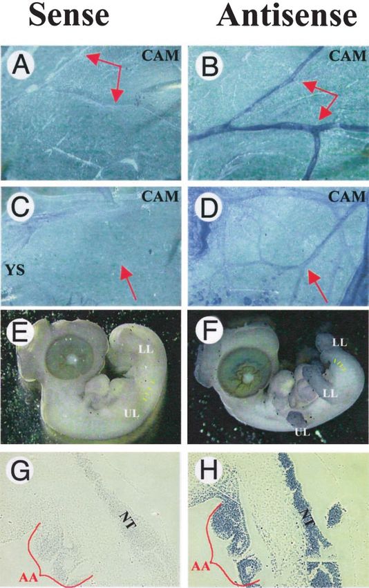
pattern of cNERF2 by in situ hybridization. Examination of cNERF2. As shown in Figure 4C and 4D, cNERF2 was able to
E11 and E14 CAMs (Figure 2B and 2D) demonstrates inhibit the ability of cELF-1 to transactivate the Tie1 and Tie2
hybridization mostly in the large vessels, with weaker levels promoters in a dose-dependent manner. cNERF was similarly
of expression in the smaller branching vessels and capillaries. able to block cELF-1 transactivation of the Tie2 gene promoter
This is in contrast to cELF-1, which is also highly expressed in transfected endothelial cells (data not shown).
in the smaller developing blood vessels and capillaries.13
Minimal staining is observed in the sense controls (Figure 2A Assessment of In Vivo Binding of cELF-1 and
and 2C). Whole mount of the chicken embryos also demon- cNERF2 During Vascular Development
strates expression of cNERF2 in the developing limb buds To extend these studies, we sought to determine whether
(Figure 2F). To examine the expression of cNERF2 in the cNERF2 binds to the conserved Ets sites of the Tie1 and Tie2
developing large vessels of the embryo, in situ hybridization promoters in vivo during development in the chicken CAM.
of paraffin-embedded embryos (E3) sections was performed. As is shown in Figure 5, in vitro–translated chicken and
cNERF2 was strongly expressed in the developing aorta human NERF and chicken ELF-1 were able to bind strongly
(Figure 2H) and was also expressed in the developing neural to the Tie1 Ets site probe (lanes 2 to 4) compared with controlGaspar et al NERF and ELF-1 Roles in Vascular Development 1109
Figure 2. Whole-mount in situ hybridization of
cNERF2 in E3 and E5 chicken embryos and in
E11 and E14 chicken CAMs. A and B, cNERF2
is evident in the larger blood vessels of per-
fused E11 CAMs but absent in the capillaries. C
and D, cNERF2 is also prominent along the
perfused blood vessels of the E14 CAMs (red
arrow). Note the punctate expression in the
yolk sac (YS). E and F, cNERF2 expression is
present in the limbs and mesenchyme (yellow
lines) of E5 chick embryos. G and H, cNERF2
expression in the developing aortic arch (AA)
and neural tube (NT) of E3 chicken embryos is
shown. LL indicates lower limb; UL; upper limb.
E3, E5, E11, and E14 correspond to Hamilton
and Hamburger stages 21, 28, 37, and 40,
respectively.27
extract (lane 1). When cell extracts derived from the chicken Discussion
CAM blood vessels (E7) were used instead of the in vitro–
translated products, a similar sized complex was formed (lane 5). The goal of the present study was to further define the role of
However, the antibody to NERF had no effect on the formation specific transcription factors in the regulation of vascular-
of a similar sized complex (lane 6). In contrast, an antibody specific gene expression during vascular development. Our
directed against ELF-1 was able to alter the formation of this particular focus has been on characterizing the role of
complex completely, resulting in a supershift (arrow, lane 7), members of the Ets transcription factor family in this process.
suggesting that cELF-1 and not cNERF2 is the Ets factor In the present study, we demonstrate that cNERF2 is highly
binding to the Tie1 Ets site in the chicken CAM. These results enriched in the developing blood vessels of the chicken CAM
suggest that although in vitro–translated cELF-1 and cNERF2 and chicken embryo.10,11 Whereas NERF2 acts as a positive
bind equally well to conserved Ets sites in the Tie1 and Tie2 regulator of transcription in humans, the chicken homologue
promoters, cELF-1 preferentially binds during the development of NERF2 acts as a competitive inhibitor of ELF-1, suggest-
of blood vessels in the chicken CAM. We similarly performed ing that the highly related Ets factors ELF-1 and NERF may
gel shifts with oligonucleotides encoding the Ets binding sites in act as positive and negative regulators of the same gene
the Tie2 promoter (data not shown). Chicken ELF-1 also bound targets in the chicken. However, over evolution, the change in
preferentially to the Tie2 Ets sites. NERF2 function from a negative to a positive regulator may1110 Arterioscler Thromb Vasc Biol. July 2002
sophila during eye development.21 The pointed gene is an
Ets factor that acts as a positive regulator of photoreceptor
determination and is activated by the Ras/mitogen-acti-
vated protein kinase pathway. In contrast, the related
Drosophila Ets factor, yan, is a negative regulator of
transcription of the same gene targets as pointed. However,
on phosphorylation by activation of the Ras/mitogen-acti-
vated protein kinase pathway, yan loses the ability to act as
a transcriptional repressor and allows pointed to bind to
DNA and activate the same gene targets. A similar process
may occur with respect to NERF2 and ELF-1 during
chicken blood vessel development. In contrast to cNERF2,
which appears to be expressed predominantly in the larger
vessels of the developing chicken CAM, cELF-1 is ex-
pressed not only in the larger vessels but also in the smaller
vessels and capillaries of the CAM.13
The Tie1 and Tie2 genes are both expressed in the early
embryonic vascular system.1 Tie2 is also upregulated in
the extraembryonic blood vessels. The results of the
present study demonstrate that cNERF2 is expressed in the
developing embryonic as well as extraembryonic vascula-
Figure 3. A, Electrophoretic mobility shift assay (EMSA) dem- ture. We have previously demonstrated that cELF-1 is
onstrating ability of in vitro–translated cNERF2 (N) to bind to similarly expressed in embryonic and extraembryonic
oligonucleotide probes encoding Tie2 (lanes 2) and Tie1 (lane blood vessels.13 Some differences in the expression of
4) Ets sites compared with in vitro–translated control (C,
lanes 1 and 3). Demonstration of the specificity of the anti-
NERF and ELF-1 are as follows: ELF-1 was expressed in
bodies directed against NERF and ELF is shown for cNERF very small as well as larger extraembryonic vessels,
(N, lanes 4 to 6) and for cELF-1(E, lanes 7 to 9) by using a whereas NERF was predominantly expressed in the larger
NERF antibody (n) or an ELF-1 antibody (e). Black arrow extraembryonic vessels. NERF and ELF-1 are both ex-
denotes supershifted cELF-1. Uppercase C, N, and E refer to
in vitro–translated control (C), chicken NERF (N), and chicken pressed in the CAM several days after primary vasculo-
ELF-1 (E) reticulolysate protein extracts. Lowercase n and e genesis has occurred, suggesting they may also be in-
represent the presence of rabbit polyclonal antibodies volved in regulating blood vessel remodeling and
directed against NERF (n) and ELF-1 (e).
maturation during later stages of development. The ability
of cELF-1 and cNERF2 to act as positive and negative
have provided important redundancy in the regulation of transcriptional regulators of the Tie1 and Tie2 genes may
critical developmental processes, such as hematopoiesis and provide an important mechanism for regulating the expres-
vascular development. sion levels of the Tie receptors during different stages of
Positive and negative regulators of developmental pro- blood vessel development and vascular remodeling. Data
cesses by Ets factors have similarly been shown in Dro- from the gel mobility shift assays demonstrating preferen-
Figure 4. A and B, Transient cotransfection,
in HEK 293 cells, of Tie1 and Tie2 promoter
luciferase reporter constructs with PCI
expression plasmids for chicken NERF2
(PCIcNERF2) compared with human NERF2
(hNERF) and empty vector (PCI). C and D,
Cotransfections of Tie1 and Tie2 promoter
luciferase reporter constructs in HEK 293
cells with PCI alone, PCIcELF-1 alone, or
PCIcELF-1 in combination with increasing
amounts of PCIcNERF2 (50, 100, 150, and
200 ng). E, Cotransfections of the Tie2 pro-
moter luciferase reporter constructs in MS-1
endothelial cells with PCI alone, PCIcELF-1
alone, or PCIcELF-1 in combination with
increasing amounts of PCIcNERF2 (50, 100,
150, and 200 ng).Gaspar et al NERF and ELF-1 Roles in Vascular Development 1111
during various stages of chicken blood vessel development
and maturation.
Acknowledgments
This study was supported by National Institutes of Health grants
RO1/HL-63008 (to P.O.) and PO1/CA72009 (to T.A.L.).
References
1. Sato TN, Qin Y, Kozak CA, Audus KL. Tie-1 and tie-2 define another
class of putative receptor tyrosine kinase genes expressed in early
embryonic vascular system [published correction appears in Proc Natl
Acad Sci U S A. 1993;90:12056]. Proc Natl Acad Sci U S A. 1993;90:
9355–9358.
2. Sato TN, Tozawa Y, Deutsch U, Wolburg-Buchholz K, Fujiwara Y,
Gendron-Maguire M, Gridley T, Wolburg H, Risau W, Qin Y. Distinct
roles of the receptor tyrosine kinases Tie-1 and Tie-2 in blood vessel
formation. Nature. 1995;376:70 –74.
Figure 5. EMSA using Tie1 Ets probe and in vitro–translated 3. Vikkula M, Boon LM, Carraway KL III, Calvert JT, Diamonti AJ,
hNERF2 (hN), cNERF2 (cN), cELF-1 (cE), or control (C) extract Goumnerov B, Pasyk KA, Marchuk DA, Warman ML, Cantley LC,
(lanes 1 to 4) or chicken CAM whole-cell extracts at E7 alone or
Mulliken JB, Olsen BR. Vascular dysmorphogenesis caused by an acti-
with the anti-NERF (n) or anti-ELF-1 (e) antibodies (lanes 5 to 7).
vating mutation in the receptor tyrosine kinase TIE2. Cell. 1996;87:
Arrow denotes supershifted complex.
1181–1190.
4. Hatva E, Kaipainen A, Mentula P, Jaaskelainen J, Paetau A, Haltia M,
Alitalo K. Expression of endothelial cell-specific receptor tyrosine
tial binding of cELF-1, a positive transactivator in the kinases and growth factors in human brain tumors. Am J Pathol. 1995;
CAM at E7, most likely reflects a proliferative phase in 146:368 –378.
5. Kaipainen A, Vlaykova T, Hatva E, Bohling T, Jekunen A, Pyrhonen S,
vascular development, whereas at later stages of relative
Alitalo K. Enhanced expression of the tie receptor tyrosine kinase mes-
quiescence, the balance could shift either in favor of senger RNA in the vascular endothelium of metastatic melanomas.
preferential binding of cNERF2 to the Tie Ets sites or Cancer Res. 1994;54:6571– 6577.
simply through a reduction in the amount of cELF-1. The 6. Korhonen J, Lahtinen I, Halmekyto M, Alhonen L, Janne J, Dumont D,
Alitalo K. Endothelial-specific gene expression directed by the tie gene
coexpression of these factors at various stages of vascular
promoter in vivo. Blood. 1995;86:1828 –1835.
development suggests that additional mechanisms of con- 7. Schlaeger TM, Bartunkova S, Lawitts JA, Teichmann G, Risau W,
trol, such as phosphorylation of the factors, may also be Deutsch U, Sato TN. Uniform vascular-endothelial-cell-specific gene
determinants of the relative activity of these transcription expression in both embryonic and adult transgenic mice. Proc Natl Acad
Sci U S A. 1997;94:3058 –3063.
factors.
8. Wakiya K, Begue A, Stehelin D, Shibuya M. A cAMP response element
Further support for the role of the Ets factor in vascular and an Ets motif are involved in the transcriptional regulation of flt-1
development and angiogenesis comes from knockout exper- tyrosine kinase (vascular endothelial growth factor receptor 1) gene.
iments of selected Ets factors. Another Ets factor, Fli-1, was J Biol Chem. 1996;271:30823–30828.
9. Kappel A, Schlaeger TM, Flamme I, Orkin SH, Risau W, Breier G. Role
also similarly shown to be critical for blood vessel develop- of SCL/Tal-1, GATA, and ets transcription factor binding sites for the
ment.22 Targeted disruption of this gene resulted in defects in regulation of flk-1 expression during murine vascular development.
vascular development and hematopoiesis. Another Ets factor, Blood. 2000;96:3078 –3085.
Tel, is involved in the development of the extraembryonic 10. Iljin K, Dube A, Kontusaari S, Korhonen J, Lahtinen I, Oettgen P, Alitalo
K. Role of ets factors in the activity and endothelial cell specificity of the
blood vessels. Targeted disruption of the gene leads to mouse Tie gene promoter. FASEB J. 1999;13:377–386.
abnormalities in vitelline vein development. The vascular 11. Dube A, Akbarali Y, Sato TN, Libermann TA, Oettgen P. Role of the Ets
defects appear to be mainly extraembryonic and restricted to transcription factors in the regulation of the vascular-specific Tie2 gene.
larger vessels with normal-appearing capillaries.23 One of the Circ Res. 1999;84:1177–1185.
12. Wasylyk B, Hahn SL, Giovane A. The Ets family of transcription factors.
recently identified targets of Tel is stromelysin, a matrix Eur J Biochem. 1993;211:7–18.
metalloproteinase (MMP). Tel acts as a transcriptional repres- 13. Dube A, Thai S, Gaspar J, Rudders S, Libermann TA, Iruela-Arispe L,
sor of the stromelysin gene.24 The Ets factor Ets-1 is Oettgen P. Elf-1 is a transcriptional regulator of the Tie2 gene during
vascular development. Circ Res. 2001;88:237–244.
upregulated in endothelial cells during angiogenesis. The
14. Chomczynski P, Sacchi N. Single-step method of RNA isolation by acid
gene targets for Ets-1 involve several MMPs, including guanidinium thiocyanate-phenol-chloroform extraction. Anal Biochem.
MMP-1 and MMP-9.25 Finally, the expression of the extra- 1987;162:156 –159.
cellular matrix glycoprotein SPARC and thrombospondin in 15. Lopez M, Oettgen P, Akbarali Y, Dendorfer U, Libermann TA. ERP, a
new member of the ets transcription factor/oncoprotein family: cloning,
endothelial cells is dependent on the expression of the Ets
characterization, and differential expression during B-lymphocyte devel-
factor Erg.26 opment. Mol Cell Biol. 1994;14:3292–3309.
In conclusion, the present study provides substantial sup- 16. Oettgen P, Carter KC, Augustus M, Barcinski M, Boltax J, Kunsch C,
port for the role of Ets factors in vascular development and Libermann TA. The novel epithelial-specific Ets transcription factor gene
ESX maps to human chromosome 1q32.1. Genomics. 1997;445:
vascular-specific gene expression. Furthermore, the present
456 – 457.
study provides evidence of the opposing roles of the chicken 17. Wilkinson DG, Bailes JA, Champion JE, McMahon AP. A molecular
Ets factors NERF2 and ELF-1 in the regulation of Tie genes analysis of mouse development from 8 to 10 days post coitum detects1112 Arterioscler Thromb Vasc Biol. July 2002
changes only in embryonic globin expression. Development. 1987;99: 23. Wang LC, Kuo F, Fujiwara Y, Gilliland DG, Golub TR, Orkin SH. Yolk
493–500. sac angiogenic defect and intra-embryonic apoptosis in mice lacking the
18. Farr A, Roman A. A pitfall of using a second plasmid to determine Ets-related factor TEL. EMBO J. 1997;16:4374 – 4383.
transfection efficiency. Nucleic Acids Res. 1992;20:920. 24. Fenrick R, Wang L, Nip J, Amann JM, Rooney RJ, Walker-Daniels J,
19. Libermann TA, Lenardo M, Baltimore D. Involvement of a second Crawford HC, Hulboy DL, Kinch MS, Matrisian LM, Hiebert SW. TEL,
lymphoid-specific enhancer element in the regulation of immunoglobulin a putative tumor suppressor, modulates cell growth and cell morphology
heavy-chain gene expression. Mol Cell Biol. 1990;10:3155–3162. of ras-transformed cells while repressing the transcription of
20. Oettgen P, Akbarali Y, Boltax J, Best J, Kunsch C, Libermann TA.
stromelysin-1. Mol Cell Biol. 2000;20:5828 –5839.
Characterization of NERF, a novel transcription factor related to the Ets
25. Oda N, Abe M, Sato Y. ETS-1 converts endothelial cells to the
factor ELF-1. Mol Cell Biol. 1996;16:5091–5106.
angiogenic phenotype by inducing the expression of matrix metallopro-
21. O’Neill EM, Rebay I, Tjian R, Rubin GM. The activities of two
Ets-related transcription factors required for Drosophila eye devel- teinases and integrin beta3. J Cell Physiol. 1999;178:121–132.
opment are modulated by the Ras/MAPK pathway. Cell. 1994;78: 26. McLaughlin F, Ludbrook VJ, Cox J, von Carlowitz I, Brown S, Randi
137–147. AM. Combined genomic and antisense analysis reveals that the tran-
22. Spyropoulos DD, Pharr PN, Lavenburg KR, Jackers P, Papas TS, Ogawa scription factor Erg is implicated in endothelial cell differentiation. Blood.
M, Watson DK. Hemorrhage, impaired hematopoiesis, and lethality in 2001;98:3332–3339.
mouse embryos carrying a targeted disruption of the Fli1 transcription 27. Hamburger V, Hamilton HL. A series of normal stages in the devel-
factor. Mol Cell Biol. 2000;20:5643–5652. opment of the chicken embryo. J Morphol. 1951;88:49 –92.You can also read