Giant Tumefactive Perivascular Spaces
←
→
Page content transcription
If your browser does not render page correctly, please read the page content below

AJNR Am J Neuroradiol 26:298–305, February 2005

                      Giant Tumefactive Perivascular Spaces
                  Karen L. Salzman, Anne G. Osborn, Paul House, J. Randy Jinkins, Adam Ditchfield,
                                       James A. Cooper, and Roy O. Weller

             BACKGROUND AND PURPOSE: The brain perivascular spaces (PVSs) are pial-lined, inter-
          stitial fluid-filled structures that accompany penetrating arteries. When enlarged, they may
          cause mass effect and can be mistaken for more ominous pathologic processes. The purpose of
          this study was to delineate the broad clinical and imaging spectrum of this unusual condition.
             METHODS: Thirty-seven cases of giant PVSs were identified from 1988 to 2004 and were
          retrospectively reviewed. Clinical data collected included patient demographics, presenting
          symptoms, and follow-up. Histopathologic data were reviewed when available. Images were
          evaluated for size and location of the giant PVSs, associated mass effect, hydrocephalus,
          adjacent white matter changes, and contrast enhancement.
             RESULTS: There were 24 men and 13 women with an age range of 6 – 86 years, (mean 46
          years). The most common presenting feature was headache (15 patients). Thirty-two cases had
          multilocular clusters of variably sized cysts. Five lesions were unilocular. All lesions had signal
          intensity comparable to CSF and did not enhance. The most common location for the giant
          PVSs was the mesencephalothalamic region (21/36). Fourteen were located in the cerebral white
          matter; two were in the dentate nuclei. Nine giant mesencephalothalamic PVSs had associated
          hydrocephalus, which required surgical intervention.
             CONCLUSION: Giant tumefactive PVSs most often appear as clusters of variably sized cysts
          that are isointense relative to CSF and do not enhance. They are most common in the
          mesencephalothalamic region and may cause hydrocephalus. Although they may have striking
          mass effect, giant PVSs should not be mistaken for neoplasm or other diseases.

The perivascular spaces (PVSs) of the brain, also                         (4). Occasionally the PVSs may become strikingly
known as Virchow-Robin spaces, are pial-lined inter-                      enlarged, causing mass effect and assuming bizarre
stitial fluid (ISF)-filled structures that accompany                      configurations that may be mistaken for a more om-
penetrating arteries and arterioles for a variable dis-                   inous disease, such as a cystic neoplasm (Figure 1).
tance as they descend into the cerebral substance                         While scattered cases of giant tumefactive PVSs have
(1–3). Recent studies have shown that the PVSs are                        been reported (5–7), no large series of these lesions
surprisingly complex entities with significant variabil-                  has been presented. The purpose of this study is to
ity in both ultrastructure and possible function. Rou-                    delineate the broad clinical and imaging spectrum of
tinely, PVSs in many areas of the brain can be iden-                      giant tumefactive perivascular spaces to further char-
tified on MR images obtained in patients of all ages                      acterize this unusual lesion.

  Received September 16, 2003; accepted after revision October                                      Methods
21, 2004.                                                                    We performed a retrospective review of all cases of giant
  From the Departments of Radiology (K.L.S, A.G.O), and Neu-              PVSs referred for imaging consultation or treated at our insti-
rosurgery (P.H.), University of Utah, Salt Lake City, Utah, the           tution over a 16-year period spanning 1988 to 2004. Most cases
Department of Radiology (J.R.J.), State University of New York,           referred for second opinion had the presumptive diagnosis of
Downstate Medical Center, Brooklyn, NY, the Department of                 cystic neoplasm.
Radiology (A.D.), Wessex Neurological Centre, Southampton                    We defined giant PVSs as equal to or greater than 1.5 cm,
General Hospital, UK, the Radiology Medical Group (J.A.C.), San
                                                                          occurring as either a solitary lesion or in clusters of multiple
Diego, CA, Clinical Neurosciences (R.O.W.), University of
Southampton School of Medicine, Southampton General Hospital,
                                                                          contiguous cysts.
UK.                                                                          Clinical data collected included patient age, sex, and pre-
  Presented in abstract form at the 39th Annual Meeting of the            senting symptoms. Histopathologic data in patients who under-
ASNR, April 23–27, 2001, Boston, MA.                                      went biopsy were collected and reviewed. The specific histo-
  Address reprint requests to Karen L. Salzman, MD., Depart-              logic stains used included: Hematoxylin and Eosin as a general
ment of Radiology, 1A71 University Hospital, 50 N. Medical Drive,         histologic stain; Hematoxylin van Gieson to stain the collagen
Salt Lake City, UT 84132.                                                 of the meninges on the outer aspect of the cyst wall; and
                                                                          reticulin to stain the collagen of the meninges on the outer
© American Society of Neuroradiology                                      aspect of the cyst wall.

                                                                    298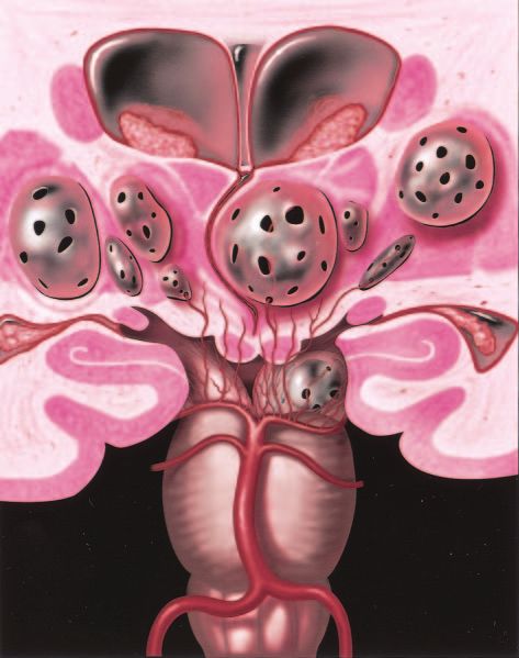
AJNR: 26, February 2005                                           GIANT TUMEFACTIVE PERIVASCULAR SPACES                   299

                                                                      TABLE 1: Presenting symptoms

                                                                             Headache                                15
                                                                             Dizziness                                3
                                                                             Dementia                                 3
                                                                             Visual changes                           3
                                                                             Post-traumatic evaluation                2
                                                                             Cranial neuropathy                       2
                                                                             Seizure                                  1
                                                                             Syncope                                  1
                                                                             Stroke                                   1
                                                                             Memory problems                          1
                                                                             Poor balance and concentration           1
                                                                             N ⫽ 33

                                                                      sion. There were 27 unilateral lesions and 10 bilateral
                                                                      lesions.

                                                                                         Clinical Findings:
                                                                         Complete clinical data were available in 33 patients
                                                                      with a partial history available in the remaining four
                                                                      patients. The most common presenting symptom was
                                                                      headache, present in 15/33 patients. Other presenting
                                                                      features included dizziness, dementia, visual changes,
                                                                      post-traumatic evaluation, seizure, syncope, stroke,
                                                                      memory problems, cranial neuropathy and poor bal-
                                                                      ance and concentration. One additional patient had a
FIG 1.    Coronal anatomic diagram depicts multiple bilateral
giant perivascular spaces (PVSs) in the mesencephalothalamic          6th nerve palsy which was found to be related to
region. There are fenestrations of the giant PVSs, which may          diabetic neuropathy and resolved on follow-up imag-
allow accumulation of interstitial fluid between the vessel and pia   ing. One patient who presented with visual changes
or within the interpial space causing enlargement of the PVSs.        was ultimately diagnosed with uveitis, which was
Note the mass effect upon the third ventricle with associated
obstructive hydrocephalus. Graphic courtesy of James Cooper,
                                                                      treated and resolved (Table 1).
MD and AMIRSYS, Inc (35).

                                                                                          Imaging Findings
   All patients were imaged with MR imaging using a variety of           CT was performed in 10 of 37 patients. All giant
1.5T MR imaging units. Typical pulse sequences included T1-           PVSs were low-attenuation lesions, isoattenuated rel-
weighted spin-echo images, fast spin-echo T2-weighted images,         ative to CSF, which showed no enhancement follow-
proton density–weighted (PD) images, and fluid attenuated             ing contrast medium administration. No calcifications
inversion recovery (FLAIR) images. Patients’ MR imaging               or other associated abnormalities were identified.
examinations included either a PD or FLAIR sequence. Gad-
olinium-enhanced T1-weighted imaging was also performed in
                                                                         All giant PVSs were isointense relative to CSF
most patients (34/36). Two studies were performed without the         signal intensity on all MR images regardless of pulse
use of contrast medium. Ten patients underwent CT performed           sequence. Contrast-enhanced images were available
with 5-mm axial sections.                                             in 35/37 cases and showed no enhancement. Diffu-
   Images were evaluated to determine the size of the giant           sion-weighted (DW) imaging was available in eight of
PVSs, establish whether cysts were unilateral or bilateral, and       37 cases and showed no diffusion restriction.
ascertain whether cysts were solitary or clustered. Images were          Lesions were subdivided by location (Table 2).
also assessed for the appearance of associated focal or gener-
alized mass effect, hydrocephalus, adjacent white matter              There were 21 patients with lesions involving the
changes, and presence of contrast enhancement.                        midbrain or thalamus (mesencephalothalamic area)
                                                                      (Fig 2) and 14 lesions predominately involved the
                                                                      hemispheric or subcortical cerebral white matter.
                            Results                                   Two lesions were present in the region of the dentate
                                                                      nuclei of the cerebellar hemispheres.
                       Demographics                                      Of the 21 mesencephalothalamic lesions, four were
   Thirty-seven patients with giant PVSs were identi-                 bilateral. No signal intensity alteration was present in
fied, including 24 men and 13 women (M:F ⫽ 1.8:1).                    the adjacent brain parenchyma. Nine of these 21
Patient age ranged from 6 to 86 years with a mean of                  mesencephalothalamic lesions had associated hydro-
46 years. Five unilocular lesions were identified rang-               cephalus and were treated with ventriculostomy (5),
ing in size from 1.5 cm to 3.5 cm. Multiple clustered                 ventriculoperitoneal shunting (3), or cystoperitoneal
cysts (32/37 cases) were present in most cases. The                   shunting (1) (Fig 3).
clusters were all greater than or equal to 1.5 cm in                     There were 14 patients with giant PVSs in the
size, ranging from 1.5 cm to 10 cm in greatest dimen-                 cerebral white matter (Fig 4). Five of these white
300 SALZMAN AJNR: 26, February 2005
TABLE 2: Lesion location and follow-up the cerebellar dentate nuclei. In one case, the lesions
were bilateral. There was no associated signal intensity
Age Sex Giant PVS Location Biopsy Follow-up Imaging
alteration in the adjacent brain parenchyma (Fig 7).
39 M Thalamus Y 2 years
76 M Thalamus N N
56 F Thalamus N 4 years Surgical and Pathologic Findings
86 M Thalamus N 12 years Surgery was performed in 12 patients. In seven
35 M Thalamus N 2 years
cases, biopsy was performed. Histopathologic results
42 F Thalamus N N
49 F Thalamus N 7 years
in most cases (6/7) disclosed a pial-lined cyst with no
35 M Midbrain Y 1 year
evidence of neoplasm or infection. One patient with a
57 M Midbrain Y N midbrain lesion had a layer of pia and arachnoid on
44 F Midbrain N N the outer aspect of the tissue but no pial lining. This
75 M Midbrain N N patient had a small fragment, approximately 1 mm
40 M Midbrain Y N cubed, of the cyst lining sent to the pathology depart-
28 M Midbrain N N ment. It was fixed in formalin, embedded in paraffin,
35 M Midbrain/Pons Y 1 year and stained with hematoxylin and eosin, hematoxylin
47 F Thalamus/Midbrain N 9 years van Gieson for collagen, and by immunocytochemis-
62 M Thalamus/Midbrain N N
try for glial fibrillary acidic protein (GFAP) for astro-
46 M Thalamus/Midbrain Y N
75 M Thalamus/Midbrain N 1 year
cytes Figure 8. Figure 8A shows the cyst lining con-
72 F Thalamus/Basal N 10 years
sists of a very thin layer of gliotic brain tissue coated
Ganglia on its outer aspects by collagenous material related to
41 M Basal Ganglia N 1 year the pia arachnoid on the surface of the midbrain. No
47 M Basal Ganglia N N lining of pia matter was identified on the inner cyst
15 M White Matter N 7 years surface. The brain tissue lining the cyst was exten-
70 M White Matter N N sively gliotic as shown in the GFAP preparation (Fig
62 F White Matter N N 8B). Close examination of the cells in the brain tissue
37 M White Matter N 1 year lining the cyst shows that it is mainly composed of
68 M White Matter N N
astrocytes (Fig 8C). The absence of a lining of pia
56 F White Matter N N
44 F White Matter N 1 year
matter is only to be expected in this case, as the cyst
21 M White Matter Y 6 years
was very large and it is unlikely that the thin delicate
36 M White Matter N 2 years lining surrounding a vessel would be stretched to this
68 F White Matter N N extent. When there is less extensive enlargement of
46 F White Matter N N perivascular spaces, the lining of pia matter may remain.
20 F White Matter N N Two patients had a ventriculoperitoneal shunt
40 F White Matter N N placed, and the size of the giant PVSs was stable in
10 M White Matter N 3 years each case. Another patient had a cystoperitoneal
17 M Cerebellum N N shunt placed, and there was a decrease in the size of
6 M Cerebellum N N
the giant PVS from 10 cm to 5 cm. In this case, the
patient’s headache also improved. However, the fluid
reaccumulated after the patient had a minor trau-
matter cases were bilateral. White matter lesions matic insult, and the giant PVS enlarged to near-
were located in the subcortical white matter (8/14), presentation size. One patient underwent PVS drain-
hemispheric white matter (4/14), or both (2/14). Most age followed by ventriculoperitoneal shunt placement
white matter lesions involved the frontal lobe (8/14) after the PVS returned to its original size. Five pa-
with other areas of involvement including the parietal tients underwent a third ventriculostomy; three of
lobe (3/14), temporal lobe (2/14), and occipital lobe these patients also had a biopsy of the cyst wall.
(1/14). There was involvement of the cingulate gyrus Follow-up MR imaging was available in seventeen
subcortical white matter in four cases and three cases patients from 1–12 years after the initial diagnosis. No
had involvement of the corpus callosum. In these interim change was seen on repeat MR imaging in
cases, the gray matter was stretched over the giant any case.
PVSs (Fig 5).
Associated white matter signal intensity alterations
occurred in seven of the cerebral white matter cases. Discussion
Two of these were focal changes, with increased T2 When PVSs become markedly expanded, they can
and FLAIR signal intensity, surrounding the giant be misinterpreted as other pathologic processes, most
PVSs, and were found in older patients (a 68-year-old often a cystic neoplasm. As most of these cysts border
woman and a 73-year-old man) (Fig 4). Five addi- a ventricle or subarachnoid space, reports of such
tional patients with lesions in the white matter had cases have offered an extensive differential diagnosis
more diffuse, confluent abnormal signal intensity al- that includes cystic neoplasms, parasitic cysts, ventric-
teration consisting of increased T2 and FLAIR signal ular diverticula, cystic infarction, non-neoplastic neu-
intensity surrounding the PVSs (Fig 6). roepithelial cysts, and deposition disorders such as
There were two lesions that involved the region of mucopolysaccharidosis (4, 7–9). The aim of this studyAJNR: 26, February 2005 GIANT TUMEFACTIVE PERIVASCULAR SPACES 301
FIG 2. Axial T2-weighted (A), postcontrast axial T1-weighted (B), and postcontrast coronal T1-weighted (C) images obtained in a
46-year-old man with headaches show a nonenhancing multiloculated cystic mass in the right midbrain, thalamus, and right medial
temporal lobe. The cysts follow CSF signal intensity on all pulse sequences and do not enhance. Follow-up imaging 13 years later
showed no change. Biopsy proved pial-lined giant perivascular spaces.
CSF, and demonstrate no enhancement following
contrast medium administration (4, 6, 8, 9). When
PVSs become enlarged, they are known as giant
PVSs, “cavernous dilatation,” or Poirier’s Type IIIb
“expanding lacunae” (6, 10, 11).
Giant PVSs are expanded PVSs that occur along
the penetrating vessels, most commonly in the mes-
encephalothalamic region in the territory of the para-
medial mesencephalothalamic artery and in the cere-
bral white matter (4, 7, 10). They differ from typical
PVSs in that they are larger in size and may have
associated focal mass effect. In addition, white matter
giant PVSs may have associated T2 and FLAIR signal
intensity alteration in the adjacent white matter.
Patients usually present with nonspecific findings
FIG 3. Coronal postcontrast T1-weighted image obtained in a
56-year-old woman with headaches shows a nonenhancing that are not attributable to the giant PVSs. In our
unilocular giant perivascular space (black arrow) in the left thal- series, headache was the most common presenting
amus with compression and displacement of the third ventricle feature and occurred in approximately 50% of our
(white arrow). Note associated hydrocephalus. Surgery dis- patients. Other presenting complaints included dizzi-
closed a smooth walled cyst with no abnormality in the adjacent
brain. The patient initially underwent a cyst fenestration with a
ness, dementia, visual changes, post-trauma, seizure,
decrease in the size of the PVS. Four months later, there was syncope, memory problems, poor balance, and poor
reaccumulation of fluid and the PVS enlarged to its original size. concentration. Several studies have found that the
A ventriculoperitoneal shunt was then placed, which relieved the clinical features of patients with this type of lesion do
hydrocephalus. Follow-up studies showed no change in cyst
size over 4 years.
not correlate with the imaging findings or have been
found incidentally at autopsy (9,12–14). However,
widespread dilatation of the PVSs has been reported
was to delineate the classic imaging appearance of in association with dementia and parkinsonism (15,
giant PVSs in an effort to prevent misdiagnosis and 16). Some authors have correlated large convexity
unnecessary biopsy. and white matter PVSs with increasing age (4). Oth-
Typical PVSs occur in many locations. The most ers have concluded that the prevalence of basal gan-
common site is along the lenticulostriate arteries, just glia PVSs in patients younger than 40 years is not
above the anterior perforated substance and adjacent significantly different from that of older patients (8).
to the anterior commissure. Less commonly, PVSs We have observed prominent PVSs on high-spatial-
occur along arteries that have penetrated the cerebral resolution MR images in patients of all ages and in all
cortex and extended into the white matter (4, 8). typical anatomical locations. The clinical symptoms
Other areas where prominent PVSs can be identified did not correlate with the giant PVSs in our series,
include the subinsular region, dentate nuclei, and except in those cases that had associated hydroceph-
cerebellum (2). PVSs have typical MR imaging fea- alus, requiring CSF diversion.
tures as previously described. They are round or oval Imaging of enlarged PVSs is characteristic (17, 18).
with a well-defined, smooth margin, occur along the All cases have associated mass effect, from mild to
path of penetrating arteries, are isointense relative to severe. The mass effect may occasionally cause ob-302 SALZMAN AJNR: 26, February 2005 FIG 4. Sagittal T1-weighted (A), axial FLAIR (B), and coronal T2-weighted (C) images obtained in a 71-year-old man with dementia show extensive involvement of the hemispheric and subcortical white matter with multilocular giant perivascular spaces. The coronal image shows the marked asymmetry of the lesions. Note the scattered focal white matter changes surrounding some of the lesions (black arrows) seen best on the FLAIR image. Case courtesy of Anthony Doyle, MD. FIG 5. Sagittal T1-weighted (A), axial FLAIR (B), and axial T2-weighted (C) images obtained in a 46-year-old woman with a visual field defect show extensive involvement of the corpus callosum and cingulate gyrus (A, white arrow) with extension to the subcortical white matter of the parietal and occipital lobes. There is slight increased signal intensity surrounding the lesions, best seen on FLAIR image (B, white arrows). The gray matter is stretched and displaced over the multiloculated giant perivascular spaces. Case courtesy of Leena Valanne, MD. FIG 6. Images obtained in a 37-year-old man with headaches. Axial T2-weighted (A) and axial FLAIR (B) images show diffuse, confluent white matter hyperintensity surrounding the giant perivascular spaces. There is mild gyral expansion over the perivascular spaces. The diffusion-weighted image (C) shows no diffusion restriction. structive hydrocephalus as was seen in eight of our thalamic region. Associated hydrocephalus has been study patients, all with lesions in the mesencephalo- reported in the literature and may require surgical
AJNR: 26, February 2005 GIANT TUMEFACTIVE PERIVASCULAR SPACES 303
FIG 7. Images obtained in a 6-year-old
boy with a history of minor trauma. Axial
T1-weighted (A), coronal T2-weighted (B),
axial FLAIR (C) and postcontrast coronal
T1-weighted (D) images show multilocu-
lated giant perivascular spaces in the left
dentate gyrus. There is mild focal mass
effect upon the fourth ventricle (white ar-
row). The clustered cysts follow CSF on all
pulse sequences.
shunt surgery (19). FLAIR imaging typically shows weighted images occurs in more than 30% of the
complete signal intensity suppression without abnor- neurologically healthy elderly population (9, 22, 23).
malities in the adjacent parenchyma. In a few re- Histopathologic study with MR imaging correlation
ported cases, small foci of high signal intensity adja- has indicated that many of these lesions are PVSs (8,
cent to the cystic spaces have been identified (20). 24 –26). One author found that PVSs greater than 2
Our study shows that giant PVSs that occur in the mm were found in 67 (8%) of 816 patients (4).
white matter may have surrounding signal intensity Giant PVSs (up to 2–3 cm in diameter) have been
abnormality seen on T2-weighted or FLAIR images, reported to occur as a normal variant (4, 27). The
as seen in half of our cases. Lesions with adjacent precise etiology of these enlarged PVSs is unknown.
white matter changes can be divided into two groups. Spiral elongation of the penetrating blood vessels has
In elderly patients in our series, the white matter been suggested as one possible cause of this phenom-
changes surrounding the dilated PVSs were discrete enon (24). Increased CSF pulsations, the ex vacuo
areas of abnormal T2 and FLAIR hyperintense signal phenomenon, or an abnormality of arterial wall per-
intensity abnormality. One possible theory is that this meability have also been cited as contributing factors
associated signal intensity alteration may represent (9, 13, 28, 29).
advanced chronic ischemic change related to mass Experimental studies suggest that the periarterial
effect of the PVSs (5). Another possibility is that spaces are the routes by which interstitial fluid (ISF)
dilated PVSs may result from chronic mechanical drains from brain tissue (30). In humans, amyloid–
stress caused by high blood pressure on the brain beta (A) appears to be entrapped within the drain-
arterioles (21). However, most hypertensive patients age pathways in the cerebral amyloid angiopathy that
imaged do not have associated giant PVSs. accompanies Alzheimer disease (AD) (31, 32). Al-
In younger patients in our series, the signal inten- though the periarterial spaces in the cortex are not
sity abnormality was more diffuse, with confluent T2 dilated in AD, periarterial spaces in the white matter
and FLAIR increased signal intensity surrounding the are expanded possibly due to obstruction of ISF
giant PVSs. Although the exact etiology is not known, drainage pathways by A in the proximal parts of
we suggest that the changes may be secondary to each artery (33). By analogy, obstruction of ISF drain-
gliosis or spongiosis. Alternatively, the signal intensity age pathways could be a major factor in the patho-
changes may be related to multiple tiny tightly clus- genesis of giant PVSs, although the exact nature of
tered PVSs that are too small to be discriminated on the obstruction is unclear.
the basis of current MR imaging findings. Abnormal Other investigators have postulated that because
hyperintensity in the cerebral white matter on T2- the pial membrane surrounding the penetrating ar-304 SALZMAN AJNR: 26, February 2005 FIG 8. Images obtained in a 35-year-old man with headache who underwent a biopsy and a third ventriculostomy procedure. Axial T1-weighted MR image (A) shows the giant perivascular space in the left midbrain. A section through the cyst wall (B) shows red-stained collagen in the leptomeninges on the outer aspect of the cyst wall coating the underlying brain tissue that forms the bulk of the cyst wall. No lining of pia matter is present on the inner aspect of the cyst. (Hematoxylin van Gieson stain, original magnification ⫻20) There is extensive gliosis in the cyst wall as demonstrated by the numerous brown stained fibrillary processes within the brain tissue (C). (Immunocytochemistry for GFAP, original magnification ⫻20) A high-power view (D) of the cyst wall near the pia arachnoid outer coating (top of picture). Reactive astrocytes are seen toward the bottom of the illustration. No neurons were identified. (Hematoxylin and eosin stain, original magnification ⫻40) tery is fenestrated, accumulation of brain interstitial appearance is virtually pathognomonic of giant PVSs. fluid between the vessel and pia or within the inter- An extensive differential diagnosis is superfluous and pial space causes the PVSs to enlarge (27). We found biopsy unnecessary (14). that giant PVSs are most common in the mesen- cephalothalamic region, which may indeed be related to the two layers of pia in this location. In addition, Conclusion the significant mass effect with associated obstructive hydrocephalus seen in the mesencephalothalamic Giant tumefactive PVSs are interstitial-fluid filled group may, in part, be related to the two pial layers. structures that accompany arteries and arterioles as Giant PVSs with mass effect may occur as single or they penetrate the brain. They have characteristic multiple clustered cysts that can be mistaken for more imaging features: round or oval, single or multilocular ominous disease (34). When the lesions in question lesions that are isointense relative to CSF regardless occur in a characteristic location along the path of a of imaging sequences and do not enhance. They are penetrating vessel, follow CSF signal intensity on all most common in the mesencephalothalamic region sequences, do not enhance with contrast material, and may have associated obstructive hydrocephalus and have normal adjacent brain parenchyma, their that may require surgical intervention. Although they
AJNR: 26, February 2005                                                  GIANT TUMEFACTIVE PERIVASCULAR SPACES                                     305

have associated mass effect, they should not be mis-                               volunteers: correlation with social and medical histories. Am J
                                                                                   Roentgenol 1993;161:855– 858
taken for neoplasm or other disease.                                         18.   Bokura H, Kobayashi S, Yamaguchi S. Distinguishing silent lacu-
                                                                                   nar infarction from enlarged Virchow-Robin spaces: a magnetic
                                                                                   resonance imaging and pathological study. J Neurol 1998;245:
                            References                                             116 –22
                                                                             19.   Mascalchi M, Salvi F, Gordano U, et al. Expanding lacunae causing
 1. Ozturk MH, Aydingoz U. Comparison of MR signal intensities of                  triventricular hydrocephalus. Report of two cases. J Neurosurg
    cerebral perivascular (Virchow-Robin) and subarachnoid spaces.                 1999;91:669 – 674
    J Comput Assist Tomogr 2002;26:902–904                                   20.   Komiyama M, Yasui T, Izumi T. Magnet resonance imaging fea-
 2. Song CJ, Kim JH, Kier EL, Bronen RA. MR imaging and histologic                 tures of unusually dilated Virchow-Robin spaces—two case re-
    features of subinsular bright spots on T2–weighted MR images:                  ports. Neurol Med Chir 1998;38:161–164
    Virchow-Robin spaces of the extreme capsule and insular cortex.          21.   Sawada M, Nishi S, Hashimoto N. Unilateral appearance of mark-
    Radiology 2000;214:671– 677                                                    edly dilated Virchow-Robin spaces. Clin Radiol 1999;54:334 –336
 3. Pollock H, Hutchings M, Weller RO, Zhang ET. Perivascular                22.   Gerard G, Weisberg LA. MRI periventricular lesions in adults.
    spaces in the basal ganglia of the human brain: their relationship             Neurology 1986;36:998 –1001
    to lacunes. J Anat 1997;191:337–346                                      23.   Drayer BP. Imaging of the aging brain. Part I. Normal findings
 4. Heier LA, Bauer CJ, Schwartz L, et al. Large Virchow-Robin                     Radiology 1988;166:785–796
    spaces: MR – clinical correlation. AJNR Am J Neuroradiol                 24.   Awad IA, Johnson PC, Spetzler RF, Hodak JA. Incidental subcor-
    1989;10:929 –936                                                               tical lesions identified on magnetic resonance imaging in the el-
 5. Shiratori K, Mrowka M, Toussaint A, Spalke G, Bien S. Extreme,                 derly II. Postmortem pathological correlations. Stroke 1986;17:
    unilateral widening of Virchow-Robin spaces: case report. Neuro-               1090 –1097
    radiology 2002;44:990 –992                                               25.   Elster AD, Richardson DN. Focal high signal on MR scans of the
 6. Kanamalla US, Calabro F, Jinkins JR. Cavernous dilatation of                   midbrain caused by enlarged perivascular spaces: MR-pathologic
    mesencephalic Virchow-Robin spaces with obstructive hydroceph-                 correlation. AJR Am J Roentgenol 1991;156:157–160
    alus. Neuroradiology 2000;42:881– 884                                    26.   Braffman BH, Zimmerman RA, Trojanowski JQ, Gonatas NK,
 7. Homeyer P, Cornu P, Lacomblez L, et al. A special form of cerebral             Hickey WF, Schlaepfer WW. Brain MR: pathologic correlation
    lacunae: expanding lacunae. J Neurol Neurosurg Psychiatr                       with gross and histopathology. 1. Lacunar infarction and Virchow-
    1996;61:200 –202                                                               Robin spaces. AJR Am J Roentgenol 1988;151:551–558
 8. Jungreis CA, Kanal E, Hirsch WL, et al. Normal perivascular              27.   Adachi M, Hosoya T, Haku T, Yamaguchi K. Dilated Virchow-
    spaces mimicking lacunar infarction: MR imaging. Radiology                     Robin spaces: MRI pathological study. Neuroradiology 1998;40:
    1988;169:101–102                                                               27–31
 9. Ogawa R, Okudera T, Fukasawa H, et al. Unusual widening of               28.   Fazekas F, Kleinert R, Roob G, et al. Histopathologic analysis of
    Virchow-Robin spaces: MR appearance. AJNR Am J Neuroradiol                     foci of signal loss on gradient-echo T2*-weighted MR images in
    1995;16:1238 –1242                                                             patients with spontaneous intracerebral hemorrhage: Evidence of
10. Poirier J, Barbizet J, Gaston A, Meyrignac C. Thalamic dementia.               microangiopathy-related microbleeds. AJNR Am J Neuroradiol
    Expansive lacunae of the thalamo-paramedian mesencephalic                      1999;20:637– 642
    area. Hydrocephalus caused by stenosis of the aqueduct of Sylvius.       29.   Derouesné C, Gray F, Escourolle R, Castaigne P. ‘Expanding
    Rev Neurol (Paris) 1983;139:349 –358                                           cerebral lacunae’ in a hypertensive patient with normal pressure
11. Poirier J, Gray F, Gherardi R, Derouesné C. Cerebral lacunae. A               hydrocephalus. Neuropath Appl Neurobiol 1987; 13:309 –320
    new neuropathological classification. J Neuropathol Exp Neurol           30.   Weller RO. Pathology of cerebrospinal fluid and interstitial fluid
    1985;44:312                                                                    of the CNS: significance for Alzheimer disease, prion disorders and
12. Ugawa Y, Shirouzu I, Terao Y, et al. Physiological analyses of a               multiple sclerosis. J Neuropathol Exp Neurol 1998;57:885– 894
    patient with extreme widening of Virchow-Robin spaces. J Neurol          31.   Weller RO, Massey A, Newman TA, Hutchings M, Kuo YM,
    Sci 1998;159:25–27                                                             Roher AE. Cerebral amyloid angiopathy: amyloid beta accumu-
13. Benhaı̈em-Sigaux N, Gray F, Gherardi R, et al. Expanding cere-                 lates in putative interstitial fluid drainage pathways in Alzheimer’s
    bellar lacunae due to dilatation of the perivascular space associ-             disease. Am J Pathol 1998;153:725–733
    ated with Binswanger’s subcortical arteriosclerotic encephalopa-         32.   Preston SD, Steart PV, Wilkinson A, Nicoll JAR, Weller RO.
    thy. Stroke 1987;18:1087–1092                                                  Capillary and arterial cerebral amyloid angiopathy in Alzheimer’s
14. Demaerel P, Wilms G, Baert AL. Widening of Virchow-Robin                       disease: defining the perivascular route for the elimination of
    spaces [letter]. AJNR Am J Neuroradiol 1996;17:800 – 801                       amyloid beta from the human brain. Neuropathol Appl Neurobiol
15. Vital C, Julian J. Widespread dilatation of perivascular spaces: A             2003;29:106 –117
    leukoencephalopathy causing dementia. Neuroradiology 1997;48:            33.   Roher AE, Kuo YM, Esh C, et al. Cortical and Leptomeningeal
    1310 –1313                                                                     Cerebro-Vascular Amyloid and White Matter Pathology in Alzhei-
16. Fénelon G, Gray F, Wallays C, et al. Parkinsonism and dilatation              mer’s Disease. Molecular Medicine 2003;9:112–122
    of the perivascular spaces (état criblé) of the striatum: A clinical   34.   Sato N, Sze G, Awad I, et al. Parenchymal perianeurysmal cystic
    magnetic resonance imaging and pathological study. Movement                    changes in the brain: Report of five cases. Radiology 200;215:
    Disorders 1995;10:754 –760                                                     229 –233
17. Yetkin FZ, Fischer ME, Papke RA, Haughton VM. Focal hyper-               35.   Osborn AG. Brain Digital Teaching File. Salt Lake City: Advanced
    intensities in cerebral white matter on MR images of asymptomatic              Medical Imaging Reference Systems; 2003
You can also read