Femoral Shaft Fractures - Andrew Chen, MD University of North Carolina Core Curriculum V5 - Orthopaedic Trauma Association
←
→
Page content transcription
If your browser does not render page correctly, please read the page content below
Femoral Shaft Fractures
         Andrew Chen, MD
     University of North Carolina

                                    Core Curriculum V5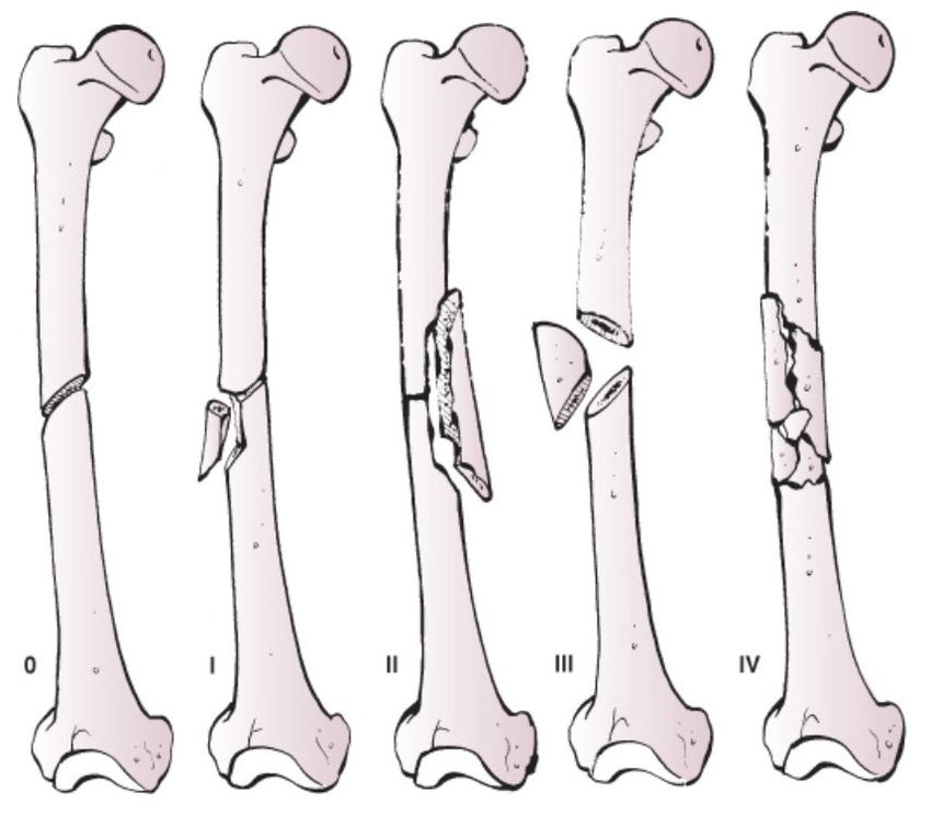
Disclosure
All figures belong to Andrew Chen, MD unless otherwise indicated

                                                             Core Curriculum V5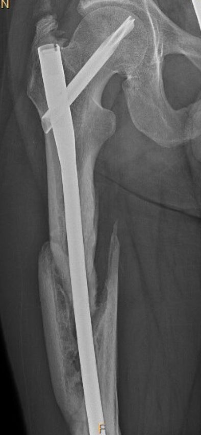
Objectives
 • Review initial management of femoral shaft fractures and possible
   concomitant injuries
 • Discuss multiple options with intramedullary nailing
    •   Antegrade/retrograde
    •   Starting point
    •   Reaming
    •   Patient positioning
 • Understand commonly associated complications

                                                                       Core Curriculum V5
Femoral Shaft Fractures
• Bimodal distribution
   • Young patients after high-energy trauma
   • Elderly patients after falls from standing
     secondary to osteopenia/osteoporosis
• MVC, MCC, pedestrian struck, fall from
  height, and gunshot wounds most
  common mechanisms
• Intramedullary nail as “gold standard”
  treatment, which has continued to
  evolve since introduction by Gerhard
  Küntscher around World War II

                                                  Core Curriculum V5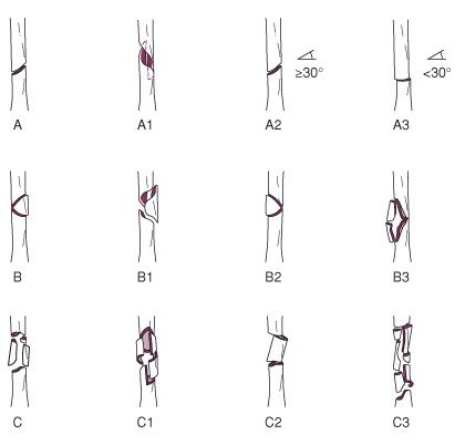
Anatomy
• Largest and strongest bone in body
• Anterior bow with radius of curvature ~120 cm1
• Blood supply from primary nutrient vessel
through linea aspera and small periosteal vessels
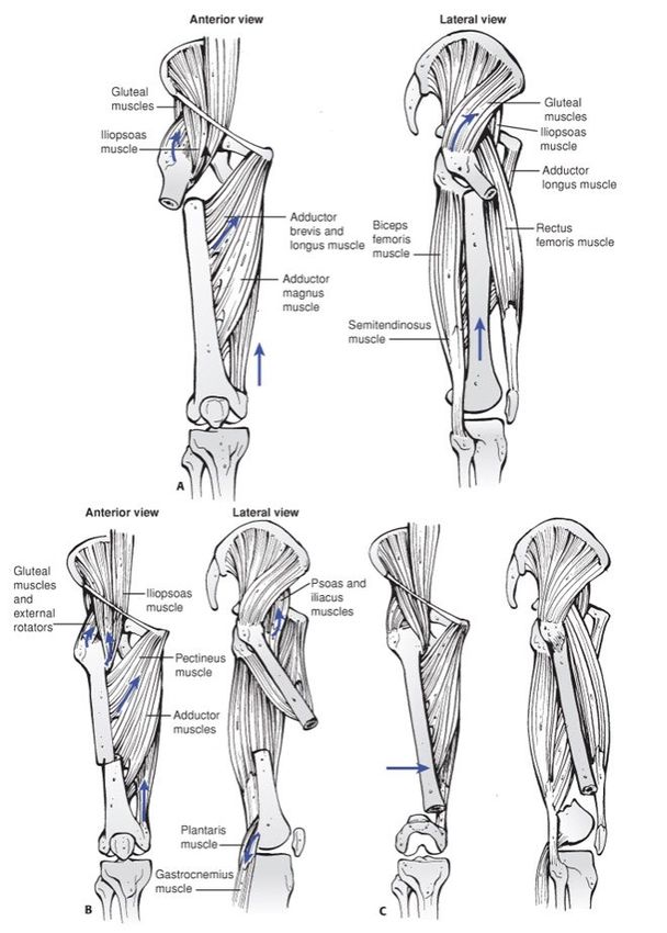
• Deformity pattern dependent on attached
musculature
• Proximal fragment
• Flexed (gluteus medius/minimus on greater trochanter)
• Abducted (iliopsoas on lesser trochanter)
• Distal fragment
• Varus (adductors inserting on medial aspect distal femur)
• Extension (gastrocnemius attaching on distal aspect of posterior femur)
Courtesy of Rockwood and Green’s Fracture in Adults2
Core Curriculum V5Femur Fracture Classification: AO/OTA
• Bone Segment 32
• Type A
• Simple
• Type B
• Wedge
• Type C
• Complex pattern
Courtesy of Rockwood and Green’s Fracture in Adults2
Core Curriculum V5Femur Fracture Classification: Winquist3
Courtesy of Rockwood and Green’s Fracture in Adults2
Core Curriculum V5Evaluation and Management
• Circumferential evaluation of thigh
for open wounds
• Full length AP/lateral films of femur
• Dedicated hip and knee films
• Average blood loss can be 1250 mL
• Vascular injury as high as 1.6%4
• Primary nerve injuries rare
• Open fracture does not preclude
compartment syndrome
Core Curriculum V5Associated Injuries
• Do not only focus on obvious shaft fracture
• Concomitant knee injury5
• Easier to diagnose in OR following fixation of femur
• Ligamentous laxity-49%
• Medial/Lateral meniscus injury-26%/28%
• Femoral neck/shaft fractures (3-10%)
• Discussed later in “special situations”
Core Curriculum V5Nonoperative Management
• Historically, traction used with months of bed rest
• High risk of pressure sores, pin infection, malunion, knee stiffness, muscle
wasting, and blood clot
• Now, traction typically used only as temporary measure for pain relief
and limit blood loss prior to surgical stabilization
Core Curriculum V5Temporary Traction
• Skin (Buck’s traction)
• Can be utilized with ease and minimal complications if
fracture can be stabilized in timely fashion (Damage Control Orthopaedics
• Select group of patients who are critically ill and not
hemodynamically stable for a long procedure
• Rapid temporary skeletal stabilization
Core Curriculum V5External Fixation
• Quick temporary stabilization as bridge to
intramedullary nailing
• Damage control orthopaedics
• Severe soft tissue contamination
• Ipsilateral arterial injury requiring repair
• Typically unilateral frame
• Pins placed anterior, anterolateral, or lateral
• At least 5 mm in size
• Can safely be converted to IMN within 2 weeks
without increased risk of deep infection7
Core Curriculum V5Plating
• More limited role given predictable results of
intramedullary nails
• Load bearing implant
• Considerations
• If extremely narrow or no canal making IMN difficult
or not possible
• Fracture adjacent to or through previous malunion
• Fracture adjacent to total arthroplasty stem
• Fracture extension into pertrochanteric or
metaphyseal region
• Fracture pattern can dictate fixation strategy
• Simple pattern with direct reduction and stable
fixation
• Comminuted pattern with bridge plating using
minimally invasive plate osteosynthesis (MIPO)
Core Curriculum V5Intramedullary Nailing
• “Gold standard”
• Minimal disruption to biology of fracture
• Usually achievable with closed reduction
• Multiple options
• Reamed vs unreamed
• Antegrade vs retrograde
• Piriformis vs trochanteric entry point
• Supine vs lateral position
Core Curriculum V5Reaming
• Potential advantages of reaming
• Larger implant and more durable construct
• Increased union
• Decrease chance of nail getting incarcerated
• Potential disadvantages of reaming
• Does reaming increase fatty emboli to lungs and increase pulmonary
complications?
Core Curriculum V5Reaming
• Reaming increases growth factors that can contribute to healing
• Can cause endosteal thermal damage and disrupt endosteal cortical
blood flow
• Reversed by 12 weeks with reamed nailing and 6 weeks with unreamed
nailing based on animal studies8
• However, increased surrounding muscle perfusion and periosteal blood flow
allows for healing9,10
Core Curriculum V5Reaming
• Canadian Orthopaedic Trauma Society11
• Multicenter prospective RCT
• 224 patients
• Risk nonunion 4.5 times greater with unreamed femoral nailing
• Bhandari et al12
• Systematic review and meta-analysis
• Reamed nailing significantly reduces rates of nonunion and implant failure
compared to unreamed nailing
Core Curriculum V5Multiply Injured Patients • Early studies showed benefit of immediate stabilization of long bone fractures in patients with multiple injuries • Johnson et al13 • 132 patients with ISS of 18 or higher • Early operative stabilization of fractures associated with decrease in ARDS • Bone et al14 • Prospective randomized study of 178 patients • Early (
Chest Injury and Femoral Shaft Fracture
• Early stabilization important, but impact of reaming with
  chest injury?
• Animal models with mixed results
     • Kropfl et al15 and Pape et al16
           •   Reaming shown to increase IM pressures and pulmonary artery
               pressures
           •   Reaming associated with fat embolization
     • Wolinsky et al17 and Duhelius et al18
           •   No adverse affect of reaming
• Clinical studies
     • Pape et al19
           •   Only clinical study to have shown detrimental effects to
               immediate reamed nailing in patients with pulmonary trauma
           •   Retrospective review 106 multiply injured patients
           •   Increased incidence of ARDS and and mortality
     • Charash et al20
           •   Retrospective study of 138 patients with blunt thoracic trauma
               and femoral shaft fracture
           •   Delayed surgical fixation (≥24 hours) associated with higher
               pulmonary complication rate

                                                                                Core Curriculum V5
Chest Injury and Femoral Shaft Fracture
• Clinical Studies
• Bosse et al21
• Center 1: reamed intramedullary nailing (95%)
• Center 2: plating (92%)
• Allowed for comparison of effects of reamed nailing
• ARDS, pneumonia, PE, multiple organ system failure, and death similar regardless of type
of treatment
• Thoracic injury is major determinant of morbidity and mortality, not IMN
• Canadian Orthopaedic Trauma Society22
• Prospective randomized multicenter study
• No difference between incidence of ARDS with reamed and unreamed nailing
Core Curriculum V5Head Injury and Femoral Shaft Fracture
• Remains controversial
• Early operative stabilization to limit pulmonary complications, but head
injured patients at risk for secondary brain injury
• Appears that early fixation itself does not lead to secondary brain injury,
but can result from hypoxemia, hypotension, and decreased cerebral
perfusion pressure
• Starr et al23
• Delay did not predict CNS complications, but pulmonary complications 45 times more likely
• McKee et al24
• No difference in early mortality, LOS, level neurologic disability, or cognitive testing
• Avoid intraoperative hypotension
Core Curriculum V5Timing of Fracture Fixation
• Brundage et al25
• “Our data show that early femur fracture fixation (< 24 hours) is associated
with an improved outcome, even in patients with coexistent head and/or
chest trauma. Fixation of femur fractures at 2 to 5 days was associated with a
significant increase in pulmonary complications, particularly with concomitant
head or chest trauma, and length of stay. Chest and head trauma are not
contraindications to early fixation with reamed intramedullary nailing.”
Core Curriculum V5Delayed IMN and Mortality
• Morshed et al26
• 3069 patients with ISS ≥ 15
• Decreased mortality by 50% with delay >12 hours
• Patients with serious abdominal trauma (AIS ≥3) benefited most with more
resuscitation
• Allow for appropriate resuscitation!
Core Curriculum V5Antegrade Nailing
• Can be used to treat majority of
femoral shaft fractures
• Surgical options
• Starting point
• Piriformis vs trochanteric entry
• Positioning
• Supine or lateral
• OR table
• Fracture table or radiolucent flat top
Core Curriculum V5Antegrade Nailing: Piriformis Entry Point
• Colinear trajectory with long axis
of femoral shaft
• Reduces risk of iatrogenic fracture
comminution and varus
malalignment
• Anterior starting point can cause
hoop stresses leading to
iatrogenic bursting through
proximal femur27
Core Curriculum V5Antegrade Nailing: Trochanteric Entry Point
• Potentially easier to identify starting point
• Tip not necessarily appropriate starting
point
• Can vary based on patient anatomy, but
typically slightly more medial entry point28
• On lateral radiograph colinear with long
axis of femur
• Avoid iatrogenic comminution
• Entry of nail should be rotated 90 degrees with
apex medial to help direct nail centrally
• Then de-rotated gradually once past fracture
Core Curriculum V5Patient Positioning
• Consider associated injuries such as spine or multiple extremity
injuries that can undergo simultaneous surgery
• Supine on fracture table
• More time consuming, but allows for consistent intraoperative traction
• Contralateral leg should be monitored to avoid compartment syndrome
• Potentially higher rate of malrotation
• Supine on radiolucent table
• +/- skeletal traction with skeletal traction pin
• Allows access to whole leg
• Starting point may be slightly more difficult, but can adduct hip to improve
access
• Lateral
• Can improve access to piriformis fossa especially in obese patients
• Longer setup time
• Harder to judge rotation
Core Curriculum V5Retrograde Nailing
• Supine on radiolucent table
• Insertion in intercondylar notch at
apex of Blumensaat line
• 1 cm anterior to PCL origin
• Collinear to long axis of femur in both
AP and lateral planes
• May be preferred for fractures close to
distal metaphysis
• Advocated with “floating knee” with
tibial shaft and femoral shaft fracture
allowing for fixation for single
percutaneous incision
Core Curriculum V5Retrograde Nailing
• Make sure distal end buried under subchondral bone to prevent
injury to patella in knee flexion29
• Only when nail is 1 mm prominent is the patellofemoral pressures increased
Core Curriculum V5Antegrade vs. Retrograde Nailing
• Ricci et al30
• Retrospective study
• 134 patients (retrograde) vs 147 (antegrade)
• Equal union rates: 88% (antegrade) and 89% (retrograde)
• Antegrade with more hip pain and retrograde with more knee pain
• Ostrum et al31
• Prospective randomized study
• Higher time to union for retrograde nail group
• Union rates similar
• Knee motion similar
• Increased symptomatic distal locking screws in retrograde group
• Tornetta et al32
• Prospective randomized study
• No difference in OR time, blood loss, technical complications, or nail size
• Time to union and rate of union same
Core Curriculum V5Knee Function
• No difference between antegrade and retrograde33
• Knee ROM
• Lysholm scores
• Isokinetic knee eval
• Secondary surgeries including hardware removal
Core Curriculum V5Static Locking
• Brumback et al34
• 98% union with statically locked nail
• Still allows for controlled motion at
fracture site while maintaining
length and rotation
Core Curriculum V5Post-Operative Weight Bearing
• Guided by multiple factors including
other injuries and location of fracture
• Brumback et al35
• Biomechanical and clinical results of
simulated and actual early weight-
bearing
• Immediate weight bearing with
segmentally comminuted mid-isthmal
fractures with statically locked nail was
safe
Core Curriculum V5Complications
• Leg length discrepancy
• Nonunion
• Malunion
• Infection
• Heterotopic Ossification
• At entry site with antegrade nailing
• Clinically symptomatic 5-10%
• Neurologic injury
• Usually secondary to patient positioning and intraoperative traction with
perineal post (pudendal nerve)
Core Curriculum V5Leg Length Discrepancy
• Can be challenging and discrepancy
noted in up to 43% cases36
• Radiographic ruler or bovie cord can
be used intraoperatively to compare
to uninjured limb
• Compare clinically immediately
after nailing
Core Curriculum V5Nonunion • Largest series of reamed antegrade nailing with
Nonunion
• Exchange Nailing
   • Results vary from retrospective studies41
   • 54-92.3%
   • Likely better for mid-shaft isthmal region and
     hyerptrophic nonunions that need more stability42
• Plate fixation ± bone grafting
   • Bellabarba et al43
       • IMN removed, indirect reduction, and plating to
         correct deformity and compress nonunion site for 23
         patients
       • Autologous bone grafting with all atrophic and 73%
         oligotrophic
       • 91% union rate after initial plating procedure
       • However, need protective weight bearing

                                                               Core Curriculum V5
Nonunion
• Augmentation plating around IMN
• Ueng et al44
• 100% union in 17 patients
• Early weight bearing allowed
• Bony union average 7 months
• Hakeos et al45
• 100% union in 7 patients
• All had autologous bone grafting
• Consider in meta-diaphyseal region
Core Curriculum V5Malunion
• Angular deformity in coronal and
sagittal plane more common in proximal
(30%) or distal (10%) fractures
• Nail fit in diaphysis usually helps prevent
this in mid-shaft (2%)
• Rotational malalignment
• Appears to be tolerated up to 15 degrees
• External deformity more symptomatic
• Braten et al46
• 110 femurs after IMN
• 19% with 15 degree deformity or more
• 38% symptomatic
Core Curriculum V5Femoral Rotation Assessment
• Clinical Exam
• Flex both hips and knees 90 degrees and check IR/ER
• Only useful after interlocks are placed
• Radiographic Exam
• Cortical thickness AP/Lateral planes47
• Femoral anteversion compared to uninjured side48
• Lesser trochanter profile compared to uninjured side49
• Inherent nail anteversion50
• Bilateral CT scan for accurate assessment post-op if
concerned51
• Axial cuts at at femoral neck and distal femur
• However, must be cautious because of native
individual bilateral differences
• Mean difference in version of 164 uninjured patients was
5.4 degrees52
Core Curriculum V5Infection
• Low rate of infection with IMN (1-3.8%)
• Sinus tract with purulent drainage signifies deep
infection
• Labs
• ESR/CRP/WBC
• Radiographic findings for sequestrum
• Infected nonunion
• Two stage
• Debridement with hardware removal followed by
temporary fixation with external fixator or antibiotic
cement fabricated in chest tube53
• Return for definitive fixation once infection eradicated
• Single stage
• Debridement followed by placement of antibiotic coated
interlocking nail54
Core Curriculum V5Special Situations
• Obesity
• Ipsilateral neck/shaft fractures
• Open fractures
• Vascular Injury
• Bilateral femur fractures
Core Curriculum V5Obesity
• Antegrade nailing can be more
difficult
• Osseous landmarks hard to palpate
• Femoral adduction limited
• Better results with trochanteric
entry point rather than piriformis
entry55
• Tucker et al56
• Retrograde nailing
• Decreased surgical time and radiation
exposure
Core Curriculum V5Ipsilateral Femoral Neck and Shaft Fracture
• 3-10% of femoral shaft fractures57
• Missed injuries 30-57% cases58,59
• Best-practice protocol60
• Dedicated IR plain radiograph of hip
• 2 mm fine cut CT scan through femoral neck
• Fluoroscopic lateral of femoral neck before fixation
• Postoperative orthogonal hip radiographs in OR
• Delayed diagnosis of femoral neck fractures reduced by 91%
• Rapid sequence MRI
• 12% of femoral neck fractures not identified on thin cut CT scan were
identified on rapid limited-sequence MRI61
• Address femoral neck/intertrochanteric fracture FIRST with
multiple lag screws or sliding hip screw62
• Femoral shaft then addressed with retrograde nail or lateral plate
• Although sequence of which to fix first – shaft versus
neck/intertrochanteric fracture fixation remains controversial
Core Curriculum V5Open Fracture
• Associated with significant soft tissue
damage even if just small skin wound
• Unless grossly contaminated, immediate
nailing after debridement is acceptable63,64
Core Curriculum V5Vascular Injury
• Rare, but usually secondary to penetrating
trauma
• Coordination between vascular team and
orthopaedic team
• Re-establish blood flow within 6 hours
• If limb perfusion needs to happen first, can
consider bony stabilization to obtain proper
length or ensure repair is made with
sufficient extra length to allow for restoration
of limb length
• Usually external fixator
• Early exchange to IMN65,66
Core Curriculum V5Bilateral Fractures
• Worse overall prognosis and
higher mortality67
• Higher ISS score and lower GCS
score68
• Nail less comminuted fracture
first to assess length/rotation
• Relative indication for
retrograde nail fixation
Core Curriculum V5Summary
• Do not miss concomitant injuries including ipsilateral femoral
neck/shaft fracture
• IMN is gold standard
• Reaming is safe and has higher union rates
• Multiple options including positioning and antegrade vs retrograde
• Many complications can be prevented!
Core Curriculum V5References
1) Egol KA, Chang EY, Cvitkovic J, Kummer FJ, Koval KJ. Mismatch of current intramedullary nails with the anterior bow of the femur. J Orthop Trauma. 2004 Aug;18(7):410-5.
2) Nork SE. (2015). Femoral Shaft Fractures. In Court-Brown CM, Heckman JD, McQueen MM, Ricci WM, Tornetta P (Eds.) Rockwood and Green for Adults. (8th ed., pp. 2150-2228). Wolters Kluwer Health.
3) Winquist RA, Hansen ST Jr, Clawson DK. Closed intramedullary nailing of femoral fractures. A report of 520 cases. J Bone Joint Surg Am. 1984;66:529–539.
4) Kluger Y, Gonze MD, Paul DB, et al. Blunt vascular injury associated with closed midshaft femur fracture: A plea for concern. J Trauma. 1994;36:222–225.
5) Vangsness CT Jr, DeCampos J, Merritt PO, et al. Meniscal injury associated with femoral shaft fractures. An arthroscopic evaluation of incidence. J Bone Joint Surg Br. 1993;75:207–209.
6) Even JL, Richards JE, Crosby CG, et al. Preoperative skeletal versus cutaneous traction for femoral shaft fractures treated within 24 hours. J Orthop Trauma. 2012; 26(10): e177-82.
7) Nowotarski PJ, Turen CH, Brumback RJ, et al. Conversion of external fixation to intramedullary nailing for fractures of the shaft of the femur in multiply injured patients. J Bone Joint Surg Am. 2000;82A:781–788.
8) Schemitsch EH, Kowalski MJ, Swiontkowski MF, et al. Cortical bone blood flow in reamed and unreamed locked intramedullary nailing: A fractured tibia model in sheep. J Orthop Trauma. 1994;8:373–382.
9) Hupel TM, Aksenov SA, Schemitsch EH. Muscle perfusion after intramedullary nailing of the canine tibia. J Trauma. 1998;45:256–262.
10) Reichert IL, McCarthy ID, Hughes SP. The acute vascular response to intramedullary reaming. Microsphere estimation of blood flow in the intact ovine tibia. J Bone Joint Surg Br. 1995;77:490–493.
11) Canadian Orthopaedic Trauma Society. Nonunion following intramedullary nailing of the femur with and without reaming. Results of a multicenter randomized clinical trial. J Bone Joint Surg Am. 2003;85A:2093–
2096.
12) Bhandari M, Guyatt GH, Tong D, et al. Reamed versus nonreamed intramedullary nailing of lower extremity long bone fractures: A systematic overview and meta-analysis. J Orthop Trauma. 2000;14:2–9.
13) Johnson KD, Cadambi A, Seibert GB. Incidence of adult respiratory distress syndrome in patients with multiple musculoskeletal injuries: Effect of early operative stabilization of fractures. J Trauma. 1985;25:375–
384.
14) Bone LB, Johnson KD, Weigelt J, et al. Early versus delayed stabilization of femoral fractures. A prospective randomized study. J Bone Joint Surg Am. 1989;71:336–340.
15) Kröpfl A, Davies J, Berger U, Hertz H, Schlag G. Intramedullary pressure and bone marrow fat extravasation in reamed and unreamed femoral nailing. J Orthop Res. 1999 Mar;17(2):261-8.
16) Pape HC, Dwenger A, Regel G, Schweitzer G, Jonas M, Remmers D, Krumm K, Neumann C, Sturm JA, Tscherne H. Pulmonary damage after intramedullary femoral nailing in traumatized sheep--is there an effect
from different nailing methods? J Trauma. 1992 Oct;33(4):574-81
17) Wolinsky PR, Banit D, Parker RE, et al. Reamed intramedullary femoral nailing after induction of an “ARDS-like” state in sheep: Effect on clinically applicable markers of pulmonary function. J Orthop Trauma.
1998;12:169–175; discussion 175–176.
18) Duwelius PJ, Huckfeldt R, Mullins RJ, Shiota T, Woll TS, Lindsey KH, Wheeler D. The effects of femoral intramedullary reaming on pulmonary function in a sheep lung model. J Bone Joint Surg Am. 1997
Feb;79(2):194-202.
19) Pape HC, Aufm'Kolk M, Paffrath T, et al. Primary intramedullary femur fixation in multiple trauma patients with associated lung contusion—a cause of posttraumatic ARDS? J Trauma. 1993;34:540–547
20) Charash WE, Fabian TC, Croce MA. Delayed surgical fixation of femur fractures is a risk factor for pulmonary failure independent of thoracic trauma. J Trauma. 1994;37:667–672.
21) Bosse MJ, MacKenzie EJ, Riemer BL, et al. Adult respiratory distress syndrome, pneumonia, and mortality following thoracic injury and a femoral fracture treated either with intramedullary nailing with reaming or
with a plate. A comparative study. J Bone Joint Surg Am. 1997;79A:799–809.
22) Canadian Orthopaedic Trauma Society. Reamed versus unreamed intramedullary nailing of the femur: Comparison of the rate of ARDS in multiple injured patients. J Orthop Trauma. 2006;20:384–387.
23) Starr AJ, Hunt JL, Chason DP, et al. Treatment of femur fracture with associated head injury. J Orthop Trauma. 1998;12:38–45.
24) McKee MD, Schemitsch EH, Vincent LO, et al. The effect of a femoral fracture on concomitant closed head injury in patients with multiple injuries. J Trauma. 1997;42:1041–1045.
25) Brundage SI, McGhan R, Jurkovich GJ, Mack CD, Maier RV. Timing of femur fracture fixation: effect on outcome in patients with thoracic and head injuries. J Trauma. 2002 Feb;52(2):299-307.
26) Morshed S, Miclau T 3rd, Bembom O, et al. Delayed internal fixation of femoral shaft fracture reduces mortality among patients with multisystem trauma. J Bone Joint Surg Am. 2009;91:3–13.
27) Johnson KD, Tencer AF, Sherman MC. Biomechanical factors affecting fracture stability and femoral bursting in closed intramedullary nailing of femoral shaft fractures, with illustrative case presentations. J
Orthop Trauma. 1987;1(1):1-11.
28) Antonelli L. Closed intramedullary nailing of diaphyseal fractures of the femur. Problems related to anatomical variations of the greater trochanter. Ital J Orthop Traumatol. 1989 Mar;15(1):67-74.
Core Curriculum V5References
29) Morgan E, Ostrum RF, DiCicco J, et al. Effects of retrograde femoral intramedullary nailing on the patellofemoral articulation. J Orthop Trauma. 1999;13:13–16.
30) Ricci WM, Bellabarba C, Evanoff B, et al. Retrograde versus antegrade nailing of femoral shaft fractures. J Orthop Trauma. 2001;15:161–169.
31) Ostrum RF, Agarwal A, Lakatos R, et al. Prospective comparison of retrograde and antegrade femoral intramedullary nailing. J Orthop Trauma. 2000;14:496–501.
32) Tornetta P 3rd, Tiburzi D. Antegrade or retrograde reamed femoral nailing. A prospective, randomised trial. J Bone Joint Surg Br. 2000;82:652–654.
33) Daglar B, Gungor E, Delialioglu OM, et al. . Comparison of knee function after antegrade and retrograde intramedullary nailing for diaphyseal femoral fractures: results of isokinetic evaluation. J Orthop Trauma.
2009 Oct;23(9):640-4.
34) Brumback RJ, Ellison TS, Poka A, et al. Intramedullary nailing of femoral shaft fractures. Part III: Long-term effects of static interlocking fixation. J Bone Joint Surg Am. 1992; 74:106–112.
35) Brumback RJ, Toal TR Jr, Murphy-Zane MS, et al. Immediate weight-bearing after treatment of a comminuted fracture of the femoral shaft with a statically locked intramedullary nail. J Bone Joint Surg Am.
1999;81:1538–1544.
36) Harris I, Hatfield A, Walton J: Assessing leg length discrepancy after femoral fracture: Clinical examination or computed tomography? ANZ J Surg 2005;75:319-321.
37) Wolinsky PR, McCarty E, Shyr Y, et al. Reamed intramedullary nailing of the femur: 551 cases. J Trauma. 1999;46:392–399.
38) Ring D, Jupiter JB, Sanders RA, et al. Complex nonunion of fractures of the femoral shaft treated by wave-plate osteosynthesis. J Bone Joint Surg Br. 1997;79:289–294.
39) Wu CC. The effect of dynamization on slowing the healing of femur shaft fractures after interlocking nailing. J Trauma. 1997;43:263–267.
40) Wu CC, Chen WJ. Healing of 56 segmental femoral shaft fractures after locked nailing. Poor results of dynamization. Acta Orthop Scand. 1997;68:537–540.
41) Webb LX, Winquist RA, Hansen ST. Intramedullary nailing and reaming for delayed union or nonunion of the femoral shaft. A report of 105 consecutive cases. Clin Orthop Relat Res. 1986;133–141.
42) Yang KH, Kim JR, Park J. Nonisthmal femoral shaft nonunion as a risk factor for exchange nailing failure. J Trauma Acute Care Surg. 2012;72:E60–E64.
43) Bellabarba C, Ricci WM, Bolhofner BR. Results of indirect reduction and plating of femoral shaft nonunions after intramedullary nailing. J Orthop Trauma. 2001;15:254–263.
44) Ueng SW, Chao EK, Lee SS, et al. Augmentative plate fixation for the management of femoral nonunion after intramedullary nailing. J Trauma. 1997;43:640–644.
45) Hakeos WM, Richards JE, Obremskey WT. Plate fixation of femoral nonunions over an intramedullary nail with autogenous bone grafting. J Orthop Trauma. 2011 Feb;25(2):84-9. doi:
10.1097/BOT.0b013e3181dfbb33. PMID: 21245710.
46) Braten M, Terjesen T, Rossvoll I. Torsional deformity after intramedullary nailing of femoral shaft fractures. Measurement of anteversion angles in 110 patients. J Bone Joint Surg Br. 1993;75:799–803.
47) Langer JS, Gardner MJ, Ricci WM. The cortical step sign as a tool for assessing and correcting rotational deformity in femoral shaft fractures. J Orthop Trauma. 2010;24:82–88.
48) Kuo TY, Skedros JG, Bloebaum RD. Measurement of femoral anteversion by biplane radiography and computed tomography imaging: Comparison with an anatomical reference. Invest Radiol. 2003;38:221–229.
49) Kim JJ, Kim E, Kim KY. Predicting the rotationally neutral state of the femur by comparing the shape of the contralateral lesser trochanter. Orthopedics. 2001;24:1069–1070.
50) Vaidya R, Dimovski R, Cizmic Z, Vaidya A, Gheraibeh P, Hudson I. Use of Inherent Anteversion of an Intramedullary Nail to Avoid Malrotation in Comminuted Femur Fractures: A Prospective Case- Control Study. J
Orthop Trauma. 2018 Dec;32(12):623-628.
51) Jaarsma RL, Pakvis DF, Verdonschot NBiert J, van Kampen A: Rotational malalignment after intramedullary nailing of femoral fractures. J Orthop Trauma 2004;18:403-409.
52) Croom WP, Lorenzana DJ, Auran RL, et al. Is contralateral templating reliable for establishing rotational alignment during intramedullary stabilization of femoral shaft fractures? A study of individual bilateral
differences in femoral version. J Orthop Trauma. 2018;32(2):61-66.
53) Paley D, Herzenberg JE. Intramedullary infections treated with antibiotic cement rods: Preliminary results in nine cases. J Orthop Trauma. 2002;16:723–729.
54) Barger J, Fragomen AT, Rozbruch SR. Antibiotic-Coated Interlocking Intramedullary Nail for the Treatment of Long-Bone Osteomyelitis. JBJS Rev. 2017 Jul;5(7):e5.
Core Curriculum V5References
55) Ricci WM, Schwappach J, Tucker M, et al. Trochanteric versus piriformis entry portal for the treatment of femoral shaft fractures. J Orthop Trauma. 2006;20:663–667.
56) Tucker MC, Schwappach JR, Leighton RK, et al. Results of femoral intramedullary nailing in patients who are obese versus those who are not obese: A prospective multicenter comparison study. J Orthop Trauma.
2007;21:523–529.
57) Wolinsky PR, Johnson KD. Ipsilateral femoral neck and shaft fractures. Clin Orthop Relat Res. 1995:81–90.
58) Yang KH, Han DY, Park HW, et al. Fracture of the ipsilateral neck of the femur in shaft nailing. The role of CT in diagnosis. J Bone Joint Surg Br. 1998;80:673–678.
59) Alho A. Concurrent ipsilateral fractures of the hip and shaft of the femur. A systematic review of 722 cases. Ann Chir Gynaecol. 1997;86:326–336.
60) Tornetta P 3rd, Kain MS, Creevy WR. Diagnosis of femoral neck fractures in patients with a femoral shaft fracture. Improvement with a standard protocol. J Bone Joint Surg Am. 2007;89:39–43.
61) Rogers NB, Hartline BE, Achor TS, et al. Improving the diagnosis of ipsilateral femoral neck and shaft fractures: a new imaging protocol. J Bone Joint Surg Am. 2020;102:309-314..
62) Swiontkowski MF, Hansen ST Jr, Kellam J. Ipsilateral fractures of the femoral neck and shaft. A treatment protocol. J Bone Joint Surg Am. 1984;66:260–268.
63) Brumback RJ, Ellison PS Jr, Poka A, et al. Intramedullary nailing of open fractures of the femoral shaft. J Bone Joint Surg Am. 1989;71:1324–1331.
64) Lhowe DW, Hansen ST. Immediate nailing of open fractures of the femoral shaft. J Bone Joint Surg Am. 1988;70:812–820.
65) Iannacone WM, Taffet R, DeLong WG Jr, et al. Early exchange intramedullary nailing of distal femoral fractures with vascular injury initially stabilized with external fixation. J Trauma. 1994;37:446–451.
66) Starr AJ, Hunt JL, Reinert CM. Treatment of femur fracture with associated vascular injury. J Trauma. 1996;40:17–21.
67) Nork SE, Agel J, Russell GV, et al. Mortality after reamed intramedullary nailing of bilateral femur fractures. Clin Orthop Relat Res. 2003;272–278
68) Copeland CE, Mitchell KA, Brumback RJ, et al. Mortality in patients with bilateral femoral fractures. J Orthop Trauma. 1998;12:315–319.
Core Curriculum V5You can also read