RESPIRATORY MANAGEMENT PROTOCOL
    OF PATIENTS WITH SARS-COV-2
             (COVID-19)
←
→
Page content transcription
If your browser does not render page correctly, please read the page content below
RESPIRATORY MANAGEMENT PROTOCOL
OF PATIENTS WITH SARS-COV-2
(COVID-19)
INTRODUCTION

       Based on what has been currently published by the Chinese, Italians,
UK, USA and the Spanish, we believe it is necessary to develop a unified action
criteria in order to optimise resources and apply the most effective therapies
for patients with COVID-19. While there have been consensus guidelines for
ventilator management with COVID-19, including those created by the
Surviving Sepsis Collaborative and the American Association of Respiratory
Care (AARC), many recommendations are based on evidence generated from
patients with more classic ARDS. From the published literature to date,
coupled with direct patient observation, we believe modifications to these
recommendations should be considered.

       It appears that in many patients, the type of hypoxemic respiratory failure
resulting from COVID-19 may differ from more classic forms of Acute
Respiratory Distress Syndrome (ARDS)(1). While many patients have
significant loss of end expiratory lung volume, compliance is often relatively
preserved with high degrees of alveolar dead-space, suggesting possible
alteration of the hypoxic pulmonary vasoconstriction (HPV) reflex (2), or other
mechanisms yet to be found.

In relation to the above we recommend that in patients with respiratory failure
related to COVID-19: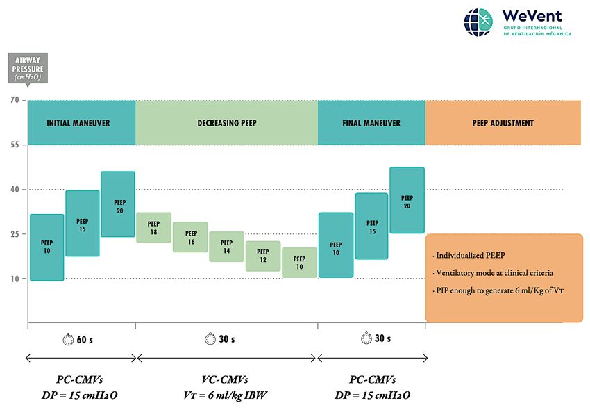
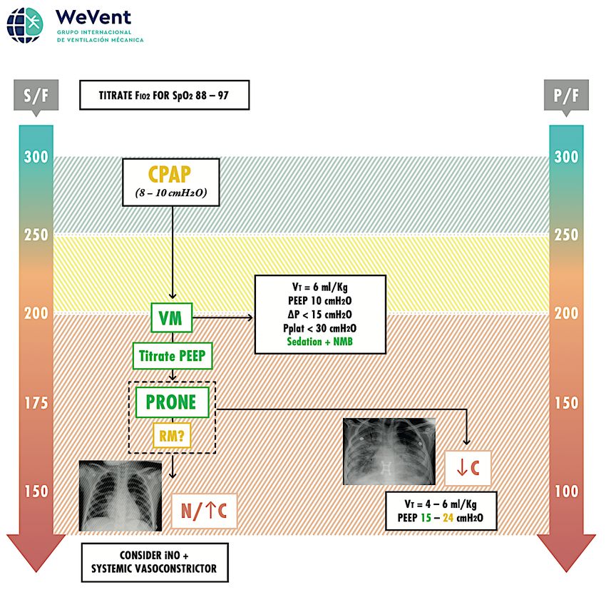
1. The degree of oxygen impairment should be measured routinely using
   pulse oximetry/inspired fraction of oxygen ratio (S/F) (3)(4)(5). S/F is
   recommended to assess patient evolution and is non-invasive, available to
   all patients. Taking into account the large number of patients to be treated,
   the S/F will be very useful as it is non-invasive. The PaO2/FiO2 ratio (P/F)
   is the gold standard(3) (6) to measure oxygen impairment but it can be
   reserved for patients with more severe disease, haemodynamic instability
   (needing invasive blood pressure monitoring), or for confirmation of S/F. It
   is important to instruct medical staff in the proper measurement of the S/F,
   which includes titration of FiO2 to achieve a saturation between 88 - 97%.
  [Figure 1]
     • In paediatric patients Oxygen Index (OI) and Oxygen Saturation Index
       (OSI) can be used to guide the treatment approach. (7)

2. High flow oxygen therapy (HFNC). High flow oxygen therapy (HFNC) can
   be considered for patients who do not have severe hypoxemia, particularly
   if the availability of ventilators is limited. However HFNC may have
   increased risks for aerosolization of the virus. The response to HFNC must
   be assessed within 30 – 60 minutes of initiation, and patients who do not
   improve significantly should not be maintained on HFNC. It is important to
   remember that HFNC does not produce significant lung recruitment.(8)(9)(10)
   If a patient on HFNC has sustained moderate/severe hypoxemia (S/F < 220;
   FiO2 > 0.4 for SpO2 > 92%) escalation to another form of respiratory support
   (NIV or intubation) should be strongly considered, depending on availability
   of resources.
       If HFNC is being used, there is risk of aerosol generation which poses an
   infection risk to the medical staff. In this sense, the use of HFNC in a
   negative pressure room with airborne precautions is highly recommended,
   if available.

     •   Oxygen therapy with mask with a reservoir. While this can deliver high
         amounts of oxygen, we believe this type of device should not be used
         since it does not generate recruitment of the lungs. Furthermore,
         administering 100% oxygen will cause an increase in PaO2 and SpO2
         with no improvement in P/F ratio (shunt, recruitment), which may lead
         to a delay in the administration of an adequate recruitment therapy,
         such as positive pressure ventilation (CPAP/BLPAP, IMV).

3. EARLY CPAP/BLPAP. Should be considered if the patient has significant
   oxygen need or high work of breathing. The response to CPAP/BLPAP
   must be assessed within 30 minutes of initiation, and those who do not
   improve significantly should be intubated. If the patient on NIV has
   sustained moderate/severe hypoxemia (S/F < 200; FiO2 > 0.4), intubation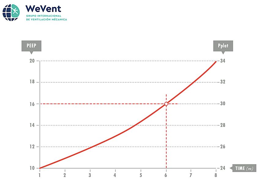
should be strongly considered, depending on availability of resources. The
   helmet(11) is recommended as the first line interface to be used, if available.
   When CPAP is provided using home care ventilators, it is important to
   remember the limitation in the administration of FiO2 (i.e. due to T piece).
   This reinforces the importance of close patient monitoring (S/F).

      It is important to consider that double limb circuits are recommended.
   However, single limb circuits can be used. In this case, it is important to
   insert a filter in between the patient and the expiratory port or directly on the
   expiratory port, depending on the different interfaces (vented interfaces and
   interfaces with anti-asphyxia valves are not recommended) available.
      As a summary, the use of non-invasive support has to be adapted to the
   local circumstances (equipment, personal, etc.).

      • Like HFNC, there is a risk for aerosolization of the virus with
        CPAP/BLPAP. This risk may be lower with the helmet interface. In
        the event that a helmet is not available, a total face mask interface
        would be the next choice. We advise the use of airborne precautions
        and negative pressure rooms if possible whenever CPAP/BLPAP is
        being used.

4. INTUBATION. If resources are available, the patient should be intubated if
   they maintain a P/F or S/F ≤ 200 (FiO2 > 0.4) after initiation of non-invasive
   therapy. If the patient is treated with NIV or HFNC and presents with high
   work of breathing (WOB) even if P/F or S/F is > 200 (FiO2 < 0.4 for SpO2 >
   92%), they should be intubated. A surrogate marker which can be used for
   guidance about work of breathing is the ROX index [(S/F) / RR] (12). If the
   patient has a ROX index ≤ 5 intubation is strongly advised. Chest X-Ray or
   lung ultrasound or chest CT should be performed to assess for ground glass
   opacities and the distribution of pulmonary opacifications. Static lung
   compliance (C) (13) should also be evaluated after intubation, with no
   spontaneous breathing present (flow zero).

5. INITIAL SETTINGS. Protective Ventilation. Since many of these patients
   have normal or high Respiratory System Compliance           (C), it is
   recommended (14):

         a. Standard sedation (controlled by SAS / RASS) + Neuromuscular
            Blockade. Continuous neuromuscular blockade should be
            considered for the first 24 – 48 hours after intubation (15), although
            intermittent neuromuscular blockade is also reasonable given
            limited availability of neuromuscular blocking medications in some
            countries.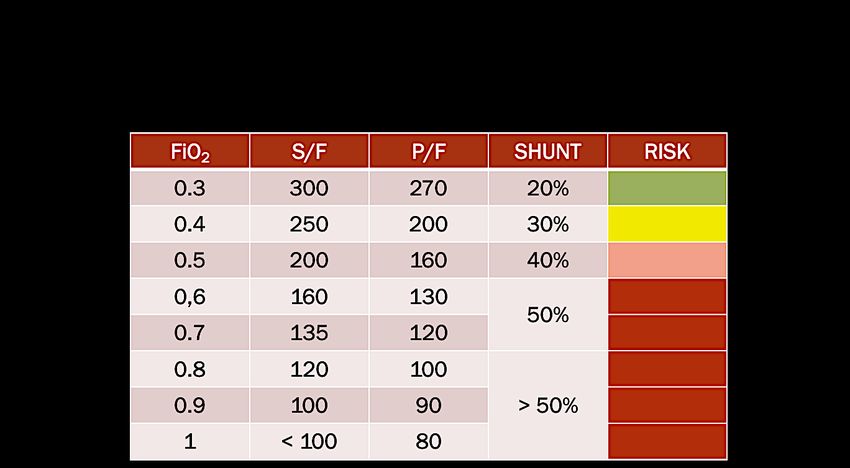
b.   Initial PEEP: 10 cmH2O. (16) (17)
        c.   VT: 6 ml/kg of IBW. (18) (19)
        d.   Driving Pressure: < 15 cmH2O. (20) (21)
        e.   Pplat: < 30 cmH2O. (22)(23)
        f.   FiO2 to achieve oxygen saturation between 88-97%

6. NO IMPROVEMENT. If P/F ratio remains < 200, consider the following:

  A. If P/F between 151 – 200 or S/F 176 – 200 (FiO2 0.4 – 0.5), perform a
     PEEP express titration (24)(25)(26)(27) [Figure 2]:

        a. Initial PEEP: 10 cmH2O. (28)
        b. Increase PEEP 2 cmH2O, every 2 minutes. Measure plateau
           pressure, and monitor oxygenation response (S/F ratio).
        c. Set the highest PEEP that maintains or improves S/F ratio and
           allows a Pplat of ≤ 30 cmH2O.

  B. If P/F ≤ 150 or S/F ≤ 175 (FiO2 > 0.5) after the express PEEP titration.
     The following therapeutic options would be recommended:

        a. PRONE POSITIONING. (29) (30) (31) (32) This should be considered
           as the first line of treatment if resources in the ICU are available.
           The evidence suggests it is most useful for patients with P/F ≤ 150,
           and is not recommended if P/F is above. Recommended approach
           (2 options):

             • Place Prone and evaluate response: If improvement in P/F – S/F
               ratio when placing prone, maintain in prone position for at least
               16 hours and until P/F or S/F ratio >200 for at least 4 hours.
               Turn supine. If patient is able to maintain P/F >150 or S/F > 175
               for at least 4 hours remain supine. Otherwise prone again for at
               least 16 hours and re-evaluate.
             • If resources are available, rotation between prone and supine
               positioning      should     be     considered     following    the
               recommendations above, with duration of prone ranging from
               16-20 hours a day.
             • It is important to considerer that most patients can suffer a
               decrease of P/F ratio after changing from prone to supine
               position.
b. RECRUITMENT MANEUVRES. (33) (34) (35) (36) This could be
           considered prior to prone positioning if resources are limited. They
           may also be considered for patients that are Prone but persist with
           P/F < 150 or S/F < 175. Careful consideration of haemodynamics
           must be considered before and during the recruitment maneuvers.
           Recruitment maneuvres should be performed under careful
           monitoring.

                 • We suggest increasing the PEEP initially to 10, then 15
                   and finally up to 20 cmH2O with 0-30 seconds at each
                   step, in PCV mode. Limit the delta pressure (Peak
                   Inspiratory Pressure-PEEP) to no more than 15 cmH20
                   during this maneuver. Then switch to volume control
                   ventilation and titrate the PEEP decrementally by the
                   lowest Driving Pressure. One option would be to follow the
                   modified Amato algorithm [Figure 3].

                 • Different methods of recruitment can be attempted as per
                   usual local practice, but no single method can be
                   recommended based on current evidence. Safety of the
                   patient has to be ensured during any RM(33). RM should
                   be used with extreme caution in patients with cardiac
                   disease or hemodynamic instability.

                •   Cardiac ultrasound in addition to lung ultrasound is highly
                    recommended when PEEP level is being titrated or during
                    recruitment maneuvres. Patients with more preserved
                    lung compliance will be more likely to suffer an increase
                    in pulmonary arterial pressure (PAP) or impairment in
                    venous return as PEEP is escalated, particularly if the
                    consolidated areas of lung are not able to be recruited.

7. If hypoxemia is refractory (P/F < 150 or S/F < 175) despite prone and RM,
   two options should be considered:

        1. ARDS with a predominance of alteration of the HPV reflex. (37) (38)
           This possibility should be considered in a patient with few alveolar-
           interstitial infiltrates (¨Black X-ray¨) and poor response to
           recruitment techniques (PEEP increments, proning and recruitment
           maneuvres). In this case, the use of iNO + systemic
           vasoconstrictors (39) (40) should be considered, particularly if there
           are signs of the pulmonary hypertension on echography.
The chest X-ray does not often reveal the extent of the problem. In
many cases the X-ray is relatively normal, but the CT is very
altered. Lung ultrasound is recommended for the diagnosis and to
guide the treatment approach.(41)
The use of ECMO as an initial treatment strategy is not
recommended; but this should be left to the evaluation by medical
staff on a case by case basis.
2. Classic ARDS. Chest X-ray with a clear bilateral alveolar-interstitial
infiltrate pattern and low C. (42)(43) A higher PEEP and lower tidal
volume strategy should be considered:
- PEEP = 12 - 24 cmH2O. (44)
- VT = 4 – 6 ml/kg IBW.
- Driving Pressure: < 15 cmH2O.
- Pplat: < 30 cmH2O.
• In these circumstances, the express PEEP titration or the
Recruitment Manuevers followed by PEEP titration
described above should be followed.
• Some patients with a typical ARDS may need levels above
15 – 18 cmH2O of PEEP. It is important to ensure that the
patient is responding favourably as PEEP is escalated. The
following criteria should be considered to gauge if higher
PEEP levels are helping the patient:
1. Improvement in oxygenation as measured by an
increase in P/F ratio by at least 25 points. If this
improvement in the P/F ratio is not observed after the
increase in the PEEP level, it would be advisable to
maintain the previous level of PEEP.(45)
2. Improvement in static compliance, as measured by a
reduction in driving pressure if volume control
ventilation is used, or improvement in tidal volume for
the same delta pressure if pressure control ventilation
is used.
3. No significant worsening of hemodynamics.S/F concept (Figure 1)
High PEEP Recommended Strategy
            Express
            (Figure 2)
Decreasing titration of PEEP
Protocol Prof. Amato (modified)
(Figure 3)Authors:
- Aurio Fajardo C
MD. Medicina Interna. Unidad de Paciente Crítico. MsC en Medicina
Intensiva. MsC en Ventilación Mecánica, Universitat de València. Grupo
Ventilación Mecánica Chile - Drive Flow Org. Viña del Mar. Chile.
- Alberto Medina V
PhD. MD. UCIP. Hospital Universitario Central de Asturias. Oviedo.
España
- Angelo Roncalli
PT. MsC. Hospital Escola Helvio Auto Maceió. Brasil
- Enrique Monares Zepeda
Médico Intensivista. Ciudad de México.
- Vicent Modesto A
MD. Jefe Clínico UCIP Hospital Universitari I Politècnic La Fe. València,
España.
- Rodrigo Adasme J
MsC, Pt, CRT. Terapia Respiratoria Hospital Clínico Red de Salud UC-
Christus. UNAB. Santiago, Chile.
- Robinder Khemani
MD, MsCI. Children´s Hospital Los Angeles, University of Southern
California; United States Of America
- Paolo Pelosi
MD. FERS. Department of Surgical Sciences and Integrated Diagnostics,
University of Genoa, Genoa, Italy
MD.FERS. Anesthesiology and Intensive Care Medicine, San Martino
Policlinico Hospital, IRCCS for Oncology and Neurosciences, Genoa,
Italy
Date: 22/03/2020
Presented to Dr. Gattinoni: 27/03/2020Co-Authors: - DR. DANIELE DE LUCA Service de Pédiatrie et Rénimation Néonatale, Hospital Antoine Béclère. Paris, France - DR. MARTÍ PONS Pediatric Intensive Care and Intermediate Care Department, Sant Joan de Déu University Hospital, Universitat de Barcelona, Esplungues de Llobregat, Spain. Critical Care Research Group, Institut de Recerca San Joan de Déu, Santa Rosa 39-57, 08950 Esplungues de Llobregat, Spain. - DRA. MIREIA GARCIA CUSCÓ MD, FRCPCH, FFICM. PICU. Bristol Royal Hospital for Children. UK - DR. GUILLERMO CHIAPPERO RICARDO Especialista en Terapia Intensiva, Neumología y Medicina Interna Director del Departamento de Docencia, Sociedad Argentina de Terapia Intensiva (SATI) Jefe de la Unidad de Ventilación Prolongada. Clínica AlterGarten. Buenos Aires Editor Libro SATI: ¨Ventilación Mecánica¨ - DR. MARTIN C.J KNEYBER MD. PhD. FCCM Chief, Division of Critical Care Medicine Chair Scientific Affairs, European Society for Paediatric and Neonatal Intensive Care Departament of Paedriatics, division of Paediatric Critical Care Medicine Beatrix Children´s Hospital University Medical Center, Groningen. Holland - DR. RAUL CARRILLO SPEARE Academia Nacional de Medicina Director de Areas Críticas Instituto Nacional de Rehabilitación. México - DR. VINKO TOMICIC FLORES MD. Medicina Interna – Terapia Intensiva Jefe Técnico Unidad De Cuidados Intensivos Hospital Regional de Antofagasta. Chile. - WILLIAM CRISTANCHO GÓMEZ Fisioterapeuta Universidad Nacional de Colombia Especialista en Docencia Universitaria Universidad El Bosque, Bogotá, Colombia - T.R.C JUAN CARLOS PÉREZ Instituto Mexicano del Seguro Social. Fundador Asociación Federal de Terapeutas Respiratorios A.C (AFTR) Presidente Federación Latinoamericana de Terapia Respiratoria (FELATERE) TRC The Latin American Board for Professional Certification in Respiratory Therapy - DR. KEVIN K. CHUNG MD, FCCM, FACP, COL, MC, USA Professor of Medicine and Surgery Chair, Department of Medicine (MED)
F. Edward Hebert School of Medicine- ¨America´s Medical School¨
 Uniformed Services University
 Bethesda, Maryland

- DRA. YOLANDA M. LOPEZ FERNANDEZ
 MD. UCIP.
 Cruces University Hospital, Bar-akaldo, Spain.

- DRA. CRISTINA CAMILO
 MD
 Pediatric Intensive Care Unit, Department of Pediatrics, Hospital Santa Maria (CHLN)
 Lisbon Academic Medical Center, Lisbon, Portugal.

- DR. CARLOS FERRANDO
 Department of Anesthesiology and Critical Care, Hospital Clínic, Institu D´investigació August Pi
 i Sunyer, Barcelona, Spain.
 CIBER de Enfermedades Respiratorias, Instituto de Salud Carlos III, Madrid, Spain.
References.

1.    Gattinoni L. Preliminary Observations on the Respiratory Behavior.
      2020;02(March):0–4.
2.    Ryan D, Frohlich S, McLoughlin P. Pulmonary vascular dysfunction in
      ARDS. Ann Intensive Care. 2014;4(1):1–11.
3.    Bilan N, Dastranji A, Ghalehgolab Behbahani A. Comparison of the Spo
      2 /Fio 2 Ratio and the Pao 2 /Fio 2 Ratio in Patients With Acute Lung
      Injury or Acute Respiratory Distress Syndrome . J Cardiovasc Thorac
      Res. 2015;7(1):28–31.
4.    Khemani RG, Thomas NJ, Venkatachalam V, Scimeme JP, Berutti T,
      Schneider JB, et al. Comparison of SpO 2 to PaO 2 based markers of
      lung disease severity for children with acute lung injury. Crit Care Med.
      2012;40(4):1309–16.
5.    Khemani RG, Patel NR, Bart RD, et al. Comparision of the Pulse Oximetric
      Saturation/Fraction of Inpired Oxygen Ratio and the PaO2/Fraction of
      Inspired Oxygen Ratio in Children. . Original research. Chest 2009; 135:
      662-668.
6.    Brown SM, Grissom CK, Moss M, Rice TW, Schoenfeld D, Hou PC, et al.
      Nonlinear Imputation of PaO2/FIO2 From SpO2/FIO2 Among Patients
      With Acute Respiratory Distress Syndrome. Chest. 2016;150(2):307–13.
7.    Kneyber MCJ, de Luca D, Calderini E, Jarreau PH, Javouhey E, Lopez-
      Herce J, et al. Recommendations for mechanical ventilation of critically ill
      children from the Paediatric Mechanical Ventilation Consensus
      Conference (PEMVECC). Intensive Care Med. 2017;43(12):1764–80.
8.    Modesto I Alapont V, Khemani RG, Medina A, Del Villar Guerra P, Molina
      Cambra A. Bayes to the Rescue: Continuous Positive Airway Pressure
      Has Less Mortality Than High-Flow Oxygen. Pediatr Crit Care Med.
      2017;18(2):e92–9.
9.    Correspondence Respiratory support for. 2020;2600(20):30110.
10.    Murthy S, Gomersall CD, Fowler RA. Care for Critically Ill Patients with
      COVID-19. JAMA - J Am Med Assoc. 2020;1–2.
11.   Patel BK, Wolfe KS, Pohlman AS, Hall JB, Kress JP. Effect of noninvasive
      ventilation delivered by helmet vs face mask on the rate of endotracheal
      intubation in patients with acute respiratory distress syndrome a
      randomized clinical trial. JAMA - J Am Med Assoc. 2016;315(22):2435–
      41.
12.   Rodriguez M, Thille AW, Boissier F, Veinstein A, Chatellier D, Robert R,
      et al. Predictors of successful separation from high-flow nasal oxygen
      therapy in patients with acute respiratory failure: a retrospective
      monocenter study. Ann Intensive Care. 2019;9(1).
13.   Beitler JR. Lung protection in acute respiratory distress syndrome: What
      should we target? Curr Opin Crit Care. 2020;26(1):26–34.
14.   Tusman G, Gogniat E, Madorno M, Otero P, Dianti J, Ceballos IF, et al.
Effect of PEEP on dead space in an experimental model of ARDS. Respir
Care. 2020;65(1):11–20.
15. Moss M, Huang DT, Brower RG, Ferguson ND, Ginde AA, Gong MN, et
al. Early neuromuscular blockade in the acute respiratory distress
syndrome. N Engl J Med. 2019;380(21):1997–2008.
16. Papazian L, Aubron C, Brochard L, Chiche JD, Combes A, Dreyfuss D, et
al. Formal guidelines: management of acute respiratory distress
syndrome. Ann Intensive Care. 2019;9(1).
17. Villar J, Kacmarek RM, Pérez-Méndez L, Aguirre-Jaime A. A high positive
end-expiratory pressure, low tidal volume ventilatory strategy improves
outcome in persistent acute respiratory distress syndrome: A randomized,
controlled trial. Crit Care Med. 2006;34(5):1311–8.
18. Pelosi P, Rocco PRM, Gama de Abreu M. Close down the lungs and keep
them resting to minimize ventilator-induced lung injury. Crit Care.
2018;22(1).
19. Gattinoni L, Pesenti A. The concept of “baby lung.” Intensive Care Med.
2005;31(6):776–84.
20. Amato MBP, Meade MO, Slutsky AS, Brochard L, Costa ELV, Schoenfeld
DA, et al. Driving pressure and survival in the acute respiratory distress
syndrome. N Engl J Med. 2014;372(8):747–55.
21. Samary CS, Santos RS, Santos CL, Felix NS, Bentes M, Barboza T, et
al. Biological impact of transpulmonary driving pressure in experimental
acute respiratory distress syndrome. Anesthesiology. 2015;123(2):423–
33.
22. Bellani G, Laffey JG, Pham T, Fan E. The LUNG SAFE study: A
presentation of the prevalence of ARDS according to the Berlin Definition!
Crit Care. 2016;20(1):268.
23. Briel M, Meade M, Mercat A, Brower RG, Talmor D, Walter SD, et al.
Higher vs lower positive end-expiratory pressure in patients with acute
lung injury and acute respiratory distress syndrome: Systematic review
and meta-analysis. JAMA - J Am Med Assoc. 2010;303(9):865–73.
24. Mercat A, Richard JCM, Vielle B, Jaber S, Osman D, Diehl JL, et al.
Positive end-expiratory pressure setting in adults with acute lung injury
and acute respiratory distress syndrome: A randomized controlled trial.
JAMA - J Am Med Assoc. 2008;299(6):646–55.
25. Bergez M, Fritsch N, Tran-Van D, Saghi T, Bounkim T, Gentile A, et al.
PEEP titration in moderate to severe ARDS: plateau versus
transpulmonary pressure. Ann Intensive Care [Internet]. 2019;9(1):81.
Available from: https://doi.org/10.1186/s13613-019-0554-3
26. Sahetya SK, Hager DN, Stephens RS, Needham DM, Brower RG. PEEP
Titration to Minimize Driving Pressure in Subjects With ARDS: A
Prospective Physiological Study. Respir Care. 2019;(C):respcare.07102.
27. Cavalcanti AB, Suzumura ÉA, Laranjeira LN, De Moraes Paisani D,
Damiani LP, Guimarães HP, et al. Effect of lung recruitment and titrated
Positive End-Expiratory Pressure (PEEP) vs low PEEP on mortality inpatients with acute respiratory distress syndrome - A randomized clinical
trial. JAMA - J Am Med Assoc. 2017;318(14):1335–45.
28. Chu EK, Whitehead T, Slutsky AS. Effects of cyclic opening and closing
at low- and high-volume ventilation on bronchoalveolar lavage cytokines.
Crit Care Med. 2004;32(1):168–74.
29. Munshi L, Del Sorbo L, Adhikari NKJ, Hodgson CL, Wunsch H, Meade
MO, et al. Prone position for acute respiratory distress syndrome: A
systematic review and meta-analysis. Ann Am Thorac Soc.
2017;14(October):S280–8.
30. Pugliese F, Babetto C, Alessandri F, Ranieri VM. Prone Positioning for
ARDS: Still misunderstood and misused. J Thorac Dis. 2018;10(Suppl
17):S2079–82.
31. Guérin C, Reignier J, Richard JC, Beuret P, Gacouin A, Boulain T, et al.
Prone positioning in severe acute respiratory distress syndrome. N Engl
J Med. 2013;368(23):2159–68.
32. Sud S, Friedrich JO, Taccone P, Polli F, Adhikari NKJ, Latini R, et al.
Prone ventilation reduces mortality in patients with acute respiratory
failure and severe hypoxemia: Systematic review and meta-analysis.
Intensive Care Med. 2010;36(4):585–99.
33. Hodgson CL, Tuxen D V., Davies AR, Bailey MJ, Higgins AM, Holland AE,
et al. A randomised controlled trial of an open lung strategy with staircase
recruitment, titrated PEEP and targeted low airway pressures in patients
with acute respiratory distress syndrome. Crit Care. 2011;15(3):1–9.
34. Meade MO, Cook DJ, Guyatt GH, Slutsky AS, Arabi YM, Cooper DJ, et
al. Ventilation strategy using low tidal volumes, recruitment maneuvers,
and high positive end-expiratory pressure for acute lung injury and acute
respiratory distress syndrome: A randomized controlled trial. JAMA - J Am
Med Assoc. 2008;299(6):637–45.
35. Medoff BD, Harris RS, Kesselman H, Venegas J, Amato MBP, Hess D.
Use of recruitment maneuvers and high positive end-expiratory pressure
in a patient with acute respiratory distress syndrome. Crit Care Med.
2000;28(4):1210–6.
36. Walkey AJ, Del Sorbo L, Hodgson CL, Adhikari NKJ, Wunsch H, Meade
MO, et al. Higher PEEP versus lower PEEP strategies for patients with
acute respiratory distress syndrome: A systematic review and meta-
analysis. Ann Am Thorac Soc. 2017;14:S297–303.
37. Nanchal RS, Truwit JD. Recent advances in understanding and treating
acute respiratory distress syndrome [version 1; referees: 2 approved].
F1000Research 2019, 8(F1000 Faculty Rev):1959 Last updated: 22 NOV
2019
38. Guérin C, Matthay MA. Acute cor pulmonale and the acute respiratory
distress syndrome. Intensive Care Med. 2016;42(5):934–6.
39. Papazian L, Bregeon F, Gaillat F, Thirion X, Roch A, Cortes E, et al.
Inhaled NO and almitrine bismesylate in patients with acute respiratory
distress syndrome: Effect of noradrenalin. Eur Respir J. 1999;14(6):1283–9.
40. Bazin JE, Mansoor O. Thierry GiUart MD, Jean E. Bazin. Combined nitric
oxide inhalation, prone positioning and almitrine infusion improve
oxygenation en severe ARDS. Canadian Journal of Anaesthesia 1998;
45(5): 402-409.
41. Singh Y, Tissot C, Fraga M V., Yousef N, Cortes RG, Lopez J, et al.
International evidence-based guidelines on Point of Care Ultrasound
(POCUS) for critically ill neonates and children issued by the POCUS
Working Group of the European Society of Paediatric and Neonatal
Intensive Care (ESPNIC). Crit Care. 2020;24(1):1–16.
42. Pintado M-C, de Pablo R, Trascasa M, Milicua J-M, Sánchez-García M.
Compliance-guided versus FiO 2 -driven positive-end expiratory pressure
in patients with moderate or severe acute respiratory distress syndrome
according to the Berlin definition. Med Intensiva (English Ed.
2017;41(5):277–84.
43. Griffiths M, Fan E, Baudouin S V. New UK guidelines for the management
of adult patients with ARDS. Thorax 2019; 74(10):931-933.
44. Brochard L, Hedenstierna G. Ten physiologic advances that improved
treatment for ARDS. Intensive Care Med. 2016;42(5):814–6.
45. Goligher EC, Kavanagh BP, Rubenfeld GD, Ferguson ND. Physiologic
responsiveness should guide entry into randomized controlled trials. Am
J Respir Crit Care Med. 2015;192(12):1416–9.You can also read