A NOVEL HIGHLY QUANTITATIVE AND REPRODUCIBLE ASSAY FOR THE DETECTION OF ANTI SARS COV 2 IGG AND IGM ANTIBODIES - NATURE
←
→
Page content transcription
If your browser does not render page correctly, please read the page content below
www.nature.com/scientificreports
OPEN A novel highly quantitative
and reproducible
assay for the detection
of anti‑SARS‑CoV‑2 IgG and IgM
antibodies
Kenta Noda1, Kouki Matsuda2, Shigehiro Yagishita3, Kenji Maeda2*, Yutaro Akiyama4,
Junko Terada‑Hirashima5,6, Hiromichi Matsushita7, Satoshi Iwata8, Kazuto Yamashita1,
Yusuke Atarashi1, Shunsuke Watanabe1, Nobuyuki Ide9, Tomokazu Yoshida1*,
Norio Ohmagari4, Hiroaki Mitsuya2,10,11 & Akinobu Hamada3*
The quantitative range and reproducibility of current serological tests for severe acute respiratory
syndrome coronavirus-2 (SARS-CoV-2) are not optimized. Herein, we developed a diagnostic test
that detects SARS-CoV-2 IgG and IgM with high quantitativeness and reproducibility and low
interference. The system was based on the high-sensitivity chemiluminescence enzyme immunoassay
(HISCL) platform and detects IgG and IgM specific to SARS-CoV-2 spike and nucleocapsid proteins.
Quantification accuracy and reproducibility were evaluated using serially diluted samples from 60
SARS-CoV-2-infected patients. Assay performance was evaluated using serum samples from the
SARS-CoV-2-infected patients and 500 SARS-CoV-2-negative serum samples collected before the
emergence of SARS-CoV-2. The system showed high quantification accuracy (range, 102), high
reproducibility (within 5%), and no cross-reaction between SARS1- and MERS-S proteins. Detection
accuracy was 98.3% and 93.3% for IgG and IgM against spike proteins and 100% and 71.7% for IgG and
IgM against nucleocapsid proteins, respectively. Mean antibody levels were > 10 times that in negative
samples upon admission and > 100 times that at convalescent periods. Clinical severity upon admission
was not correlated with IgG or IgM levels. This highly quantitative, reproducible assay system with
high clinical performance may help analyze temporal serological/immunological profiles of SARS-
CoV-2 infection and SARS-CoV-2 vaccine effectiveness.
The epidemic triggered by the severe acute respiratory syndrome coronavirus 2 (SARS-CoV-2) that originated
in China has rapidly spread worldwide. SARS-CoV-2 causes severe, acute—and in some cases—fatal corona-
virus disease in humans, named COVID-19, and is considered a global public threat1–4. However, no specific
therapeutic agents for COVID-19 are currently available. Moreover, SARS-CoV-2 may persist in some conva-
lescent COVID-19 survivors, and its infection could continue to recur, causing a continued pandemic. In such
circumstances, developing a high-performance and cost-effective diagnostic tool for COVID-19 is a high priority.
Currently, polymerase chain reaction (PCR) testing based on the detection of the SARS-CoV-2 genome
has been widely employed in clinical settings and used as a gold-standard to confirm positive and negative
1
Central Research Laboratories, Sysmex Corporation, Kobe, Hyogo, Japan. 2Department of Refractory Viral
Infections, National Center for Global Health and Medicine (NCGM) Research Institute, Tokyo, Japan. 3Division
of Molecular Pharmacology, National Cancer Center Research Institute, Tokyo, Japan. 4Disease Control and
Prevention Center (DCC), NCGM, Tokyo, Japan. 5Center for Clinical Science, NCGM, Tokyo, Japan. 6Respiratory
Medicine, NCGM Center Hospital, Tokyo, Japan. 7Department of Laboratory Medicine, National Cancer
Center Hospital, Tokyo, Japan. 8Department of Infectious Diseases, National Cancer Center Hospital, Tokyo,
Japan. 9Bio‑Diagnostic Reagent Technology Center, Sysmex Corporation, Kobe, Hyogo, Japan. 10Experimental
Retrovirology Section, HIV and AIDS Malignancy Branch, National Cancer Institute, National Institutes of Health,
Bethesda, MD, USA. 11Department of Clinical Sciences, Kumamoto University Hospital, Kumamoto, Japan. *email:
kmaeda@ri.ncgm.go.jp; Yoshida.Tomokazu@sysmex.co.jp; akhamad@ncc.go.jp
Scientific Reports | (2021) 11:5198 | https://doi.org/10.1038/s41598-021-84387-3 1
Vol.:(0123456789)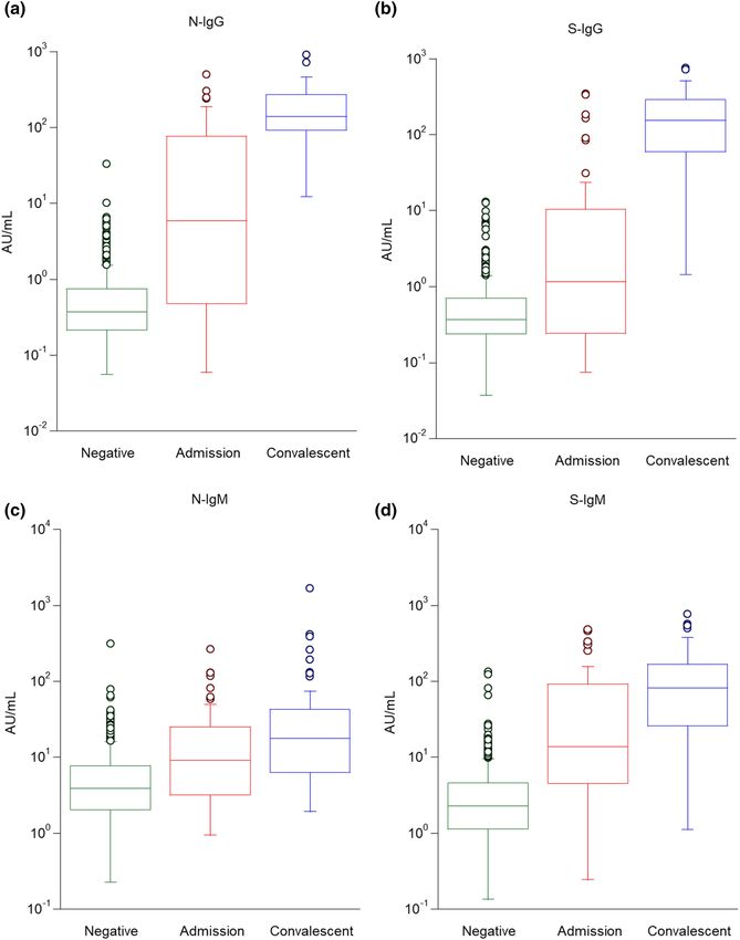
www.nature.com/scientificreports/
infections5,6. More recently, antigen testing has also been used, although it is slightly less sensitive and precise
than PCR t esting7.
Antibody testing aimed at detecting SARS-CoV-2-related immunity of patients is believed to be associated
with the clinical history of the infected patients and their virus-neutralizing immune r esponse8. Blood-based
antibody diagnostic analysis commonly assesses IgG and IgM titers9–11. It has been reported that IgM levels
increase early after infection in common viral infections, as well as in COVID-19, followed by an increase in IgG
levels12. However, it has been reported that in the early stage of SARS-CoV-2 infection IgG levels increase rather
than IgM levels13,14. Additionally, several methods have been developed for measuring the titers of antibodies
against SARS-CoV-2 proteins, such as nucleocapsid proteins and receptor proteins (Spike protein, S1 domain,
and receptor binding domain)15–19. Because the relationship between antibody levels and clinical response is
still unclear20–22, it is necessary to identify the SARS-CoV-2 protein, which can be used as a target in diagnos-
tic tests with a better diagnostic performance. Recently, some antibody-test kits for SARS-CoV-2 have been
made available for research; however, their performance is poor, and they generate unreliable and low-quality
results17,23–25. Currently available immunochromatographic COVID-19 antibody testing gives rise to qualitative
detection; thus accounting for false-negative results in clinical practice. Although there are quantitative detection
kits using ELISA for research purposes, the measurement accuracy and range are limited. In addition, commer-
cially available detection reagents that are used for an automatic immunoassay instrument are only qualitative
determinations26,27. In future, a high-precision quantitative assay is required not only for the purpose of posi-
tive qualitative determination but also for monitoring the antibody titer of vaccine administration and setting a
threshold value. Additionally, to observe antibody titers over time, a monitoring system that is quantitative and
has a wide measurement range is required. Therefore, a high-quality serological test with appropriate analytical
standards that is available at a reasonable cost is warranted.
This study describes a novel quantitative assay using a fully automated immunochemistry analyzer that
employs chemiluminescence enzyme immunoassay (CLEIA) m ethodology28,29, HISCL (Sysmex Corporation,
Kobe, Japan), to simultaneously detect IgG and IgM antibodies against two SARS-CoV-2 antigens, the spike (S)
protein and the nucleocapsid (N) protein (N-IgG, S-IgG, N-IgM, and S-IgM). HISCL is widely used in several
clinical fields due to its rapid reaction (17 min), wide dynamic ranges, and high reproducibility compared with
standard enzyme-linked immunosorbent assay (ELISA). The analytical performance of the HISCL-based serolog-
ical assay was evaluated with respect to its sensitivity, specificity, and reproducibility. Moreover, given the clinical
nature of this pilot study, the levels of N-IgG, S-IgG, N-IgM, and S-IgM in SARS-CoV-2-infected patients at the
time of hospital admission and during convalescence were also evaluated using the validated analytical method.
Results
Analytical performance. Development of an analytical method for detecting SARS CoV‑2 antigen. Stand-
ard curve. A standard curve was plotted based on the chemiluminescence intensity of the diluted SARS-CoV-
2-positive plasma to investigate the relationship between the emission intensity and the antibody concentration
(Fig. 1).
Reproducibility. The precision of the assay was determined using two SARS-CoV-2 concentrations. Within-
assay coefficient of variation (CV) was determined using 10 replicates for each sample. The within-run CVs for
N-IgG, S-IgG, N-IgM, and S-IgM were less than 2.1%, 3.3%, 2.1%, and 1.2%, respectively (Supplemental Table 1).
Interferences. The interference of common blood components was assessed by adding interfering substances
into plasma samples. The changes between samples with and without all types of interfering substances were less
than 15%.
Cross‑reactivity. In the assay using the nucleocapsid protein, the addition of the SARS-CoV-S antigen resulted
in the same level of inhibition in all positive specimens as the addition of the SARS-CoV-2 antigen. The inhibi-
tion rates of the NL63 and 229E antigens were 40–70% higher than those of the SARS-CoV-2 antigens, depend-
ing on the specimens. Furthermore, in the assay using the S protein, little cross-reactivity was observed in all
specimens (Supplemental Figure 1). When the antigen was added to the negative specimens, the quantitative
values were all less than 0.5 U/mL.
Clinical performance. Sensitivity, specificity, and AUC assessment. The ROC curves for N-IgG, S-IgG,
N-IgM, and S-IgM in the positive and negative subjects are shown in Fig. 2. The AUC for N-IgG, S-IgG, N-IgM,
and S-IgM were 0.9998, 0.9984, 0.8391, and 0.9671, respectively. The sensitivity and specificity of N-IgG and S-
IgG were over 98%. The sensitivity and specificity of N-IgM were 71.7% and 83.4%, whereas that for S-IgM were
93.3% and 93.6%, respectively (Supplemental Table 1).
The number of SARS-CoV-2 binding antibodies (IgG and IgM) was evaluated in all patients at the time of
admission as well as during convalescence. The IgG and IgM levels in blood samples from 500 non-infected
patients were also assessed as a negative control. As shown in Fig. 3, the antibody amount in the serum samples
from convalescent patients was higher than that at the time of admission for all tested antibodies (N-IgG, S-IgG,
N-IgM, and S-IgM). Overall, the on-admission N-IgM and S-IgM values were higher than N-IgG and S-IgG
(Table 1), which was in agreement with the general understanding that the levels of IgM increase more rapidly
than those of IgG after antigen exposure in patients with a viral infection. However, even though the levels of
Scientific Reports | (2021) 11:5198 | https://doi.org/10.1038/s41598-021-84387-3 2
Vol:.(1234567890)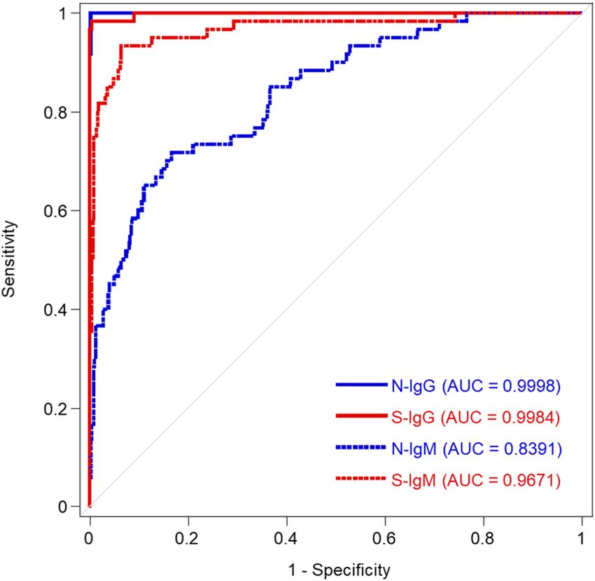
www.nature.com/scientificreports/

                                  Figure 1.  Relationship between chemiluminescence and the levels of each antibody. Working curve of N-IgG
                                  (a), S-IgG (b), N-IgM (c), and S-IgM (d).

                                  N-IgM and S-IgM were higher in patients with COVID-19, this is not a practical approach for the diagnosis of
                                  COVID-19 as the blood samples from healthy donors exhibit a high rate of false positives.
                                      Supplemental Figure 2 shows a comparison between the HISCL analysis system and the Euroimmun SARS-
                                  CoV-2 IgG ELISA kit available for research purposes. A total of 19 cases between "on admission" and "conva-
                                  lescence" were evaluated using both the assays, and both revealed significant differences between the admission
                                  and convalescence samples (Supplemental Figure 2). Nevertheless, the HISCL system seemed to be better at
                                  detecting the differences between the two time points.

Scientific Reports |   (2021) 11:5198 |               https://doi.org/10.1038/s41598-021-84387-3                                               3

                                                                                                                                          Vol.:(0123456789)
www.nature.com/scientificreports/
Figure 2. Clinical performance of SARS-CoV-2 antibodies. Receiver operating characteristic (ROC) curves for
convalescent and negative samples. Blue line, N-IgG; red line, S-IgG; blue dotted line, N-IgM; red dotted line,
S-IgM estimated by the logistic regression model. Thin gray line represents the random classification.
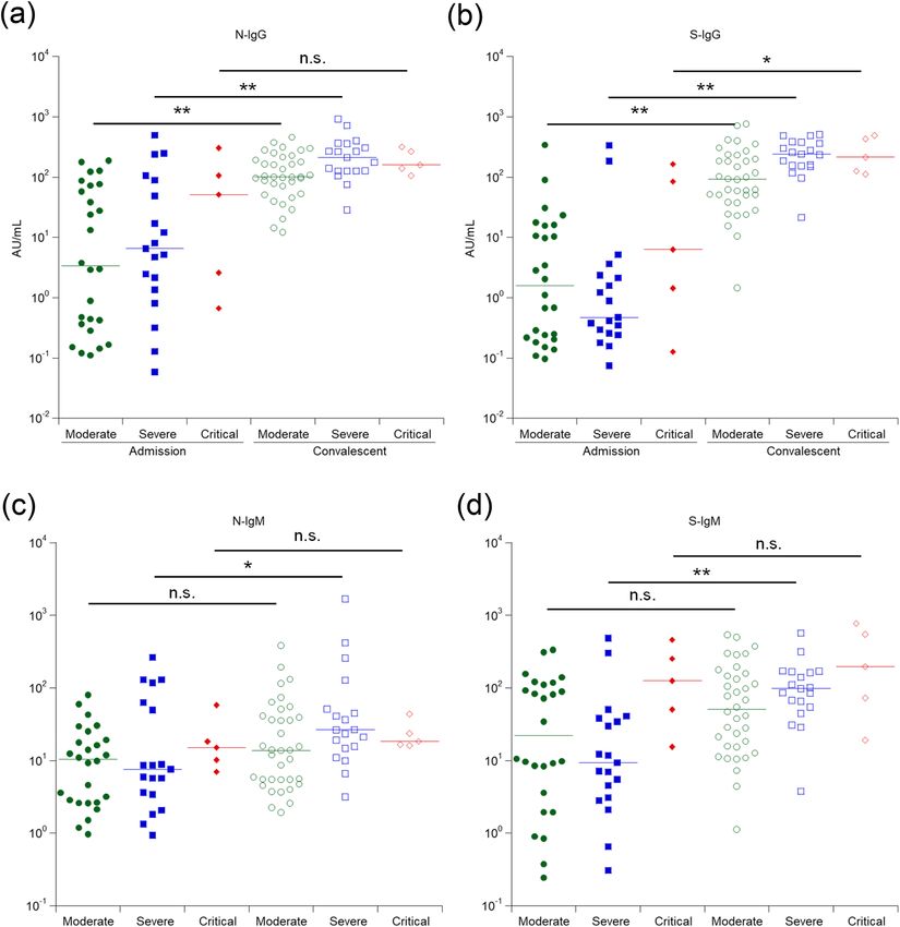
Clinical information for all patients was obtained and analyzed (Table 2). All patients were divided into
three groups (Moderate, Severe, and Critical) based on the symptoms and treatments during the disease course
(for definition, see the footnote in Table 2). Next, it was investigated whether the severity of each patient could
influence the antibody level. Regarding the titer of antibodies at the time of admission, patients in critical con-
dition exhibited the highest levels of all four antibodies among the three groups; however, the difference was
significant (P < 0.05) only for S-IgM (Table 1). Additionally, during convalescence, patients with severe/critical
disease course exhibited higher antibody levels than those with moderate disease. This result may suggest that
the duration of exposure to high titers of a virus (in patients in severe and critical conditions) is an important
factor for acquiring potent immunity against SARS-CoV-2.
Discussion
Serological testing to detect anti-SARS-CoV-2‒specific antibodies is an important approach to understand the
extent of COVID-19 spread in the community. These specific antibodies are immunological evidence of exposure
to the virus. SARS-CoV-2-triggered immunity is characterized by an early increase in IgM levels, followed by
an increase in IgG levels in the first few days post-infection. Subsequently, these antibodies are immunologi-
cal biomarkers that indicate the infection experience, even after the virus has been eliminated. Immunological
chromatography has been used as a qualitative test to distinguish between positive and negative results. However,
there is a need for a rapid and easy method that can quantitatively measure the antibody levels in the blood.
This study presents a highly sensitive quantitative test that can not only detect SARS-CoV-2 infection but also
quantifies antibody levels that define "acquired immunity".
The method developed herein aimed to detect IgG and IgM antibodies against the SARS-CoV-2 receptor-
binding domain (RBD). In particular, quantification of the S and N proteins and evaluating the levels of antibod-
ies associated with acquired immunity, are expected to be useful indicators for the development of a vaccine.
This current assay qualification builds on previously described E LISAs30 that focused on capturing antibodies
against the RBD, due to its critical role as the primary target of neutralizing antibodies31. Additionally, unlike
the trimeric stabilized S protein, which is more challenging to produce, RBD is a highly stable structure that
can be produced and scaled-up for large-scale ELISA testing. Given the immunogenic nature of the RBD, it
likely represents a reasonable target to quantify population-level exposure and define antibody levels, which
may ultimately be associated with immunity. However, additional viral components, including the membrane
protein‒the N or envelope protein—that are generated in large amounts during infection32,33 could provide
enhanced diagnostic value. This is of particular interest during early infection, when antigen concentration may
influence the kinetics of antibody evolution.
Furthermore, quantitative testing could help diagnose the infection at its early stages, as antigen concentra-
tions may affect antibody production. Besides, the quantification of antibody levels and the immune response
can enhance our understanding of SARS-CoV-2. Although some antibody testing methods have been reported,
it is unclear whether the results of qualitative antibody tests reflect immunity against SARS-CoV-2. Moreover,
antibodies that target alternate sites on the spike antigen or with the ability to drive additional antibody effector
functions may also contribute to immunity. Thus, next-generation assay development, using alternate antigens,
Scientific Reports | (2021) 11:5198 | https://doi.org/10.1038/s41598-021-84387-3 4
Vol:.(1234567890)www.nature.com/scientificreports/
Figure 3. Relationship between antibody levels at negative, admission, and convalescent patients. A total of
500 negative samples, 50 admission samples, and 60 convalescent samples were analyzed using each antibody
detection reagent. N-IgG (a), S-IgG (b), N-IgM (c), and S-IgM (d) levels.
may provide early diagnostic value in the absence of RNA testing and might provide additional insights into
antibody levels that reflect the immune response.
Herein, we described a rapid and high-reproducible diagnostic system for N-IgG, S-IgG, N-IgM, and S-IgM
using the HISCL platform. The developed assay exhibited good reproducibility and a wide dynamic range. Fur-
thermore, the antibody levels measured using these assays were not influenced by standard blood components,
and the specimens harboring coronavirus protein showed the highest antibody titer. As a result, the S protein
assay specifically detected the SARS-CoV-2 S protein but did not detect antibodies that targeted the proteins of
the common cold coronavirus (Supplemental Figure 1). To avoid potential cross reactivity of S protein-targeting
antibodies, the new system was designed based on the recognition of the three-dimensional structure of the
Scientific Reports | (2021) 11:5198 | https://doi.org/10.1038/s41598-021-84387-3 5
Vol.:(0123456789)www.nature.com/scientificreports/
P-value: admission vs
Admission Convalescent convalescent
Antibodies Moderate Severe Critical P-value Moderate Severe Critical P-value Moderate Severe Critical
N-IgG 3.4 6.6 51.5 M vs S, n.s. 101.8 214.7 159.2 M vs S, < 0.01 < 0.001 < 0.001 n.s.
S vs C, n.s. S vs C, n.s.
(AU/mL) (0.4–72.8) (1.6–78.8) (2.1–156.4) (62.4–205.3) (127.2–345.8) (132.2–282.6)
M vs C, n.s. M vs C, n.s.
S-IgG 1.6 0.5 6.3 M vs S, n.s. 93 242.6 216.4 M vs S, < 0.01 < 0.001 < 0.001 < 0.05
S vs C, n.s. S vs C, n.s.
(AU/mL) (0.2–15.8) (0.3–2.3) (1.1–104.5) (44.6–217.0) (154.9–375.9) (123.6–448.7)
M vs C, n.s. M vs C, n.s.
N-IgM 10.4 7.6 15.2 M vs S, n.s. 13.9 26.5 18.6 M vs S, n.s. n.s. < 0.05 n.s.
S vs C, n.s. S vs C, n.s.
(AU/mL) (2.7–19.3) (3.5–59.5) (9.4–28.4) (5.5–40.5) (15.0–49.6) (16.6–28.7)
M vs C, n.s. M vs C, n.s.
S-IgM 22.3 9.4 125.8 M vs S, n.s. 51.1 98 196.1 M vs S, n.s. n.s. < 0.001 n.s.
S vs C, < 0.05 S vs C, n.s.
(AU/mL) (3.6–111.1) (3.5–37.5) (41.7–304.5) (15.6–153.7) (57.4–166.3) (59.3–607.6)
M vs C, n.s. M vs C, n.s.
Table 1. Changes in IgG and IgM antibodies in response to the presence of SARS-CoV-2 antigen during
hospital admission and convalescence. Antibody concentrations are expressed as medians (distribution range).
P-values were obtained by Mann–Whitney U test and Steel–Dwass test. C critical, M moderate, S severe, n.s.
not significant.
Variables Early stage Convalescent
Sample collection date From 2/24/2020 to 5/3/2020 From 4/11/2020 to 6/2/2020
Total number 50 60
Sex (M/F) 39 / 11 48 / 12
Age, median (range) 50 (23–85) 53 (25–85)
Days since symptoms, median (range) 9 (1–25) 36 (20–95)
Disease severity1 (moderate/severe/critical) 26/19/5 36/19/5
Table 2. Patients and sample collection information. 1 Disease severity definitions—Moderate: febrile or
fatigue, and with/without pneumonia, and no oxygen inhalation required; Severe: febrile, fatigue, dyspnea,
severe pneumonia identified, and oxygen inhalation required; Critical: febrile, fatigue, severe dyspnea, critical
pneumonia identified, and positive pressure ventilation plus extracorporeal membrane oxygenation (ECMO)
required.
targeted antigen34. In contrast, assays targeting the N protein may also detect proteins of the cold coronavirus,
probably due to the high homology of the N proteins among the coronavirus f amily35,36. However, COVID-19
is clinically different from the common cold coronavirus-infection37, thus cross-reactivity with SARS-CoV-2 S
protein might be considered acceptable for the diagnosis of COVID-19. Therefore, the analytical performance
of the proposed novel antibody assay has sufficient for clinical application.
Although IgG antibody tests have recently become available as unapproved diagnostic kits, many have poor
performance as clinical diagnostic tools. The strategy of using the HISCL analysis system can serve as a robust
approach to quantify the levels of IgG and IgM antibodies against SARS-CoV-2. To fully explore the potential
of the new antibody assay, its detection results were compared with those obtained with commercially available
ELISA. This improved detection capacity may be attributed to the wider dynamic range of the HISCL-based
analysis system compared to that of reference ELISA assay, which is dependent on the absorbance, OD ratio
value, and detection system. These results demonstrate the superiority of the HISCL method for measuring a
wide range of antibody titers.
Maeda et al. confirmed that the concentration of neutralizing antibodies varies in patients, and the neutral-
izing activity and total antibody levels did not always correlate (manuscript in preparation). It is predicted that
the SARS-CoV-2 RBD may be an important target for such neutralizing antibodies38–41. However, assessing these
associations requires the accurate detection of the antibody levels at the following time points: at the beginning of
the infection, at the start of treatment, during recovery, and after treatment. Herein, it was shown that is possible
to detect some differences in the blood IgG levels among patients in the convalescent stage who exhibited moder-
ate or severe/critical disease conditions (Fig. 4), suggesting the importance of a highly quantitative SARS-CoV-2
antibody detection system to accurately evaluate the medical status. Additionally, this study showed that the
anti-SARS-CoV-2 IgM levels increase in the early phase of the disease in many patients; the earliest IgM detection
was on day three after onset (data not shown). Indeed, significantly higher S-IgM levels were detected on admis-
sion in patients with the critical disease. To further explore these differences, interleukin (IL)-4 levels, which are
considered to be important for the class-switching from IgM to IgG, were also examined, revealing that patients
who later entered a critical condition had significantly higher IL-4 levels in the blood on admission (Supplemental
Scientific Reports | (2021) 11:5198 | https://doi.org/10.1038/s41598-021-84387-3 6
Vol:.(1234567890)www.nature.com/scientificreports/
Figure 4. Relationship between severity status and antibody levels. Comparison of antibody titers on admission
and convalescent for N-IgG (a), S-IgG (b), N-IgM (c), and S-IgM (d). *P > 0.05, **P > 0.01.
Figure 3). Even though the mechanism underlying IL-4 increase in patients with critical COVID-19 course is
unknown, the data suggest that the IL-4 levels combined with the anti-SARS-CoV-2-S-IgM data at the onset of
the disease may help predict whether patients will enter a critical status in the future, requiring assisted ventila-
tion or extracorporeal membrane oxygenation support. In addition, the collected data also suggest that highly
sensitive anti-SARS-CoV-2 IgM detection may be useful for diagnosing and predicting disease severity.
There are a few limitations to this study. First, as the objective of the study was to establish a quantitative
antibody test, only a few blood serum samples were obtained from patients infected with COVID-19, as well as
data from patients who have been cured of the disease as stored samples were analyzed. Therefore, it is necessary
to evaluate the medical implications of this examination method for prospective tests in the future. Furthermore,
the change in antibody levels prior to and following vaccine administration is expected to be an important factor
during analysis. As our quantitative antibody assay is the preferred method for monitoring the response of the
immune system to COVID-19 following vaccination, the Japanese government is planning to begin vaccina-
tions in March 2021. Thus, we are currently planning to conduct an observational study on the infection rate
Scientific Reports | (2021) 11:5198 | https://doi.org/10.1038/s41598-021-84387-3 7
Vol.:(0123456789)www.nature.com/scientificreports/
and severity in clinical practice by monitoring antibody levels in healthcare workers, office staff, and patients
following vaccination.
In summary, we developed a novel anti-SARS-CoV-2 antibody detection system to accurately and robustly
measure the levels of IgG and IgM antibodies in patients infected with SARS-CoV-2. This new system may play
a valuable role in clinical practice and in understanding the ongoing COVID-19 epidemic.
Methods
Human clinical specimens. Sixty patients who were clinically diagnosed with COVID-19 and admitted to
the National Center for Global Health and Medicine (NCGM) in Tokyo were enrolled in the study. All patients
were confirmed to be SARS-CoV-2-positive using RNA-quantitative PCR on nasopharyngeal swab samples at
the time of enrollment in the study (Table 2 and Supplementary Table 2). A total of 500 serum samples from
cancer patients (337/163 male/female patients; age range: 7–87 years old) were provided by the National Cancer
Center Biobank, Japan and used as the negative control samples. The Ethics Committee from the NCGM and
the National Cancer Center approved this study (NCGM-G-003472-02, NCC 2020-026). Each patient provided
written informed consent, and this study abided by the Declaration of Helsinki principles.
Recombinant antigen production. The recombinant nucleocapsid proteins were produced based on
the following accession number sequences: SARS-CoV-2, YP_009724397; SARS-CoV, YP_009825061; MERS-
CoV, YP_009047211; HCoV-HKU1, YP_173242; HCoV-OC43, YP_009555245; HCoV-NL63, YP_003771; and
HCoV-229E, NP_073556. The recombinant S1 regions of the spike proteins were produced based on the fol-
lowing accession number sequences: SARS-CoV-2, YP_009724390; SARS-CoV, YP_009825051; MERS-CoV,
YP_009047204; HCoV-HKU1, YP_173238; HCoV-OC43, NP_937950; HCoV-NL63, YP_003767; and HCoV-
229E, NP_073551. Each sequence was cloned into a pcDNA3.4 vector (Thermo Fisher Scientific, Waltham, MA,
USA) with a C-terminus His-tag and transfected into Expi293 cells (Thermo Fisher Scientific) according to the
manufacturer’s protocol. The supernatants were harvested 6 days post-transfection. Recombinant antigens were
purified using a HisTrap HP column (Cytiva, Marlborough, MA, USA) and HiLoad 26/600 Superdex 200 pg
column (Cytiva). Purified SARS-CoV-2 nucleocapsid protein and S1 protein were respectively coupled with
magnetic beads using 1-ethyl-3-(3-dimethylaminopropyl)carbodiimide (Dojindo Molecular Technologies Inc.,
Kumamoto, Japan) and N-hydroxysuccinimide (Sigma-Aldrich, St. Louis, MO, USA).
Assay description. HISCL anti-SARS-CoV-2 immunoassay was developed to quantify the titer of IgG
and IgM antibodies in human serum or plasma. HISCL was operated in a fully automatic manner using the
chemiluminescent sandwich principle. In this system, the serum or plasma sample first reacted with SARS-CoV-
2-specific recombinant antigens bound to magnetic beads. After bound/free separation, the antigen–antibody
complex was incubated with an alkaline phosphatase-conjugated antibody against human IgG or IgM to form a
sandwich immunocomplex. After a second bound/free separation, a luminescent substrate was added into the
solution to allow for luminescence measurement. Chemiluminescence intensity was acquired within 17 min fol-
lowing the addition of the substrate. The temperature of the reaction chamber was maintained at 42 °C through-
out the procedure.
Analytical performance. Calibrator and control preparation. Calibrators and controls were prepared
using commercially available SARS-CoV-2 positive samples from Cantor Bioconnect (Toronto, Canada) and
TRINA BIOREACTIVES (Naenikon, Switzerland). The calibrators were prepared by serial dilutions of SARS-
CoV-2 positive samples with phosphate buffer. Each calibrator was measured three times. The assigned value of
the calibrator was defined based on the cut-off values. The assay value of the antibody was calculated from the
standard curve obtained from the logistic regression analysis.
Interferences. Potential interference materials were added to SARS-CoV-2 patient plasma up to the following
concentrations: free-form bilirubin (up to 200 mg/L), conjugated-form bilirubin (up to 200 mg/L), chyle (up
to 1600 FTU), hemoglobin (up to 500 mg/L), and rheumatoid factor (up to 4000 IU/L). These reagents were
obtained from Interference Check A Plus and Interference Check Rheumatoid Factor (Sysmex Corporation).
Cross‑reactivity. The specificity of the novel assay was evaluated by measuring samples with recombinant pro-
teins from coronaviruses strains (SARS-CoV-2, SARS1, MERS, OC43, HKU1, 229E, NL63). Each antigen was
added to the commercially available SARS-CoV-2 antibody-positive and negative samples so that the final con-
centration of the antigen in the measured sample was 20 µg/mL. Cross-reactivity was assessed based on the rate
of inhibition by antigen addition.
Clinical performance. Determination of sensitivity, specificity, and area under the curve (AUC). To deter-
mine the AUC, sensitivity, and specificity, 500 negative and 60 positive serum samples were measured with each
assay. AUC was determined by analyzing a receiver operating characteristic (ROC) curve. The sensitivity and
specificity were calculated using a threshold point defined based on the Youden Index.
Statistical analysis. The overall diagnostic accuracies of each antibody marker were evaluated by ROC
analysis and non-parametric pairwise comparisons were evaluated by Mann–Whitney U test using StatFlex v.7.0
software (Artech Co. Ltd., Osaka, Japan). R version 3.6.3 (The R Foundation for Statistical Computing, Vienna,
Scientific Reports | (2021) 11:5198 | https://doi.org/10.1038/s41598-021-84387-3 8
Vol:.(1234567890)www.nature.com/scientificreports/
Austria) was used to perform the Steel–Dwass test for non-parametric multiple comparisons. Differences were
considered significant if P < 0.05.
Data availability
The datasets generated during and/or analyzed during the current study are available from the corresponding
author on reasonable request.
Received: 30 November 2020; Accepted: 12 February 2021
References
1. Zhou, P. et al. A pneumonia outbreak associated with a new coronavirus of probable bat origin. Nature 579, 270–273 (2020).
2. Zhu, N. et al. A novel coronavirus from patients with pneumonia in China, 2019. N. Engl. J. Med. 382, 727–733 (2020).
3. World Health Organization. Coronavirus disease (COVID-19) outbreak situation. https://www.who.int/emergencies/diseases/
novel-coronavirus-2019 (Accessed June 2020).
4. Li, Q. et al. Early transmission dynamics in Wuhan, China, of novel coronavirus-infected pneumonia. N. Engl. J. Med. 382,
1199–1207 (2020).
5. Chu, D. K. W. et al. Molecular diagnosis of a novel coronavirus (2019-nCoV) causing an outbreak of pneumonia. Clin. Chem. 66,
549–555 (2020).
6. Wang, X. et al. Limits of detection of 6 approved RT-PCR kits for the novel SARS-coronavirus-2 (SARS-CoV-2). Clin. Chem. 66,
977–979 (2020).
7. Ogata, A. F. et al. Ultra-sensitive serial profiling of SARS-CoV-2 antigens and antibodies in plasma to understand disease progres-
sion in COVID-19 patients with severe disease. Clin. Chem. https://doi.org/10.1093/clinchem/hvaa213 (2020).
8. Tang, F. et al. Lack of peripheral memory B cell responses in recovered patients with severe acute respiratory syndrome: A six-year
follow-up study. J. Immunol. 186, 7264–7268 (2011).
9. Woo, P. C. et al. Longitudinal profile of immunoglobulin G (IgG), IgM, and IgA antibodies against the severe acute respiratory
syndrome (SARS) coronavirus nucleocapsid protein in patients with pneumonia due to the SARS coronavirus. Clin. Diagn. Lab.
Immunol. 11, 665–668 (2004).
10. Yu, F. et al. Recombinant truncated nucleocapsid protein as antigen in a novel immunoglobulin M capture enzyme-linked immu-
nosorbent assay for diagnosis of severe acute respiratory syndrome coronavirus infection. Clin. Vaccine Immunol. 14, 146–149
(2007).
11. He, Q. et al. Development of a Western blot assay for detection of antibodies against coronavirus causing severe acute respiratory
syndrome. Clin. Diagn. Lab. Immunol. 11, 417–422 (2004).
12. Woo, P. C. et al. Detection of specific antibodies to severe acute respiratory syndrome (SARS) coronavirus nucleocapsid protein
for serodiagnosis of SARS coronavirus pneumonia. J. Clin. Microbiol. 42, 2306–2309 (2004).
13. To, K. K. et al. Temporal profiles of viral load in posterior oropharyngeal saliva samples and serum antibody responses during
infection by SARS-CoV-2: An observational cohort study. Lancet Infect. Dis. 20, 565–574 (2020).
14. Guo, L. et al. Profiling early humoral response to diagnose novel coronavirus disease (COVID-19). Clin. Infect. Dis. 71, 778–785
(2020).
15. Nicol, T. et al. Assessment of SARS-CoV-2 serological tests for the diagnosis of COVID-19 through the evaluation of three immu-
noassays: Two automated immunoassays (Euroimmun and Abbott) and one rapid lateral flow immunoassay (NG Biotech). J. Clin.
Virol. 129, 104511 (2020).
16. Algaissi, A. et al. SARS-CoV-2 S1 and N-based serological assays reveal rapid seroconversion and induction of specific antibody
response in COVID-19 patients. Sci. Rep. 10, 16561 (2020).
17. Tang, M. S. et al. Clinical performance of two SARS-CoV-2 serologic assays. Clin. Chem. 66, 1055–1062 (2020).
18. Tang, M. S. et al. Association between SARS-CoV-2 neutralizing antibodies and commercial serological assays. Clin. Chem. https
://doi.org/10.1093/clinchem/hvaa211 (2020).
19. Shrock, E. et al. Viral epitope profiling of COVID-19 patients reveals cross-reactivity and correlates of severity. Science 370,
eabd4250 (2020).
20. Espejo, A. P. et al. Review of current advances in serologic testing for COVID-19. Am. J. Clin. Pathol. 154, 293–304 (2020).
21. Long, Q. X. et al. Antibody responses to SARS-CoV-2 in patients with COVID-19. Nat. Med. 26, 845–848 (2020).
22. Sun, B. et al. Kinetics of SARS-CoV-2 specific IgM and IgG responses in COVID-19 patients. Emerg. Microbes Infect. 9, 940–948
(2020).
23. Liu, W. et al. Evaluation of nucleocapsid and spike protein-based enzyme-linked immunosorbent assays for detecting antibodies
against SARS-CoV-2. J. Clin. Microbiol. 58, e00461-20 (2020).
24. Bundschuh, C. et al. Evaluation of the EDI enzyme linked immunosorbent assays for the detection of SARS-CoV-2 IgM and IgG
antibodies in human plasma. Clin. Chim. Acta 509, 79–82 (2020).
25. Li, Z. et al. Development and clinical application of a rapid IgM–IgG combined antibody test for SARS-CoV-2 infection diagnosis.
J. Med. Virol. 92, 1518–1524. https://doi.org/10.1002/jmv.25727 (2020).
26. Lippi, G. et al. Preliminary evaluation of Roche Cobas Elecsys Anti-SARS-CoV-2 chemiluminescence immunoassay. Clin. Chem.
Lab. Med. 58, e251–e253 (2020).
27. Bryan, A. et al. Performance characteristics of the Abbott Architect SARS-CoV-2 IgG assay and seroprevalence in Boise, Idaho.
Clin. Microbiol. 58, e00941-e1020 (2020).
28. Jekarl, D. W. et al. Analytical and clinical evaluation of chemiluminescent carcinoembryonic antigen (CEA) by HISCL-5000
immunoanalyzer. Ann. Clin. Lab. Sci. 50, 417–422 (2020).
29. Feng, S. et al. Evaluation of the novel HISCL chemiluminescence enzyme immunoassay for laboratory screening of hepatitis C
virus. Clin. Vaccine Immunol. 23, 652–654 (2016).
30. Amanat, F. et al. A serological assay to detect SARS-CoV-2 seroconversion in humans. Nat. Med. 26, 1033–1036 (2020).
31. Quinlan, B. D. et al. The SARS-CoV-2 receptor-binding domain elicits a potent neutralizing response without antibody-dependent
enhancement. bioRxiv https://doi.org/10.1101/2020.04.10.036418v1 (2020).
32. Kim, D. et al. The architecture of SARS-CoV-2 transcriptome. Cell 181, 914-921.e10 (2020).
33. Hofmann, H. & Pöhlmann, S. Cellular entry of the SARS coronavirus. Trends Microbiol. 12, 466–472 (2004).
34. Ladner, J. T. et al. Epitope-resolved profiling of the SARS-CoV-2 antibody response identifies cross-reactivity with an endemic
human CoV. bioRxiv https://doi.org/10.1101/2020.07.27.222943v1 (2020).
35. Grifoni, A. et al. A sequence homology and bioinformatic approach can predict candidate targets for immune responses to SARS-
CoV-2. Cell Host Microbe 27, 671-680.e2 (2020).
36. Klompus, S. et al. Cross-reactive antibody responses against SARS-CoV-2 and seasonal common cold coronaviruses. medRxiv
https://doi.org/10.1101/2020.09.01.20182220v2 (2020).
Scientific Reports | (2021) 11:5198 | https://doi.org/10.1038/s41598-021-84387-3 9
Vol.:(0123456789)www.nature.com/scientificreports/
37. Murchu, E. O. et al. Immune response following infection with SARS-CoV-2 and other coronaviruses: A rapid review. Rev. Med.
Virol. https://doi.org/10.1002/rmv.2162 (2020).
38. Peterhoff, D. et al. A highly specific and sensitive serological assay detects SARS-CoV-2 antibody levels in COVID-19 patients that
correlate with neutralization. Infection https://doi.org/10.1007/s15010-020-01503-7 (2020).
39. Deshpande, G. R. et al. Neutralizing antibody responses to SARS-CoV-2 in COVID-19 patients. Indian J. Med. Res. 152, 82–87
(2020).
40. Tan, C. W. et al. A SARS-CoV-2 surrogate virus neutralization test based on antibody-mediated blockage of ACE2-spike protein-
protein interaction. Nat. Biotechnol. 38, 1073–1078 (2020).
41. He, Y., Lu, H., Siddiqui, P., Zhou, Y. & Jiang, S. Receptor-binding domain of severe acute respiratory syndrome coronavirus spike
protein contains multiple conformation-dependent epitopes that induce highly potent neutralizing antibodies. J. Immunol. 174,
4908–4915 (2005).
Acknowledgements
The authors thank Dr. Noriko Kinoshita for providing information regarding the clinical samples, and Takeshi
Imoarai and Tatsuya Narikawa for supporting this activity.
Author contributions
A.H., K.Mae., and T.Y. conceived the project. K.N., A.H,. and K.Mae. designed the study concept and wrote the
manuscript. K.N., K.Y., Y.A., S.W., and N.I. established assays for N-IgG, S-IgG, N-IgM, and S-IgM. K.Mat., S.Y.,
K.Mae., Y.A., J.T.-H., H.Ma., S.I., N.O., H.Mi., and A.H. collected and interpreted the data. All authors read and
approved the final version of the manuscript.
Funding
This study was supported by a research fund from the Japan Health Research Promotion Bureau Research Fund
(2020-A-01) granted to AH and HMi, and a grant from the Japan Agency for Medical Research and Develop-
ment (JP20fk0108160) granted to KMae. This research was also funded by the Sysmex Corporation. The National
Cancer Center Biobank is supported by the National Cancer Center Research and Development Fund, Japan.
Competing interests
KN, KY, YA, SW, NI, and TY are salaried employees of Sysmex Corporation. AH. and HMi have received research
grants from Sysmex Corporation. The other authors declare no potential conflict of interest.
Additional information
Supplementary Information The online version contains supplementary material available at https://doi.
org/10.1038/s41598-021-84387-3.
Correspondence and requests for materials should be addressed to K.M., T.Y. or A.H.
Reprints and permissions information is available at www.nature.com/reprints.
Publisher’s note Springer Nature remains neutral with regard to jurisdictional claims in published maps and
institutional affiliations.
Open Access This article is licensed under a Creative Commons Attribution 4.0 International
License, which permits use, sharing, adaptation, distribution and reproduction in any medium or
format, as long as you give appropriate credit to the original author(s) and the source, provide a link to the
Creative Commons licence, and indicate if changes were made. The images or other third party material in this
article are included in the article’s Creative Commons licence, unless indicated otherwise in a credit line to the
material. If material is not included in the article’s Creative Commons licence and your intended use is not
permitted by statutory regulation or exceeds the permitted use, you will need to obtain permission directly from
the copyright holder. To view a copy of this licence, visit http://creativecommons.org/licenses/by/4.0/.
© The Author(s) 2021
Scientific Reports | (2021) 11:5198 | https://doi.org/10.1038/s41598-021-84387-3 10
Vol:.(1234567890)You can also read