Calcific spurs at the insertion of the Achilles tendon: a clinical and histological study - MLTJ
←
→
Page content transcription
If your browser does not render page correctly, please read the page content below
Original article

Calcific spurs at the insertion of the Achilles tendon:
a clinical and histological study

Kristian Jarl Johan Johansson,                                mon type of disorders along with partial Achilles tendon
Janne Julius Sarimo,                                          tear and tendinosis3.
Lasse Lennart Lempainen,                                      Calcific spurs occasionally grow to the calcaneal insertion
Tiina Laitala-Leinonen,                                       of the Achilles tendon. They are seen in various rheumatic
Sakari Yrjö Orava                                             and articular diseases but also in physically active individ-
                                                              uals4,5. A symptomatic tendon with a confirmed inser-
                                                              tional spur may be referred to as an insertional calcific
Department Paavo Nurmi Center, Hospital NEO,                  tendinopathy (CT), and may be present with or without
Turun Yliopisto, Turku, Finland                               calcific deposits of the main body of the tendon, referred
                                                              to as CT of the main body6.
                                                               Surgery for distal tendinopathy of the Achilles tendon may
Corresponding author:                                         sometimes become necessary to eliminate the disturbing
Kristian Jarl Johan Johansson                                 symptoms7,8. Due to the rare nature of insertional CT, a
Department Paavo Nurmi Center                                 good understanding of effective surgical treatments and
Hospital NEO, Turun Yliopisto                                 the presence of the possible outcome related factors is
Savikkakatu 1 A 2, Turku, Finland                             lacking. Also, the cellular distribution in the spurs, which
e-mail: kristian.johansson@utu.fi                             can be assessed by histological staining, found in the in-
                                                              sertional CT of the Achilles tendon is unclear.
                                                              We present a series of 36 cases treated surgically due to
Summary                                                       insertional CT of the Achilles tendon, most of which is in
                                                              conjunction with a Haglund’s deformity.
In active people, insertional calcific tendinopathy
(CT) of the Achilles tendon is rare. We evaluated the
results of surgical treatment for Achilles tendon CT          Materials and methods
and analyzed post-surgery Achilles tendon histolog-
ical features. The study included 36 operations in 34         Study design
patients. Twenty-eight (78%) cases had a resection of
a Haglund’s deformity performed. The mean age of              The study consists of material gathered retrospectively
the patients was 42 years (range=23 to 68). Thirteen          from 36 operations conducted on 34 patients in Turku,
of the patients were professional athletes and 20             Finland and Rome, Italy during the years 1999 to 2007.
recreational athletes. In twenty-five (69%) cases, the        In two of the patients the finding was bilateral. The 36
result of surgery was rated good, in nine cases (25%)         cases of Achilles tendon insertional CT were selected
moderate and in two (6%) cases poor. The mean age             from materials of a total of 1425 operations done for
of those with a good result was 10 years lower (40            Achilles tendon over-use injuries by the senior author
versus 50 years) than those with a moderate result            (SO). The incidence of insertional CT of the Achilles ten-
(p=0.0239). Higher athletic activity was also related to      don was thus 2.5% in our material. All patients were eval-
a better outcome (p=0.0205). Histology samples                uated at the senior authors’ office.
showed fast remodellation and stem-cell activation.
Surgery seemed to result in a good outcome in pa-
tients with or without a Haglund’s deformity which            Inclusion criteria
failed conservative treatment.
                                                              Clinical symptoms of CT included exercise related pain
Key words: Achilles tendon, achillodynia, calcific            and swelling and warmth in the back of the foot. Clinical
tendinopathy, enthesophyte, Haglundʼs deformity, sur-         findings included tenderness during palpation, with bur-
gery.                                                         sitis specifically at the upper posterior calcaneus, swelling
                                                              in the heel and also a thickened distal Achilles tendon.
                                                               The diagnosis of CT at the Achilles insertion was con-
Introduction                                                  firmed radiologically on average of 10 months after the ap-
                                                              pearance of the first symptoms and the patients were
Achilles tendinopathy is common in athletes and physi-        treated conservatively for an average of 8 months after
cally active people1,2. Approximately one-third of all        the diagnosis. In addition to radiography, ultrasound and
Achilles tendinopathies are distal with retrocalcaneal bur-   MRI were used to either confirm the diagnosis or rule out
sitis and Haglund’s heel deformity being the most com-        other causes for the symptoms.

Muscles, Ligaments and Tendons Journal 2012; 2 (4): 273-277                                                            273
K.J.J. Johansson et al.
Preoperative treatment and 2006. Two of the specimens were contained in
50% EtOH and the rest in 10% fosfatic buffered forma-
The preoperative conservative treatment used in these lin. All of the specimens were transferred to 70% EtOH
patients included anti-inflammatory pain medication, cor- at the beginning of this study. The spurs were cut into
ticosteroid injections, rest, physiotherapy with eccentric two equal parts. One piece was stored in the 70%
exercise, heel elevation and modified shoes. EtOH and the other decalcified for 16 days in a 14%
solution of EDTA. The decalcified spurs were washed
in tap water for 4 hours and dehydrated in graded al-
Surgical technique and post-operative treatment cohols, cleared in xylene, then embedded in paraffin
and cut with microtome. Samples were stained with
The surgery was performed in spinal anaesthesia, isolat- hematoxyline-eosine, Weigert-van Gieson and TRACP
ing the leg from the circulation by tourniquet and with the for immunohistochemistry and analysed under light
patient lying in prone position. An incision was made lat- microscopy.
erally, vertically, and continued in L-form medially distal to
the Achilles tendon. After exposure of the retrocalcaneal
space the hypertrophic retrocalcaneal bursa was re- Statistical analysis
sected. When a Haglund’s deformity had been diag-
nosed, the prominent part of the superior corner of calca- The patients were tabled in the statistical program SAS
neus was excised with a chisel. The calcific spur was Enterprise Guide 4 by 8 variables: sex, age, athletic ac-
palpated at the insertion of the Achilles tendon and a lon- tivity, spur size, duration of conservative treatment, dura-
gitudinal incision was made in the tendon insertion paral- tion of symptoms before operative treatment, surgical
lel to the tendon fibres. The calcification was then care- procedure (spur resection with or without resection of a
fully prepared, free from the surrounding tendon, with a Haglund’s deformity), and outcome. In the processing of
small scalpel and removed from its base with a chisel. The the material, we used one-way ANOVA and table analy-
tendon defect was closed using 3-0 PDS suture. If the de- sis with p-values calculated with the Pearson chi-square-
fect in the tendon was over 1-cm in width or almost test. Significant differences were rated as p-value < 0.05.
through the tendon in depth, reinforcing sutures were
applied by drilling holes through the calcaneus. In cases
of CT of the main body of the tendon, the calcified frag- Results
ments were also carefully freed and excised.
All patients had the same postoperative course. At first, an Twenty of the patients were Finnish, twelve Italian and two
elastic bandage was used for 2 weeks. Partial weight bear- from other countries. Eight of the patients were women
ing was allowed immediately and crutches were used usu- and 26 were men. Twelve of the patients were profes-
ally for 2 to 3 days. Heel-elevation of approximately 3-cm sional athletes and 21 recreational athletes. One patient
was used for 3 weeks. During the first postoperative month was not engaged in any sports activities, however, the oc-
activity was limited to walking, cycling and swimming. cupation of this patient was farming and required intense
Physical activity was then gradually increased, but heavy physical activity. The sports activities of the patients are
strain such as running was not allowed until 2 to 3 months listed in Table 1. In Figures 1 and 2, different types of in-
from the operation. Postoperative evaluation was made by sertional calcifications are shown.
the operating surgeon. Follow-up evaluation was done at The mean age of the patients, at surgery, was 42 years
the office until patients were free from symptoms. Those pa- (range=23 to 68). In the operation, the calcific spur at
tients unable to attend the office were contacted by phone the calcaneal insertion of the Achilles tendon was re-
for evaluation of the result approximately 1 year after the sected in all cases. In 29 of the 36 cases (81%) a con-
surgery. Painful patients were seen at the office more fre- comitant Haglund’s deformity of the calcaneus was si-
quently and for an extended period of time. A postoperative multaneously resected. Four patients had also in
radiograph was only done for symptomatic patients. addition to insertional calcific tendinopathy, CT of the
The grading of the results of surgery was made at the fi- main body of the tendon, and excisions of separate cal-
nal follow-up. If the patient was free of symptoms and had cific deposits of the Achilles tendon was performed. One
returned to same level of activity, prior to the onset of the patient had three separate calcific insertional spurs in
symptoms, the result was rated good. When there were the same heel that were all resected at the same oper-
symptoms in athletic activities which prevented the return ation.
to the same level of activity as prior to the onset of the The outcome of surgery was rated good in 25 (69%)
symptoms, but there were no symptoms in activities of cases, moderate in 9 (25%) and poor in 2 (6%) cases with
daily living, the result was rated moderate. The result was an average follow-up of 35 months (range=6-108
rated poor when the patient had disturbing symptoms, months). The patients with poor outcome were re-oper-
even in activities of daily living. ated, 1 and 4 years respectively, after the primary oper-
ation, because of a regrowth of a spur in the same loca-
tion, and this resulted in a successful outcome. All athletes
Histology did eventually resume their training successfully. Those
patients who had a moderate outcome had minor symp-
The histological material consisted of 7 specimen of toms in sports activities, usually in strenuous exercise
calcaneal spurs resected from patients between 1999 such as fast running.
274 Muscles, Ligaments and Tendons Journal 2012; 2 (4): 273-277Surgery for Achilles calcific tendinopathy
Table 1. Sports activities of the 34 patients with a calcific spur at the distal Achilles tendn insertion.
Sports n (professional athlete) %
Running 16 (4) 47.1
Football 5 (5) 14.7
Jogging 6 17.6
Floorball 1 (1) 2.9
Orienteering 2 (2) 5.8
Aerobics 1 (1) 2.9
Walking 1 2.9
Hiking 1 2.9
No specific sport 1 2.9
Total 34 (13) 100 (38.2)
Table 2.
Dependent variables for outcome p-value
Duration of symptoms 0.2604
Age 0.0239
Athletic activity 0.0205
Sex 0.2009
Spur size 0.9543
Haglund’s deformity 0.0613
this difference was not statistically significant
(p=0.2604). In the group with a good outcome the mean
age was 10.2 years lower (39.6 years, standard devia-
tion (SD)=10.7) than of those in the moderate group
Figure 1. Radiograph of a pointy calcaneal spur at the (49.8 years, SD=11.8) and this difference was statisti-
Achilles tendon insertion. cally significant (p=0.0239). A correlation between the
outcome and athletic activity existed -higher activity
was related with a better outcome (p=0.0205). Gender
differences occurred in the mean size of the calcific
spur. The mean size in women was 19.8-mm and 25.2-
mm in men, however, no significant difference in the out-
come between men and women (p=0.2009) occurred.
The size of spurs varied from 1.5- to 4-cm. No correla-
tion was seen between spur size and outcome
(p=0.9543). Good outcome associated with a concomi-
tant resection of a Haglund’s deformity but our study was
too limited to bring forth significant differences
(p=0.0613).
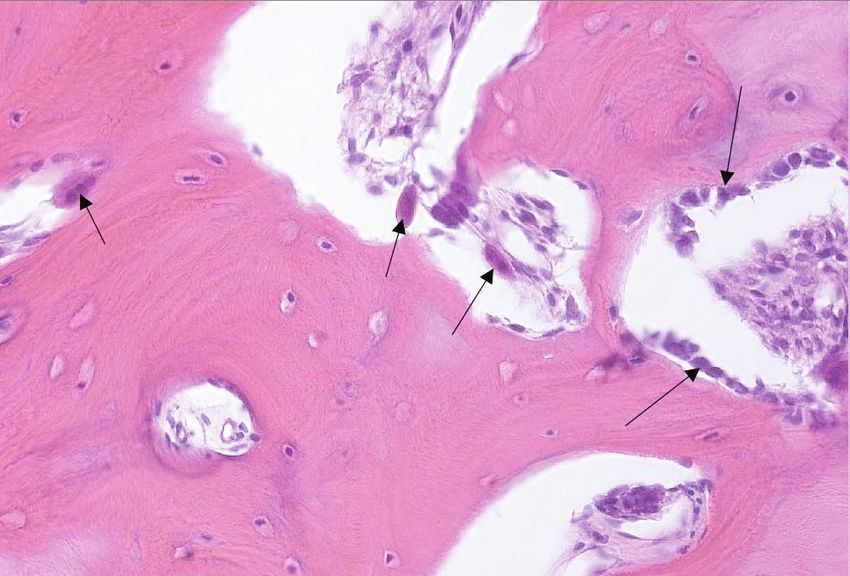
All histological samples showed trabecular bone and
medullary cavities. In comparison to ordinary tendon-
bone junctions, the samples showed a relatively large
amount of fibrocartilage and occasional, diffusely organ-
ized chondrocytes in collagen- rich medium. In some of
Figure 2. Radiograph of a round calcaneal spur at the the samples, a large number of bone-absorbing osteo-
Achilles tendon insertion. clasts and osteoblast-like cells were observed at the sur-
face of the bone.
The results from statistical analysis are summarised in The cell population in the medullary cavity looked ab-
Table 2. The average duration of conservative treat- normal, resembling mostly fetal mesenchymal tissue.
ment was 8 months, 8.1 months and 5 months in the Histologically, all of the 7 samples resembled rapid
group with good, moderate and poor outcome, respec- remodellation in the bone tissue. Because of decalci-
tively after the patients had come to the observation of fication, an estimate of the amount of osteoids could
the treating surgeon. The patients with good outcome not be done. Presentation of cartilage cells was atyp-
were operated on average 6.8 months earlier after the ical. They were located anatomically in an unusual
onset of symptoms than those with moderate outcome way, further the matrix did not fit to either hyaline-, elas-
(14.5 vs. 21.3 months of symptoms prior to surgery), but tic- or fibrocartilage. The presence of the mesenchy-
Muscles, Ligaments and Tendons Journal 2012; 2 (4): 273-277 275K.J.J. Johansson et al.
of simultaneous resection of the upper posterior cal-
caneal corner to success rates. While a statistically sig-
nificant connection was not shown, partly due to the lim-
its of the study, between the outcome and the resection
of a concomitant Haglund’s heel, it is still interesting to
speculate whether the vaguely perceived beneficial con-
nection could be due to the mere elimination of a promi-
nent upper corner of the calcaneal bone or, to the expo-
sure to growth factors within the cancellous bone.
Conservative treatments for main body or insertional cal-
cifying Achilles tendinopathy are largely derived from
studies for non-calcific tendinopathy, although a different
degree of malady is suspected. The best evidence-based
conservative treatments for tendinopathy include eccen-
tric exercises and extracorporeal shock wave therapy
(ESWT)11, although our patients did not receive ESWT
Figure 3. Light microscophy of resected spur in haema-
due to limited availability.
toxylin and eosin staining with 400x magnification, showing
osteoclasts (three leftside arrows) and osteoblasts (two
The etiology of insertional CT of the Achilles tendon re-
rightmost arrows) lining the bone cavities. mains disputed and the incidence is unknown. The preva-
lence of calcific spurs was, however, surprisingly high in
a cadaver study. In an investigation of Achilles tendons
mal-like tissue in the bone cavities gave the impression from 50 elderly corpses, 16 small insertional entheso-
that the stem-cells would have activated, proliferating phytes were found12. In these subjects, this does not in-
cartilage cells and osteoblasts (Fig. 3). The source of dicate that clinical tendinopathy would be present. In ra-
the osteoclasts remains unknown but vascularisation diological studies, approximately 25% of population is
was observed. estimated to develop a spur, either in the plantar fascia or
Achilles tendon5. The majority of the present patients
had a simultaneous Haglund’s heel, which is a typical de-
Discussion formity in exercise related tendinopathy3. It is possible that
part of the population may have an undiagnosed spur
Surgical treatment for insertional CT of the Achilles ten- which, if not aggravated by simultaneous Haglund’s de-
don with or without a concomitant Haglund’s deformity re- formity and physical exercise, remains asymptomatic.
sulted in good outcomes in most patients with our proce- Trauma is widely believed to have a primary role in the de-
dure. Occasionally, regrowth of the insertional calcification velopment of CT in the main body of the tendon12,13. How-
occured. In these cases, re-operation lead to a success- ever in our study, no distinct trauma preceded the onset
ful outcome. In the histological examination, the general of the disabilitating symptoms associated with insertional
appearance of the spur showed active remodellation and CT. The correlation between insertional CT and the main
some sections featured signs of stem-cell activity. body CT of the Achilles is not known. In our study, these
Within the power of our study, there was not a significant two were concomitant in 4 cases of 36 (11%).
association of sex, spur size, Haglund’s deformity or du- Studies have confirmed a correlation between athletic ac-
ration of symptoms before surgery to the clinical out- tivity and incidence of calcific spurs14,15. This indicates that
come. When we compare the groups with good and mod- repeated tensional stress and/or microtrauma are predis-
erate outcome, we find that younger patients and higher posing factors. This theory is in line with our study as well,
athletic activity is related to better outcome with statisti- as all the patients but one were engaged with some
cal significance. Because of limited observations with sports activity, mostly running. Speculation has also ex-
poor outcomes, we cannot show the same significant tended to physiological mechanics, suggesting that an in-
difference when a calculation is done for all three levels creased tendon-bone junction area might be an adapta-
of outcome. tional process in response to the increased mechanical
This study included only one surgical procedure with a rel- stress, because spurs can develop without inflammatory
atively large number of subjects and an encouraging fol- or microtraumatic conditions16. Distal insertional Achilles
low-up time, therefore it should add confidence in the un- enthesopathy develops in similar circumstances, usually
derstanding of the surgical treatment of insertional CT of without calcification.
the Achilles tendon. We also distinguish shortcomings of More detailed studies as to when the insertional CT is ma-
this study that challenge our conclusions. This is merely ture for surgery would be valuable, as some spurs tend
a retrospective study and the procedure of assessing to regrow. The true impact of posterior calcaneoplasty to
the outcome does not include written pre- and postoper- surgical outcome needs investigation in a larger study.
ative assessment, independent of the surgeon. The lack Also, further histological investigations are necessary to
of rating of symptoms before surgery is due to the retro- evaluate our microscopical findings, while the reason for
spective nature of this study. There are only a few small disarranged cartilage cells and atypical matrix and con-
sized, population-based studies on the subject, but the re- firmation of the presence of osteoblasts would be of spe-
sults are optimistic, most recently by Johnson et al.9 and cial interest.
Maffulli et al.10. Previous studies do not show the relation This investigation shows that a good clinical outcome to in-
276 Muscles, Ligaments and Tendons Journal 2012; 2 (4): 273-277Surgery for Achilles calcific tendinopathy
sertional CT may be attributed to younger age and profes- 6. Oliva F, Via AG, Maffulli N. Physiopathology of intra-
sional athleticism when compared to factors related to tendinous calcific deposition. BMC Med. 2012; 10:
moderate outcomes. Also, the resection of a prominent 95.
Haglund’s heel is possibly a positive outcome factor. This 7. Krahe MA, Berlet GC. Achilles tendon ruptures, re
investigation may guide future treatments to insertional CT. rupture with revision surgery, tendinosis, and inser-
tional disease. Foot Ankle Clin 2009; 14: 247-275.
8. Oshri Y, Palmanovich E, Brin YS. Chronic insertional
Acknowledgments Achilles tendinopathy: surgical outcomes. MLTJ
2012; 2: 91-95.
We wish to acknowledge Hans Helenius, Biostatistics, 9. Johnson KW, Zalavras C, Thordarson DB. Surgical
University of Turku and TBDP. We also wish to thank management of insertional calcific achilles tendi-
Robert M. Badeau for linguistic checking of this manu- nosis with a central tendon splitting approach. Foot
script. Ankle Int 2006; 27: 245-250.
10. Maffulli N, Testa V, Capasso G, Sullo A. Calcific inser-
tional Achilles tendinopathy: reattachment with bone
References anchors. Am J Sports Med 2004; 32: 174-182.
11. Loppini M, Maffulli N. Conservative management of
1. Williams JPG. Achilles tendon lesions in sport. Re- tendinopathy: an evidence-based approach. MLTJ
view. Sports Med 1986;3:114-135. 2011; 1: 133-136.
2. Orava S, Leppilahti J, Karpakka J. Operative treat- 12. Rufai A, Benjamin M, Ralphs JR. Structure and
ment of typical overuse injuries in sport. Annales histopathology of the insertional region of the human
Chirurgiae et Gynaecologiae 1991;80:208-211. achilles tendon. J Orthop Res 1995; 13: 585-593.
3. Leppilahti J, Karpakka J, Gorra A, Puranen J, Orava 13. Jozsa L, Balint BJ, Reffy A. Calcifying tendinopathy.
S. Surgical treatment of overuse injuries to the Arch Orthop Trauma Surg 1980; 97: 305-307.
achilles tendon. Clinical J of Sport Med 1994; 4:100- 14. Krahl H, Pieper HG, Quack G. Die Knochenhypertro-
107. phie als Trainingseffekt. Orthopade 1995; 24: 441-
4. Bassiouni M. Incidence of calcaneal spurs in osteo- 445.
arthrosis and rheumatoid arthritis, and in control pa- 15. Tyrdal S, Finnanger AM. Osseous manifestations in
tients. Ann Rheum Dis 1965: 24:490-493. handball goalie´s elbow. Scand J Med Sci 1999; 9:
5. Resnick D, Feingold ML, Curd J, Niwayama G, Geor- 92-97.
gen TG. Calcaneal abnormalities in articular disor- 16. Benjamin M, Rufai A, Ralphs JR. The mechanism of
ders: rheumatoid arthritis, ankylosing spondylitis, formation of bony spurs (enthesophytes) in the
psoriatic arthritis, and Reiter’s syndrome. Radiology achilles tendon. Arthritis & Rheumatism 2000; 43:
1977; 125: 355-366. 576-583.
Muscles, Ligaments and Tendons Journal 2012; 2 (4): 273-277 277You can also read