Superselective anterior temporal resection in mesial temporal lobe epilepsy
←
→
Page content transcription
If your browser does not render page correctly, please read the page content below
Clinical commentary
                                             Epileptic Disord 2011; 13 (3): 284-90

                                      Superselective anterior
                                      temporal resection in mesial
                                      temporal lobe epilepsy
                                      Joacir Graciolli Cordeiro 1,2 , Kathrin Wagner 3 ,
                                      Michael Trippel 1 , Josef Zentner 4 , Andreas Schulze-Bonhage 3
                                      1 Department of Stereotactic Neurosurgery, University Hospital Freiburg, Freiburg,

                                      Germany
                                      2 Department of Neurosurgery, Hospital de Clínicas, Federal University of Paraná,

                                      Paraná, Brazil
                                      3 Epilepsy Center, University Hospital Freiburg, Freiburg, Germany
                                      4 Department of General Neurosurgery, University Hospital Freiburg, Freiburg, Germany

                                      Received April 16, 2011; Accepted August 5, 2011

                                      ABSTRACT – We report the case of a patient with pharmacoresistant mesial
                                      temporal lobe epilepsy presenting psychomotor seizures with onset at
                                      early childhood. MRI showed a blurred internal structure of the right hip-
                                      pocampus and right mammillary body atrophy. Neuropsychological testing
                                      revealed deficits in selective attention and visual planning. Non-invasive
                                      recording was not sufficient to precisely detect the seizure onset zone. Inva-
                                      sive recording showed seizure onset in the temporo-polar neocortex, with
                                      spread to the amygdalum and hippocampus. A superselective resection of
                                      the temporal pole and amygdalum was performed with preservation of the
                                      hippocampus. Histology revealed the presence of focal cortical dysplasia
                                      (Palmini type Ib). Seizure frequency was reduced after surgery, and seizure
                                      freedom for two years was achieved with optimisation of the antiepileptic
                                      drug regime. Memory functions were preserved, and selective attention
                                      and visual planning improved following limited resection. This case sug-
                                      gests that, in selected cases, highly targeted resections with preservation
                                      of memory-relevant structures may be the best choice considering both
                                      seizure control and unimpaired cognitive functioning.
                                      Key words: epilepsy surgery, resection, mesial, temporal lobe, epilepsy,
                                      focal cortical dysplasia

                                         Temporal lobe epilepsy (TLE) is the         (Vermathen et al., 2002). Since many
                                         most common form of epilepsy in             patients are resistant to pharmaco-
                                         the adult population, often caused          logical treatment, surgical resection
                                         by mesial temporal generators               of the epileptogenic area may be
                                         which are reported to be present            indicated (Semah et al., 1998; Volcy
                                                                                                                                            doi:10.1684/epd.2011.0460

Correspondence:
                                         in around 60-70% of the patients            Gomez, 2004). There is an ongo-
J. Graciolli Cordeiro                    (Commission, 1989; Watson et al.,           ing debate, however, regarding the
Department of Stereotactic Neurosurgery, 1992). Epileptogenic structures can         extent of resection necessary to
Uniklinik,                               comprise the hippocampus itself,            achieve seizure control. Progressive
Breisacherstrasse 64,
79106 Freiburg im Breisgau, Germany      the parahippocampal gyrus, the              advances in diagnostic evaluation
  amygdala, and adjacent structures           allow a more precise determination

284                                                                                      Epileptic Disord, Vol. 13, No. 3, September 2011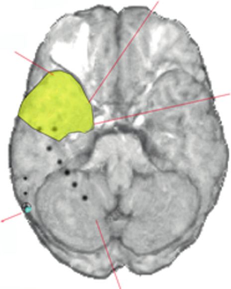
Superselective temporal resection in MTLE
of the ictal focus which may open options for supers- cognitive functions were average or even above aver-
elective resections. age (including verbal and visual declarative memory).
We present the case of a patient with mesiotempo- Clinical psychological examination showed depressive
ral epilepsy who underwent a highly selective surgical symptoms and increased anxiety.
procedure sparing the hippocampus and achieved High resolution MRI (3T) showed a blurred inter-
favourable seizure outcome and improvement in cog- nal structure of the right hippocampus without clear
nitive performance. atrophy or signal change (figure 1) and right mam-
millary body atrophy. An interictal FDG-PET was
non-localising.
Case report EEG revealed interictal sharp waves recorded with
maximum amplitude over the right sphenoidal
A right-handed 52-year-old man was admitted for electrodes extending to adjacent temporo-anterior
presurgical evaluation of his epilepsy. Since the age of contacts. Ictal recordings showed a widespread rhyth-
two years he suffered from complex partial seizures mic activity over the right temporal lobe with later
characterised by behavioural arrest, staring, impair- propagation to left temporal and bifrontal areas. Pre-
ment of consciousness, oroalimentary and manual cise focus localisation within the right temporal lobe
automatisms and head deviation to the left side with was not possible. In order to define the role of
preserved language functions, and secondary gene- the hippocampus in seizure generation, the patient
ralised tonic-clonic seizures. Seizure frequency was underwent stereotactic implantation of two depth
from one to three per month. There was comorbi- electrodes with a lateral approach targeting the right
dity of severe depression and hypothyroidism. The first amygdala and hippocampal head. Moreover, temporo-
epileptic episodes occurred in his early childhood; in basal and temporo-lateral subdural strip electrodes
the course of the disease, pharmacoresistance against were implanted (figure 2). Invasive recordings showed
phenytoin, oxcarbamazepine, topiramate, lamotrigine extended interictal spiking in the amygdala, hippocam-
and clobazam was documented. pus, as well as in the temporo-basal and temporo-polar
Therapy at the time of admission was 300 mg/d lamo- areas (figure 3). Ictal recordings showed seizure onset
trigine, 1,200 mg/d oxcarbazepine, 10 g/d vitamin with low-amplitude fast activity over the polar neo-
B12 and 125 g/d levothyroxin. Physical examination cortex and rhythmic spiking in the amygdalum, with
was unremarkable. Neurological examination showed spread to the hippocampus after a few seconds.
normal findings except for facial hypoaesthesia due Since the hippocampus was not the primary seizure
to a former facial trauma. Routine laboratory testing generator, and since the patient was unwilling to risk
showed no relevant abnormalities. memory deterioration following surgery, the patient
He was working as an international manager in a was offered a superselective surgical procedure pre-
large German company. Neuropsychological evalua- serving the hippocampus which was assumed to carry
tion revealed above average general intellectual abil- relevant function in memory processing. Therefore,
ities. There were specific deficits in the domains of selective resection of the right amygdala, uncus and
selective attention and visual planning while the other temporal pole with preservation of the hippocampus
R
R
10mm 10mm
Figure 1. High resolution MRI (3T) shows a blurred internal structure of the right hippocampus without clear atrophy or signal change.
Epileptic Disord, Vol. 13, No. 3, September 2011 285J.G. Cordeiro, et al.
Figure 2. MRI showing the positions of depth electrodes targeting the right amygdala and hippocampus
as well as the temporo-basal and temporo-lateral strips.
R R TBa1
HRa5 TLa1
TBb1
TLb1
TLa1
TLb1
Seizure Onset
Frequent Interictal Spiking
Interictal propagation TBc1
Subclinical seizures
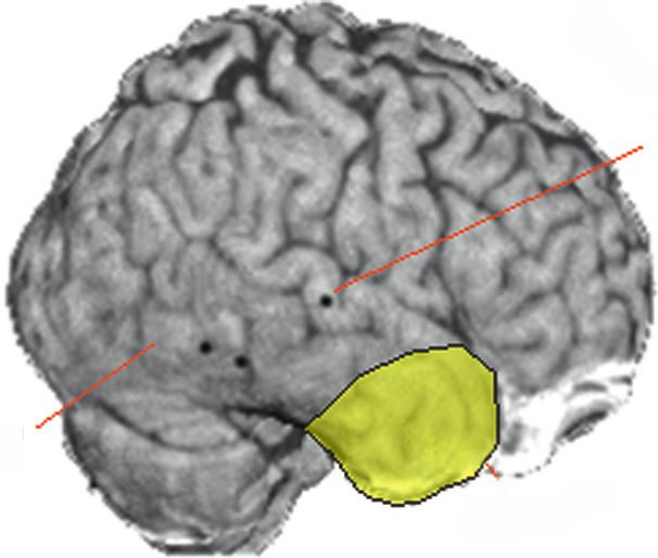
Figure 3. Results of invasive recordings showing ictal onset in the right temporal pole and amygdala, and delay between anterior ictal
onset and hippocampal involvement; superselective resection preserving the hippocampus was planned (yellow shaded area).
and parahippocampal gyrus was performed. Histolo- right amygdala, uncus and temporal pole with mild sur-
gical analysis revealed the presence of focal cortical rounding gliosis (figure 4). Routine interictal scalp EEG
dysplasia (Palmini type Ib). was free of epileptic activity.
Within the first three months after surgery, the With add-on introduction of levetiracetam in combi-
patient continued to have complex partial seizures nation with oxcarbazepine, complete seizure freedom
at a lower frequency. Visual fields were unchanged. was achieved 11 months after the surgical procedure;
Neuropsychological performance concerning selec- the patient has now remained completely seizure-
tive attention and visual planning improved to an free (Engel 1a, Wieser 1) for 26 months. Depressive
average level, and declarative memory performance symptoms have resolved over this period, and due to
remained unchanged (figure 5). Depressive symptoms his professional performance he was promoted to a
were still present. MRI showed the resection of the higher position.
286 Epileptic Disord, Vol. 13, No. 3, September 2011Superselective temporal resection in MTLE
A H
Figure 4. Postoperative MRI demonstrating the superselective resection of the temporal pole, uncus and amygdala with preservation
of the hippocampus and parahippocampal gyrus.
(Gloor, 1991) is frequently the structure responsible
Presurgical Postsurgical
for seizure generation, the amygdala (Zentner et al.,
9
1999) and neocortical abnormalities, particularly focal
cortical dysplasias (also as dual pathology) (Fauser and
8
Schulze-Bonhage, 2006; Levesque et al., 1991), may also
7 be seizure generators in TLE.
Remembered items
6 In general, intracerebral recordings have demons-
5 trated that ictal patterns in mesial TLE (MTLE) are
frequently first recorded from the hippocampus or
4
simultaneously from the hippocampus and the amyg-
3 dala. Although latencies between involvement of
2 hippocampus and amygdala are often brief, an ori-
1
gin confined to the amygdala with some delay to
the propagation to the hippocampus (e.g. five sec-
0
onds) suggests an amygdalar seizure onset, which
Trial 1 Trial 2 Trial 3 Trial 4 Trial 5
Visuospatial memory test
is found in only approximately 5% of patients with
appropriate intracerebral recordings (Wieser, 2000). In
a subsequent study, the Montreal group used phase-
Figure 5. Pre- and postsurgical visual memory performance; lines coherence analysis (Gotman and Levtova, 1996) and
show similar learning increment across the five trials and overall
found that the amygdala was leading in 21% of focal
average visual memory performance at both time points.
mesial and in 53% of regional temporal lobe seizures.
The hippocampus was leading in 48.5% of focal mesial
and in 27% of regional temporal lobe seizures. In the
Discussion remaining seizures, discharges were synchronous in
these two structures.
TLE is not a homogenous disease. Different neo- In our patient, seizure onset in the amygdala and tem-
cortical and archicortical areas may contribute to poral pole clearly preceded hippocampal involvement,
epileptogenesis, corresponding to a large spectrum of although both structures were part of the irritative
histological abnormalities. Although the hippocampus zone and zone of early propagation. Involvement of
Epileptic Disord, Vol. 13, No. 3, September 2011 287J.G. Cordeiro, et al. the hippocampus was thus considered to be secondary electrocorticography or extraoperative recordings to the epileptogenic region in the anterior temporal using intracranial electrodes (Kanner et al., 1995). lobe. The selective surgery performed in this patient Patients with epilepsy secondary to temporo-apical with non-specific MRI and PET findings demonstrates lesions visible on MRI may also benefit from highly that targeting the seizure onset zone and leaving a selective approaches. Elsharkawy et al. (2011) reported functional hippocampus intact may not only be effi- related findings in a series of 61 patients with lesions in cacious but may also improve cognitive performance. the apex of the temporal lobe and normal hippocampal In the case presented, the patient was highly depen- aspect on MRI as well as intact memory function. These dent on unimpaired cognitive functioning and was subjects underwent apical temporal resection, sparing not willing to take any risk associated with removal the mesial temporal structures and contributed to the of a presumably normal functioning hippocampus. In 80.9% of cases of Engel Class 1 at two years of follow-up. the end, the epileptogenic zone was not completely Memory outcome was favourable in most of patients removed during the limited resection, nevertheless, while worsening was observed in only two patients with the introduction of a second antiepileptic drug, (3.2%) (Elsharkawy et al., 2011). the patient has now been rendered seizure-free for Regarding lateralisation of cognitive functions, most more than two years, and according to his unimpaired authors describe patients with right-sided MTLE to cognitive functioning, he is able to meet the demands show frequently impaired visuospatial memory perfor- of an international management position in his mance (Fauser and Schulze-Bonhage, 2006; Giovagnoli company. Consistent with the report of Fauser et al. et al., 1995; Ladavas et al., 1979; Majdan et al., (2008), the brief duration of epilepsy was likely to have 1996), caused by the underlying structural damage offered good seizure outcome. and epilepsy or after resection of the hippocam- Selective approaches to TLE have been reported in pus. Glikmann-Johnston et al. (2008) reported on a recent case series. Takaya et al. (2009) described the series of TLE patients in which an extended assess- effects of subtemporal SAH (selective amygdalohip- ment of spatial memory and hippocampal volumetry pocampectomy) in cognition and cerebral metabolism was performed. This analysis revealed that visu- by means of FDG-PET in MTLE patients. A postop- ospatial memory impairment was neither dependent erative improvement was observed in delayed recall on the side of resection in ATL patients (anterior and attention/concentration scores, regardless of the temporal lobectomy) nor on the side of hippocam- resected side. PET analysis suggested that the selec- pal atrophy in unilateral non-operated TLE patients tive subtemporal resection improved cerebral glucose (Glikmann-Johnston et al., 2008). In order to explore metabolism in areas receiving projections from the the relationship between visuospatial memory and affected mesial temporal lobe. These results would hippocampal metabolism, PET scans were performed support the notion that cognitive improvement might in healthy participants during navigation-related result from seizure control and minimised postopera- tasks. As result, bilateral hippocampal activation was tive functional impairment (Takaya et al., 2009). Kanner observed. Predominance at the right hippocampus et al. (1995) reported 24 patients with anterotemporal was only detected by accurate navigation (Maguire seizure focus, referred for tailored anterior temporal et al., 1998). These findings suggest that visuospa- lobectomy. In this series, the extent of resection was tial memory may relate to the integrity of both tailored by intraoperative electrocorticographic find- temporal lobe structures and the concept of task speci- ings and functional mapping of eloquent cortex. In six ficity could represent a useful description of patterns patients, the amygdala and hippocampus were spared, of lateralisation of visuospatial memory (Glikmann- nine patients had partial to total resection of the amyg- Johnston et al., 2008; Saling, 2009). dala, eight patients had resection of the amygdala and In our case there was no impairment of visuospa- anterior third of the hippocampus and one patient tial memory prior to or after surgery which suggests underwent resection of the amygdalum and anterior functional hippocampal integrity. An extensive litera- two thirds of the hippocampus. Complete or almost ture review of clinical aspects after epilepsy surgery complete seizure relief (Engel class I) was achieved in revealed an average rate of verbal memory decline 21 of the 24 patients; three patients had rare persis- in TLE patients of 44% for those operated on the left ting seizures (Engel class II). These results suggested side versus 20% operated on the right side. For visual that sparing or limiting resection of the mesial tempo- memory, the risk of loss was similar for both right and ral structures in appropriately selected patients with left temporal resections (23% and 21%, respectively) anterotemporal seizure focus is not necessarily asso- (Sherman et al., 2011). Even if the risk of visual mem- ciated with a poor seizure outcome, provided that the ory loss is lower than that for verbal memory, super- decision not to resect the hippocampus is based on an selective approaches may further decrease the risk of absence of epileptiform activity during intraoperative impairment. Postsurgical maintenance of hippocampal 288 Epileptic Disord, Vol. 13, No. 3, September 2011
Superselective temporal resection in MTLE

functional integrity of our patient was evident from       Ravagnati, 1987; Siegfried and Wieser, 1988). Research
neuropsychological assessment three months after           is ongoing to define the number of patients who are
surgery, which revealed a stable general cognitive per-    completely “cured” (i.e. seizure-free for several years
formance as well as an improvement of attention and        without taking antiepileptic drugs [AEDs]) by epilepsy
visual planning. This postoperative functional recovery    surgery versus a switch to pharmacoresponsiveness. In
may have been promoted by a reduction of interic-          the study of Schmidt et al. (2004), following temporal
tal spiking and complex partial seizures during the        lobe surgery, approximately one in four adult patients
daytime. The standardised neuropsychological follow-       were reported to become seizure-free for five years
up was relatively short, however, at two years after       without AEDs. However, as 55% of patients preferred
surgery the patient did not report any deterioration       not to discontinue their medication despite seizure
in declarative memory functions and was successful in      freedom, it is impossible to know if they were cured.
his professional life. Our patient had normal functional   It is possible that more selective approaches may
memory and a hippocampus with normal appearance;           be associated with a higher dependency of phar-
this patient group has only recently been shown to be      macotherapy, as well as later relapse. Wieser (2000)
at particular risk of memory decline following resec-      reported the case of a patient with amygdalar epilepsy
tion of the hippocampus (Helmstaedter et al., 2011).       who was referred for stereotactic amygdalotomy. After
Current evidence supports the utility of highly selec-     a seizure-free outcome of 11 years, he required
tive surgical approaches in specific patients, aiming at    selective amygdalohippocampectomy due to recur-
the prevention of progressive cognitive deterioration      rent seizures. Therefore, the long-term outcome of
due to poor seizure control. In contrast, total resec-     our patient concerning his seizures as well as memory
tion of the temporal lobe including the hippocampus        functions should be observed. However, even consid-
may be associated with a higher risk of memory             ering these risks, particular aspects, such as the high
decline (Clusmann et al., 2002) and larger neocortical     cognitive demand necessary for the patient’s profes-
resections may correlate with cognitive impairment,        sional position reported here, may turn the balance in
which has been shown, at least, on the language            favour of a limited resection which has allowed him to
dominant side (Alpherts et al., 2008). Material-specific    advance in his career in the years following epilepsy
memory aspects after transsylvian SAH were com-            surgery.
pared to those after temporal pole resection with          Epileptic foci can be very limited (Fauser and Schulze-
amygdalohippocampectomy (TPR+). Figural memory             Bonhage, 2004) and even electrode impalement
outcome was worse after right-sided surgery, mainly        can have a curative effect (Schulze-Bonhage et al.,
when the temporal pole was included (TPR+). Atten-         2010). Thus, additional options of even more focused
tion improved, independent of side or type of surgery,     approaches may be chosen for appropriate patients.
and language functions showed some improvement             Constant advances in the methodology available for
after right-sided surgery (Helmstaedter et al., 2008).     presurgical investigation allow for a more refined iden-
This further points to the positive role of super-         tification of targets for epilepsy surgery. Further studies
selective surgical approaches; preservation of mem-        with longer follow-up are required to define the role
ory in our patient suggests that hippocampal sparing       of superselective approaches in patients with a very
had a positive effect, whereas resection of the pole       localised epileptogenic zone.
and amygdala alone may be well tolerated. In addition
to the preservation of functional tissue, the effect on    Disclosure.
networks (Frings et al., 2009), including the contralat-   None of the authors has any conflict of interest or financial
                                                           support to disclose.
eral temporal lobe, may contribute to postoperative
improvement in cognition. Spectroscopic investiga-
tions may demonstrate these effects (Vermathen et al.,     References
2002).
With the goal to use minimally invasive surgical           Alpherts WCJ, Vermeulen J, van Rijen PC, da Silva FHL, van
modalities, several authors have performed stereo-         Veelen CWM. Standard versus tailored left temporal lobe
tactic lesioning of limbic targets and in particular,      resections: differences in cognitive outcome? Neuropsy-
the amygdala, as treatment for TLE. Reviewing the          chologia 2008; 46: 455-60.
literature, Ojemann and Ward (1975) and Ravagnati          Clusmann H, Schramm J, Kral T, et al. Prognostic factors and
(1987) concluded that overall results of stereotac-        outcome after different types of resection for temporal lobe
tic approaches were inferior compared to resective         epilepsy. J Neurosurg 2002; 97: 1131-41.
surgery. Regarding the treatment of TLE due to amyg-       Commission on classification and terminology of inter-
dalar epilepsy, only limited data on long-term efficacy     national league against epilepsy: proposal of revised
of amygdalotomy are available (Hood et al., 1983;          classification of epilepsies and epileptic syndromes. Epilepsia
Narabayashi and Mizutani, 1970; Narabayashi, 1976;         1989; 30: 389-99.

Epileptic Disord, Vol. 13, No. 3, September 2011                                                                    289
J.G. Cordeiro, et al.
Elsharkawy AE, Pannek H, Woermann FG, et al. Apical tempo- Majdan A, Sziklas V, Jones-Gotman MJ. Performance of
ral lobe resection; “tailored” hippocampus-sparing resection healthy subjects and patients with resection from the anterior
based on presurgical evaluation data. Acta Neurochir (Wien) temporal lobe on matched tests of verbal and visuopercep-
2011; 153: 231-8. tual learning. J Clin Exp Neuropsychol 1996; 18: 416-30.
Fauser S, Schulze-Bonhage A. How large must an epileptic Narabayashi H. Lessons from amygdaloid surgery in long-
focus be to cause an electrographic status epilepticus - a case term observation. In: Gillingham FJ, Hitchcock ER, Nádvornik
report. Clin Neurophysiol 2004; 115: 2274-9. P. Stereotactic treatment of epilepsy. Acta Neurochir 1976; 23:
241-5.
Fauser S, Schulze-Bonhage A. Epileptogenicity of cortical
dysplasia in temporal lobe dual pathology: an electrophysio- Narabayashi H, Mizutani T. Epileptic seizures and the stereo-
logical study with invasive recordings. Brain 2006; 129: 82-95. tactic amygdalotomy. Confinia Neurol 1970; 32: 289-97.
Fauser S, Bast T, Altenmüller D, et al. Factors influencing Ojemann GA, Ward Jr. AA. Stereotactic and other procedures
surgical outcome in patients with focal cortical dysplasia. for epilepsy. In: Purpura DP, Penry JK, Walter RD. Adv Neurol
J Neurol Neurosurg Psychiatr 2008; 79: 103-5. 1975; 8: 241-63.
Frings L, Schulze-Bonhage A, Spreer J, Wagner K. Remote Ravagnati L. Stereotactic surgery for epilepsy. In: Wieser HG,
effects of hippocampal damage on default network connec- Elger CE. Presurgical evaluation of epileptics. Heidelberg,
tivity in the human brain. J Neurol 2009; 256: 2021-9. New York: Springer, 1987: 361-71.
Giovagnoli AR, Casazza M, Avanzini G. Visual learning on a Saling MM. Verbal memory in mesial temporal lobe epilepsy:
selective reminding procedure and delayed recall in patients beyond material specificity. Brain 2009; 132: 570-82.
with temporal lobe epilepsy. Epilepsia 1995; 36: 704-11. Schmidt D, Baumgartner C, Löscher W. The chance of cure
Glikmann-Johnston Y, Saling MM, Chen J, et al. Structural following surgery for drug-resistant temporal lobe epilepsy.
and functional correlates of unilateral mesial temporal lobe What do we know and do we need to revise our expectations?
spatial memory impairment. Brain 2008; 131: 3006-18. Epilepsy Res 2004; 60: 187-201.
Gloor P. Mesial temporal sclerosis: historical background Schulze-Bonhage A, Dennig D, Wagner K, et al. Seizure
and an overview from a modern perspective. In: Lüders H. control resulting from intrahippocampal depth electrode
Epilepsy surgery. New York: Raven, 1991: 689-703. insertion. J Neurol Neurosurg Psychiatr 2010; 81: 352-3.
Gotman J, Levtova V. Amygdala-hippocampus relationships Semah F, Picot MC, Adam C, et al. Is the underlying cause of
in temporal lobe seizures: a phase-coherence study. Epilepsy epilepsy a major prognostic factor for recurrence? Neurology
Res 1996; 25: 51-7. 1998; 51: 1256-62.
Helmstaedter C, Richter S, Röske S, et al. Differential effects Sherman EMS, Wiebe S, Fay-McClymont TB, et al. Neu-
of temporal pole resection with amygdalohippocampectomy ropsychological outcomes after epilepsy surgery: systematic
versus selective amygdalohippocampectomy on material- review and pooled estimates. Epilepsia 2011; 52: 857-69.
specific memory in patients with mesial temporal lobe
epilepsy. Epilepsia 2008; 49: 88-97. Siegfried J, Wieser HG. The actual role of stereotactic opera-
tions on deep brain structures in the treatment of medically
Helmstaedter C, Petzold I, Bien CG. The cognitive con- refractory epilepsies. In: Broggi G. The rational basis of
sequence of resecting nonlesional tissues in epilepsy the surgical treatment of epilepsies (Current Problems in
surgery-Results from MRI- and histopathology-negative Epilepsy 5). London, Paris: John Libbey, 1988: 113-9.
patients with temporal lobe epilepsy. Epilepsia 2011; 52:
1402-8. Takaya S, Mikuni N, Mitsueda T, et al. Improved cerebral
function in mesial temporal lobe epilepsy after subtemporal
Hood TW, Siegfried J, Wieser HG. The role of stereotactic amygdalohippocampectomy. Brain 2009; 132: 185-94.
amygdalotomy in the treatment of temporal lobe epilepsy
associated with behavioural disorders. Appl Neurophysiol Vermathen P, Ende G, Laxer KD, et al. Temporal lobectomy
1983; 46: 19-25. for epilepsy: recovery of the contralateral hippocampus mea-
sured by (1)H MRS. Neurology 2002; 59: 633-6.
Kanner AM, Kaydanova Y, de Toledo-Morrell L, et al.
Tailored anterior temporal lobectomy. Relation between Volcy Gomez M. Mesial temporal lobe epilepsy: its
extent of resection of mesial structures and postsurgical physiopathology, clinical characteristics, treatment and prog-
seizure outcome. Arch Neurol 1995; 52: 173-8. nosis. Rev Neurol 2004; 38: 663-7.
Ladavas E, Umilta C, Provinciali L. Hemisphere-dependent Watson C, Andermann F, Gloor P, et al. Anatomic basis of
cognitive performances in epileptic patients. Epilepsia amygdaloid and hippocampal volume measurement by mag-
1979; 20: 493-502. netic resonance imaging. Neurology 1992; 42: 1743-50.
Levesque MF, Nakasato N, Vinters HV, Babb TL. Surgical Wieser HG. Mesial temporal lobe epilepsy versus amygdalar
treatment of limbic epilepsy associated with extrahippocam- epilepsy: late seizure recurrence after initially successful
pal lesions: the problem of dual pathology. J Neurosurg amygdalotomy and regained seizure control following hip-
1991; 75: 364-70. pocampectomy. Epileptic Disord 2000; 2: 141-52.
Maguire EA, Burgess N, Donnett JG, et al. Knowing where Zentner J, Wolf HK, Helmstaedter C, et al. Clinical relevance
and getting there: a human navigation network. Science of amygdala sclerosis in temporal lobe epilepsy. J Neurosurg
1998; 280: 921-4. 1999; 91: 59-67.
290 Epileptic Disord, Vol. 13, No. 3, September 2011You can also read