Cardiac reserve by 6- minute walk stress echocardiography in systemic sclerosis
←
→
Page content transcription
If your browser does not render page correctly, please read the page content below
Open access Special populations
Open Heart: first published as 10.1136/openhrt-2020-001559 on 19 February 2021. Downloaded from http://openheart.bmj.com/ on December 18, 2021 by guest. Protected by copyright.
Cardiac reserve by 6-minute walk stress
echocardiography in systemic sclerosis
Miharu Arase,1,2 Kenya Kusunose ,2 Sae Morita,3 Natsumi Yamaguchi,3
Yukina Hirata,3 Susumu Nishio,3 Yuichiro Okushi,2 Takayuki Ise,2 Takeshi Tobiume,2
Koji Yamaguchi,2 Daiju Fukuda,4 Shusuke Yagi,2 Hirotsugu Yamada ,5
Takeshi Soeki,2 Tetsuzo Wakatsuki,2 Masataka Sata2
►► Additional material is ABSTRACT
published online only. To view, Key questions
Objectives There is a high prevalence of left ventricular
please visit the journal online
diastolic dysfunction (LVDD) in systemic sclerosis (SSc)
(http://dx.doi.org/10.1136/ What is already known on this subject?
which is associated with high mortality. Thus, early
openhrt-2020-001559). ►► The myocardial involvement of systemic sclerosis
detection of LVDD could be important in management of
(SSc) clinically causes cardiac dysfunction, espe-
To cite: Arase M, Kusunose K, SSc. We hypothesised that exercise echocardiography
cially left ventricular diastolic dysfunction (LVDD).
Morita S, et al. Cardiac reserve in SSc patients with normal resting haemodynamics
Several studies have shown that LVDD in SSc is
by 6-minute walk stress may expose early phase LVDD, which could affect its
highly prevalent and is associated with increased
echocardiography in systemic prognosis, defined as cardiovascular death and unplanned
sclerosis. Open Heart risk of clinical worsening. In addition, LVDD may oc-
hospitalisation for heart failure.
2021;8:e001559. doi:10.1136/ cur before clinical manifestation.
Methods Between January 2014 and December 2018,
openhrt-2020-001559 we prospectively enrolled 140 patients with SSc who What does this study add?
underwent 6-minute walk (6MW) stress echocardiographic ►► ΔmPAP/ΔCO and ΔmPAWP/ΔCO in the event group
Received 22 December 2020 studies with normal range of estimated mean pulmonary were significantly increased compared with the
Revised 23 January 2021 arterial pressure (mPAP) (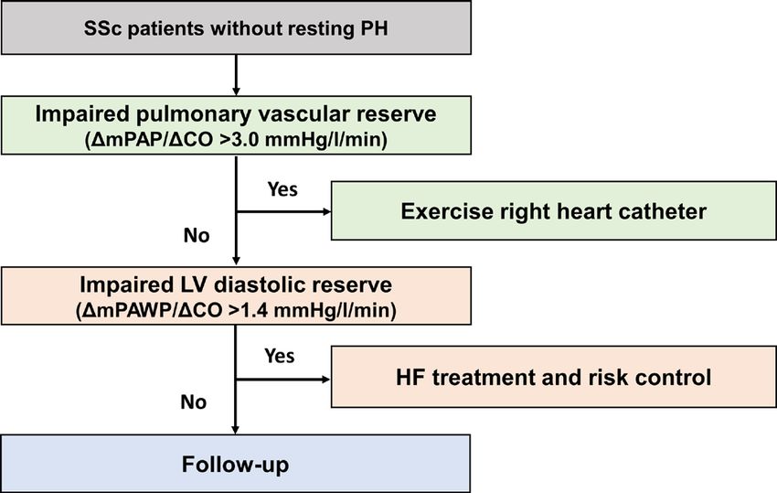
Open Heart
Open Heart: first published as 10.1136/openhrt-2020-001559 on 19 February 2021. Downloaded from http://openheart.bmj.com/ on December 18, 2021 by guest. Protected by copyright.
Figure 1 Patient selection. PAH, pulmonary arterial hypertension; SSc, systemic sclerosis.
early stage of LVDD, despite showing normal resting
Table 1 Clinical characteristics filling pressure.8 Several stress methods are exten-
All Event (+) Event (-) P value sively used in the clinical setting, and among them, a
6-minute walk (6MW) test is a simple, easy, inexpensive
Number 140 25 115 –
and widely used method. In fact, our previous studies
Age 61±12 62±10 60±12 0.54
showed that measuring Δmean pulmonary artery pres-
Male, % 14 (10) 1 (4) 13 (11) 0.11 sure (mPAP)/Δcardiac output (CO) by 6MW stress
Body mass index 22±3 22±3 22±3 0.88 echocardiography can be associated with development
WHO Class I/II/III/IV 27/82/31/0 5/16/4/0 22/66/27/0 0.54 of pulmonary hypertension (PH); thus, it is effec-
History tive in assessing right-sided haemodynamic responses
Disease duration, 1.7 1.5 1.7 0.75 in SSc.9 In contrast, the balance of mean pulmonary
months arterial wedge pressure (mPAWP) and cardiac output
Medication (CO) is essential for the assessment of early LVDD.10
Antihypertensive drugs, 2 (1) 0 (0) 2 (2) 0.16
From the viewpoint of the Frank-Starling mechanism,
% CO increases as LV filling pressure (eg, PAWP) elevates
Diuretic, % 3 (2) 1 (4) 2 (2) 0.59 in normal subjects. However, patients with severely
impaired diastolic dysfunction are unable to augment
Anticoagulants, % 0 (0) 0 (0) 0 (0) –
their cardiac output effectively even though LV filling
Respiratory function
pressure is significantly elevated. We hypothesised that
%EFV1, % 80±4 82±6 79±4 0.82 SSc patients with normal resting haemodynamics may
%FVC, % 109±13 113±15 107±12 0.34 present an early phase of LVDD measured by mPAWP
%DLCO 80±13 81±14 79±11 0.53 and CO with exercise echocardiography, and lead to
Baseline a model to predict long-term outcomes. We sought to
haemodynamics (1) evaluate early phase LVDD with a 6MW stress echo-
HR, bpm 68±12 68±10 67±12 0.67 cardiographic study in our SSc group; and (2) assess
Systolic BP, mm Hg 123±20 122±22 123±19 0.93 the prognosis in those patients using variables of 6MW
Diastolic BP, mm Hg 68±10 68±10 69±10 0.68
stress echocardiography.
SpO2, % 98±1 97±2 98±1 0.53
Post-6-minute walk haemodynamics METHODS
HR, bpm 90±17 92±18 89±16 0.54 Data sharing statement
Individual anonymised data supporting the analyses
Systolic BP, mm Hg 128±25 129±22 128±25 0.79
contained in the manuscript will be made available
Diastolic BP, mm Hg 66±15 70±19 65±14 0.22
on reasonable written request from researchers whose
SpO2, % 96±3 96±2 96±3 0.80 proposed use of the data for a specific purpose has
6MW distance, m 432±89 451±94 428±87 0.26 been approved.
Data are presented as number of patients (percentage) and mean±SD.
BP, blood pressure; %DLCO, diffusing capacity for carbon monoxide; Patient and public involvement
%FEV1, percent forced expiratory volume in 1 s; %FVC, percent Patients or the public were not involved in the design,
forced vital capacity; HR, heart rate; MCTD, mixed connective tissue or conduct, reporting or dissemination plans of our
disease; SpO2, percutaneous oxygen saturation; SSc, systemic
sclerosis. research.
2 Arase M, et al. Open Heart 2021;8:e001559. doi:10.1136/openhrt-2020-001559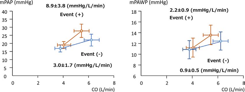
Special populations
Open Heart: first published as 10.1136/openhrt-2020-001559 on 19 February 2021. Downloaded from http://openheart.bmj.com/ on December 18, 2021 by guest. Protected by copyright.
Table 2 Haemodynamic parameters
All Event (+) Event (-) P value
Rest echocardiographic variables
LVEDVi, mL/m2 49±11 49±14 49±10 0.87
2
LVESVi, mL/m 17±4 17±6 17±4 0.59
LVEF, % 65±4 66±3 65±4 0.15
LVMi, g/m2 75±17 72±19 76±16 0.31
LAVi, mL/m2 29±9 29±12 28±9 0.79
e’, cm/s 10±3 11±3 10±3 0.27
E/e' 7±3 8±4 7±3 0.09
2
RVEDA, cm 14±3 13±3 14±3 0.10
RVESA, cm2 8±2 8±2 8±2 0.78
RVFAC, % 44±10 41±12 45±10 0.14
TAPSE, mm 22±4 21±4 22±4 0.50
GLS, % 19±3 18±2 19±3 0.19
RVLS, % 25±6 25±7 25±6 0.87
Exercise haemodynamics
Mean PAP, mm Hg 17±3 18±3 17±3 0.29
Mean PAWP, 11±4 12±4 11±4 0.09
CO, L/min 3.8±1.2 4.1±1.7 3.8±1.1 0.41
PVR, wood unit 1.9±1.1 1.9±0.8 1.9±1.2 0.57
Exercise mean PAP, mm Hg 23±5 25±4 23±5 0.05
Exercise mean PAWP, mm Hg 13±4 14±5 12±4 0.04
Exercise CO, L/min 6.1±2.1 5.6±2.1 6.2±2.1 0.19
Exercise PVR, wood unit 1.9±1.0 2.2±1.1 1.9±0.9 0.17
ΔmPAP/ΔCO, mm Hg/L/min 4.1±2.5 8.9±3.8 3.0±1.7 0.008
ΔmPAWP/ΔCO, mm Hg/L/min 1.1±0.6 2.2±0.9 0.9±0.5 0.004
Δe’, cm/s 0.9±3.1 0.5±3.6 1.0±3.1 0.63
Data are presented as number of patients (percentage) and mean±SD.
CO, cardiac output; e’, early diastolic mitral annular motion; E, early diastolic transmitral flow velocity; LAVi, left atrial volume index; LVEDVi, left ventricular end-
diastolic volume index; LVEF, left ventricular ejection fraction; LVESVi, left ventricular end-systolic volume index; LVMi, left ventricular mass index; mPAP, mean
pulmonary artery pressure; mPAWP, mean pulmonary artery wedge pressure; PVR, pulmonary vascular resistance; RVEA, right ventricular end-diastolic area;
RVESA, right ventricular end-systolic area; RVFAC, right ventricular functional area change; TAPSE, tricuspid annular plane systolic excursion.
Study population severe valvular disease (n=2), atrial fibrillation/flutter
We prospectively enrolled consecutive patients with SSc (n=3), left ventricular ejection fractionOpen Heart
Open Heart: first published as 10.1136/openhrt-2020-001559 on 19 February 2021. Downloaded from http://openheart.bmj.com/ on December 18, 2021 by guest. Protected by copyright.
Table 3 Univariate associations of outcomes
Six-minute walk stress echocardiography
Online supplemental clip shows the 6MW stress echocar-
Univariate model
diography. The 6MW tests were performed according to
HR 95% CI P value
the American Thoracic Society guidelines.16 The trans-
Age 1.02 0.98 to 1.05 0.54 cutaneous arterial oxygen saturation was determined by
Male, % 0.41 0.01 to 9.17 0.25 pulse oximetry. As we performed in our previous study,9
Disease duration 0.99 0.99 to 1.01 0.75 post-6MW (within 30 s), early diastolic transmitral flow/
HR, bpm 1.00 0.97 to 1.04 0.87 annular velocity and the peak tricuspid regurgitation
Systolic BP, mm Hg 0.99 0.98 to 1.02 0.72 jet were obtained immediately. CO was also obtained
SpO2, % 0.93 0.73 to 1.19 0.57
from electric cardiometry (Aesculon Electrical Veloci-
metry, Osypka Medical GmbH, Berlin, Germany) at the
Rest echocardiographic variables
same time in order to check the changes of CO imme-
LVEF, % 1.01 0.95 to 1.07 0.86
diately after the 6MW test.17 Because it was difficult to
LAVi, mL/m2 1.01 0.97 to 1.05 0.73 measure many indices immediately after the 6MW test,
E/e' 1.09 1.01 to 1.19 0.04 we used electric cardiometry to obtain CO. We calculated
GLS 0.88 0.71 to 1.10 0.26 the slope of mPAWP/CO and mPAP/CO in individual
RVLS 0.97 0.87 to 1.09 0.67 patients (ΔmPAWP/ΔCO and ΔmPAP/ΔCO).
Exercise haemodynamics In 14 patients, exercise right heart catheterisation
mPAP, mm Hg 1.09 0.95 to 1.26 0.22 was performed using a Swan-Ganz catheter to assess the
actual haemodynamics. Pressure measurements were
mPAWP, mm Hg 1.07 1.00 to 1.15 0.04
obtained at rest and during supine bicycle ergometery.
CO, L/min 1.14 0.84 to 1.55 0.41
The workload was increased at 25 W increments every 3
Exercise mPAP, mm Hg 1.06 1.00 to 1.13 0.07
min. Average peak workload was 50 W in our cohort. The
Exercise mPAWP, mm Hg 1.08 1.02 to 1.15 0.012 following haemodynamic parameters were recorded at
Exercise CO, L/min 0.84 0.66 to 1.06 0.14 the peak workload: PAWP, mPAP and CO. Thermodilu-
ΔmPAWP 1.27 1.01 to 1.61 0.049 tion CO were analysed after averaging the sum of three
ΔCO 0.58 0.38 to 0.87 0.009 measurements at rest and during exercise.
ΔmPAP/ΔCO, mm Hg/L/min 1.06 1.02 to 1.10 0.002
ΔmPAWP/ΔCO, mm Hg/L/min 1.38 1.17 to 1.63 40 frame/s. All strains were anal- Statistical analysis
ysed offline using speckle tracking vender-independent Data are presented as mean±SD. Statistical significance
software (EchoInsight, Epsilon Imaging, Ann Arbor, of differences between the groups was assessed using the
Michigan, USA). Global longitudinal strain (GLS) was Student’s t-test for data with normal distribution, and
obtained by averaging all segmental strain values from the Mann-Whitney U test was used for data that were not
the apical 4-chamber, 2- chamber and long- axis views. normally distributed. For categorical variables, the Fish-
In RV longitudinal strain analysis from the RV focused er’s exact test was used. The association of clinical vari-
apical 4-chamber view, the interventricular septum was ables with outcome was identified by Cox proportional
included in the region- of-
interest for speckle-
tracking hazards models in univariate and multivariate analyses. A
echocardiography, but only the free wall strain values HR with a 95% CI was calculated for each variable. The
were included and the septal strain values were discarded scaled Schoenfeld residuals for each independent vari-
to avoid LV interaction. GLS and RV longitudinal strain able were plotted against time to assess the assumption
were obtained at rest. of proportional hazards; these correlations were found
4 Arase M, et al. Open Heart 2021;8:e001559. doi:10.1136/openhrt-2020-001559Special populations
Open Heart: first published as 10.1136/openhrt-2020-001559 on 19 February 2021. Downloaded from http://openheart.bmj.com/ on December 18, 2021 by guest. Protected by copyright.
Figure 2 Multipoint for mPAWP-mPAP and cardiac output. (A) Multipoint mPAP-CO plots at baseline and post-6MW. ΔmPAP/
ΔCO with events (the red line) is significantly greater than ΔmPAP/ΔCO without events (the blue line) (8.9±3.8 mm Hg/L/min vs
3.0±1.7 mm Hg/L/min; p=0.002). (B) Multipoint mPAWP-CO plots at baseline and post-6MW. ΔmPAWP/ΔCO with events (the
red line) is significantly greater than ΔmPAWP/ΔCO without events (the blue line) (2.2±0.9 mm Hg/L/min vs 0.9±0.5 mm Hg/L/
min; pOpen Heart
Open Heart: first published as 10.1136/openhrt-2020-001559 on 19 February 2021. Downloaded from http://openheart.bmj.com/ on December 18, 2021 by guest. Protected by copyright.
shown in table 5. Patients with impaired PV reserve and
Table 4 Multivariate associations of outcomes
LV reserve had the highest risk for outcomes among the
Multivariate model
groups (HR 35.4; 95% CI 4.6 to 271, p3.0 mm Hg/L/
min) or ΔmPAWP/ΔCO (>1.4 mm Hg/L/min) are strongly associated with shorter event-free survival. Moreover, patients with
both impaired LV and PV function had worst outcomes.
6 Arase M, et al. Open Heart 2021;8:e001559. doi:10.1136/openhrt-2020-001559Special populations
Open Heart: first published as 10.1136/openhrt-2020-001559 on 19 February 2021. Downloaded from http://openheart.bmj.com/ on December 18, 2021 by guest. Protected by copyright.
Table 5 Clinical characteristics in categorised groups by LV and PV reserve
Impaired PV and Impaired LV and Impaired LV and PV
Norma LV and PV reserve normal LV reserve normal PV reserve reserve
Number 49 36 27 28
Age 57±12 61±12 64±10 63±11
Male, % 5 (10) 6 (17) 1 (4) 2 (7)
Rest echocardiographic variables
LVEDVi, mL/m2 50±10 50±14 48±11 48±9
LVESVi, mL/m2 17±4 17±5 17±4 17±4
LVEF, % 65±4 66±3 64±3 66±4
2
LVMi, g/m 76±17 76±15 74±20 74±15
LAVi, mL/m2 27±8 27±10 32±7 29±11
e’, cm/s 11±3 11±3 10±2 10±3
E/e' 6±2 8±3 8±4 7±3
RVEDA, cm2 14±3 15±4 14±3 14±3
RVESA, cm2 8±2 8±2 8±2 8±2
RVFAC, % 44±11 46±9 43±10 44±10
GLS, % 19±2 19±4 19±2 19±3
RVLS, % 24±5 26±6 24±7 25±7
Exercise haemodynamics
Mean PAP, mm Hg 17±2 17±4 18±2 18±3
Mean PAWP, mm Hg 10±2 11±5 12±4 11±4
CO, L/min 3.9±1.1 3.9±1.6 3.7±1.0 3.7±0.9
PVR, wood unit 1.9±0.9 1.8±0.9 1.8±0.8 2.4±1.8
Exercise mean PAP, mm Hg 21±4 24±5* 21±3 26±6*
Exercise mean PAWP, mm Hg 11±3 12±5 15±4* 14±5*
Exercise CO, L/min 7.3±2.5 5.5±1.7* 5.8±1.8* 4.9±0.9*
Exercise PVR, wood unit 1.6±0.6 2.4±0.8* 1.3±0.6 2.7±1.4*
ΔmPAP/ΔCO, mm Hg/L/min 1.6±0.7 5.0±2.0* 1.8±0.7 9.5±6.0*
ΔmPAWP/ΔCO, mm Hg/L/min 0.3±0.2 0.7±0.4 1.5±0.5* 3.0±1.7*
HR for outcomes Reference 9.9 (1.2–83.1)* 8.4 (1.1–72.8)* 35.4 (4.6–271)*
*Versus normal LV and PV, pOpen Heart
Open Heart: first published as 10.1136/openhrt-2020-001559 on 19 February 2021. Downloaded from http://openheart.bmj.com/ on December 18, 2021 by guest. Protected by copyright.
in connective tissue diseases including SSc.9 Our results
confirmed that ΔmPAP/ΔCO measured by the 6MW test
predicts a worse outcome in patients with SSc. However,
the possibility remains that LVDD leads to an elevated left
atrial pressure, resulting in PH (WHO Group 2) which
affects prognosis. In fact, it has been reported that PH
related to SSc can sometimes be associated with occult
left-sided diastolic dysfunction.24 Also, SSc patients with
PH due to early LVDD showed a twofold increased risk
of death, compared with isolated patients with SSc-
PAH.25 Furthermore, LVDD might affect mortality inde-
pendently in patients with SSc. Thus, the assessment of
both PV function and early diastolic dysfunction is neces-
sary to measure the prognosis of patients with SSc. In
Figure 4 Potential approach to exercise haemodynamics in
addition, the RV-PA coupling index may be useful as an systemic sclerosis (SSc). We propose a potential approach to
early prognostic indicator in early SSc.26 It could be an identify a high-risk group in SSc. CO, cardiac output; mPAP,
important indicator in this area in the future. mean pulmonary artery pressure; mPAWP, mean pulmonary
In our analysis, although most of the resting echocar- artery wedge pressure.
diographic measurements, including GLS and RVLS,
did not have predictive values in SSc, the value of 6MW
stress echocardiography related to clinical worsening missing tricuspid regurgitant velocity (65%). In this test,
in both LV cardiac reserve (ΔmPAWP/ΔCO) and PV observers must check the tricuspid regurgitant jet using
reserve (ΔmPAP/ΔCO). Importantly, CO change was a multiplane scanning and arrange the machine setting,
more prominent factor associated with adverse outcomes including the velocity range to overcome this issue. The
in univariate analysis. CO is a key diagnostic parameter 6MW test does not require any equipment such as an
and a major prognostic factor in PAH. We should take ergometer, and exercise tolerance can be assessed by the
CO into account when we assess pulmonary artery pres- 6MW distance. This method is cost-effective and simple
sure. In the setting of impaired PV reserve, 6MW stress to screen in SSc anywhere. Our outcomes had relatively
echocardiography revealed that SSc patients with LVDD soft endpoints because we focused on the very early stage
had worse prognoses than those without LVDD, consis- of cardiac dysfunction in patients with SSc who have, in
tent with previous studies. Interestingly, in the setting of general, few hard endpoints. Previous data showed a line-
normal PV reserve, SSc patients with LVDD had worse arity in ΔmPAP/ΔCO; however, there were limited data
prognosis than those without LVDD. These results indi- on this physiology.30 With these limitations, we believe
cate that impaired LV cardiac reserve in SSc, enhanced that larger prospective multicentre studies are warranted.
by 6MW stress echocardiography, can be an independent
prognostic factor prior to development of complications,
CONCLUSIONS
including PH, or treatment, since our cohort was exam-
Impairments in both the PV and the LV reserve assessed
ined at an initial naïve state. Therefore, it is essential to
by 6MW stress echocardiography were associated with
assess LV function as soon as SSc is diagnosed.
significant increase in the risk of the composite event
(heart failure hospitalisation and cardiovascular death).
Clinical implications
Thus, 6MW stress echocardiography can be applied to
It is clinically important to monitor high-risk patients
treat naïve patients with SSc.
and treat those who show an abnormal pulmonary arte-
rial bed. Figure 4 shows a potential approach to exercise Acknowledgements The authors acknowledge Kathryn Brock, BA, for editing the
haemodynamics in SSc. The 6MW stress echocardiog- manuscript.
raphy should be considered to assess PV and LV cardiac Contributors Design of the work: MA, KK and MS. Conduct of the work and data
reserve and used as a guide for definition in this high-risk acquisition: SM, NY, YH, SN, TI, TT, KY, DF, SY, HY, TS and TW. Data analysis and
cohort. interpretation: MA, KK and MS. Drafting the work: MA, KK and MS. Reviewing the
work and providing input: all authors. Final approval: all authors.
Limitations Funding This work was supported by the Japan Society for the Promotion of
Science Kakenhi Grants (Number 20K17084 to YO), Takeda Science Foundation
This is a single centre study that included a selected patient
(to KK), Public Trust Cardiovascular Research Fund (to KK), and Japan Agency for
population. The data are a little limited by a large number Medical Research and Development under Grant Number JP19lk1010035 to KK.
of patients early in the study without useable echocardi- Disclaimer The funding source had no role in the design and conduct of the
ographic data.27–29 Early in the study, many patients had study; collection, management, analysis and interpretation of the data; preparation,
incomplete echocardiographic study data (2014–2016: review or approval of the manuscript; and decision to submit the manuscript for
n=37, 2017–2018: n=12). Possibly the amount of missing publication.
data decreased as the observers gained experience. A Competing interests None declared.
major reason for incomplete echocardiography was Patient consent for publication Not required.
8 Arase M, et al. Open Heart 2021;8:e001559. doi:10.1136/openhrt-2020-001559Special populations
Open Heart: first published as 10.1136/openhrt-2020-001559 on 19 February 2021. Downloaded from http://openheart.bmj.com/ on December 18, 2021 by guest. Protected by copyright.
Ethics approval This study was approved by the local ethics committee and 14 Chemla D, Castelain V, Humbert M, et al. New formula for predicting
Institutional Review Board (protocol: 1095-2). mean pulmonary artery pressure using systolic pulmonary artery
pressure. Chest 2004;126:1313–7.
Provenance and peer review Not commissioned; externally peer reviewed. 15 Nagueh SF, Middleton KJ, Kopelen HA, et al. Doppler tissue
Data availability statement The individual anonymised data supporting the imaging: a noninvasive technique for evaluation of left ventricular
analyses contained in the manuscript will be made available upon reasonable relaxation and estimation of filling pressures. J Am Coll Cardiol
written request from researchers whose proposed use of the data for a specific 1997;30:1527–33.
16 ATS Committee on Proficiency Standards for Clinical Pulmonary
purpose has been approved.
Function Laboratories. Ats statement: guidelines for the six-minute
Open access This is an open access article distributed in accordance with the walk test. Am J Respir Crit Care Med 2002;166:111–7.
Creative Commons Attribution 4.0 Unported (CC BY 4.0) license, which permits 17 Bernstein DP. A new stroke volume equation for thoracic electrical
others to copy, redistribute, remix, transform and build upon this work for any bioimpedance: theory and rationale. Crit Care Med 1986;14:904–9.
purpose, provided the original work is properly cited, a link to the licence is given, 18 Steen VD, Medsger TA. Severe organ involvement in
systemic sclerosis with diffuse scleroderma. Arthritis Rheum
and indication of whether changes were made. See: https://creativecommons.org/
2000;43:2437–44.
licenses/by/4.0 /.
19 Mascherbauer J, Zotter-Tufaro C, Duca F, et al. Wedge pressure
rather than left ventricular end-diastolic pressure predicts outcome
ORCID iDs
in heart failure with preserved ejection fraction. JACC Heart Fail
Kenya Kusunose http://o rcid.org/0 000-0002-4909-754X 2017;5:795–801.
Hirotsugu Yamada http://orcid.org/0000-0003-3741-5560 20 Burgess MI, Jenkins C, Sharman JE, et al. Diastolic stress
echocardiography: hemodynamic validation and clinical significance
REFERENCES of estimation of ventricular filling pressure with exercise. J Am Coll
1 Smith V, Scirè CA, Talarico R, et al. Systemic sclerosis: state of the Cardiol 2006;47:1891–900.
art on clinical practice guidelines. RMD Open 2019;4:e000782. 21 Holland DJ, Prasad SB, Marwick TH. Prognostic implications of left
2 Lambova S. Cardiac manifestations in systemic sclerosis. World J ventricular filling pressure with exercise. Circ Cardiovasc Imaging
Cardiol 2014;6:993. 2010;3:149–56.
3 Parks JL, Taylor MH, Parks LP, et al. Systemic sclerosis and the 22 Williams SG, Cooke GA, Wright DJ, et al. Peak exercise cardiac
heart. Rheum Dis Clin North Am 2014;40:87–102. power output; a direct indicator of cardiac function strongly
4 Ferri C, Giuggioli D, Sebastiani M, et al. Heart involvement and predictive of prognosis in chronic heart failure. Eur Heart J
systemic sclerosis. Lupus 2005;14:702–7. 2001;22:1496–503.
5 Hinchcliff M, Desai CS, Varga J, et al. Prevalence, prognosis, and 23 Galiè N, Humbert M, Vachiery J-L, et al. 2015 ESC/ERS guidelines
factors associated with left ventricular diastolic dysfunction in for the diagnosis and treatment of pulmonary hypertension: the
systemic sclerosis. Clin Exp Rheumatol 2012;30:S30. joint Task force for the diagnosis and treatment of pulmonary
6 Faludi R, Költő G, Bartos B, et al. Five-Year follow-up of left hypertension of the European Society of cardiology (ESC) and the
ventricular diastolic function in systemic sclerosis patients: European respiratory Society (ERS): endorsed by: association for
determinants of mortality and disease progression. Semin Arthritis European paediatric and congenital cardiology (AEPC), International
Rheum 2014;44:220–7. Society for heart and lung transplantation (ISHLT). Eur Heart J
7 Tennøe AH, Murbræch K, Andreassen JC, et al. Left ventricular 2016;37:67–119.
diastolic dysfunction predicts mortality in patients with systemic 24 Fox BD, Shimony A, Langleben D, et al. High prevalence of occult
sclerosis. J Am Coll Cardiol 2018;72:1804–13. left heart disease in scleroderma-pulmonary hypertension. Eur
8 Borlaug BA, Nishimura RA, Sorajja P, et al. Exercise hemodynamics Respir J 2013;42:1083–91.
enhance diagnosis of early heart failure with preserved ejection 25 Bourji KI, Kelemen BW, Mathai SC, et al. Poor survival in patients
fraction. Circ Heart Fail 2010;3:588–95. with scleroderma and pulmonary hypertension due to heart failure
9 Kusunose K, Yamada H, Hotchi J, et al. Prediction of future overt
with preserved ejection fraction. Pulm Circ 2017;7:409–20.
pulmonary hypertension by 6-min walk stress echocardiography
26 Tello K, Dalmer A, Axmann J, et al. Reserve of right ventricular-
in patients with connective tissue disease. J Am Coll Cardiol
arterial coupling in the setting of chronic overload. Circ Heart Fail
2015;66:376–84.
10 Kusunose K. Clinical application of stress echocardiography in 2019;12:e005512.
management of heart failure. Heart Fail Clin 2020;16:347–55. 27 Argiento P, Vanderpool RR, Mulè M, et al. Exercise stress
11 Hochberg MC. Updating the American College of rheumatology echocardiography of the pulmonary circulation: limits of normal and
revised criteria for the classification of systemic lupus sex differences. Chest 2012;142:1158–65.
erythematosus. Arthritis Rheum 1997;40:40. 28 Rudski LG, Gargani L, Armstrong WF, et al. Stressing the
12 Lang RM, Badano LP, Mor-Avi V, et al. Recommendations for cardiac cardiopulmonary vascular system: the role of echocardiography. J
chamber quantification by echocardiography in adults: an update Am Soc Echocardiogr 2018;31:e11:527–50.
from the American Society of echocardiography and the European 29 Naeije R, Saggar R, Badesch D, et al. Exercise-induced pulmonary
association of cardiovascular imaging. J Am Soc Echocardiogr hypertension: translating pathophysiological concepts into clinical
2015;28:e14:1–39. practice. Chest 2018;154:10–15.
13 Milan A, Magnino C, Veglio F. Echocardiographic indexes for the 30 Lewis GD, Bossone E, Naeije R, et al. Pulmonary vascular
non-invasive evaluation of pulmonary hemodynamics. J Am Soc hemodynamic response to exercise in cardiopulmonary diseases.
Echocardiogr 2010;23:225–39. Circulation 2013;128:1470–9.
Arase M, et al. Open Heart 2021;8:e001559. doi:10.1136/openhrt-2020-001559 9You can also read