Comparison of the effect on postoperative pain between instrumentation with and without connected electronic apex locator: a randomized clinical ...
←
→
Page content transcription
If your browser does not render page correctly, please read the page content below
F1000Research 2021, 10:868 Last updated: 27 AUG 2021

RESEARCH ARTICLE

Comparison of the effect on postoperative pain between
instrumentation with and without connected electronic apex
locator: a randomized clinical trial [version 1; peer review:
awaiting peer review]
Khoa Van Pham               , Cuong Hoang
Department of Operative Dentistry and Endodontics, University of Medicine and Pharmacy at Ho Chi Minh City, Ho Chi Minh,
700000, Vietnam

v1   First published: 27 Aug 2021, 10:868                                      Open Peer Review
     https://doi.org/10.12688/f1000research.70645.1
     Latest published: 27 Aug 2021, 10:868
     https://doi.org/10.12688/f1000research.70645.1                            Reviewer Status AWAITING PEER REVIEW

                                                                               Any reports and responses or comments on the

Abstract                                                                       article can be found at the end of the article.
Background: The aim of the present study was to evaluate the
postoperative pain between root canal instrumentation with
unconnected electronic apex locator and instrumentation with
connected electronic apex locator.
Methods: Forty-two patients were randomly divided into two groups
(n=21). Group 1 was treated using the traditional endodontic motor
with unconnected electronic apex locator (EAL) and group 2 was
treated using the endodontic motor with connected EAL. All teeth
were treated in single-visit endodontic therapy. Postoperative pain
levels at 6, 24, 48, 72 h and 1 week were recorded by patients. The
data were collected and analyzed using the χ2, and Mann-Whitney U
tests with significance at 0.05.
Results: Postoperative pain levels were significantly reduced by half at
6 hours in both experimental groups; however, no significant
differences were found in postoperative pain levels between the two
groups at all considered times. The postoperative pain levels using a
percussion test were reduced on day 7 in both groups, and there was
no significant difference in this variable between two groups.
Conclusions: Both groups have a similar effect on reduction of the
postoperative pain for endodontic patients undergoing root canal.

Keywords
nickel-titanium, electronic apex locator, single-visit endodontic,
postoperative pain

                                                                                                                        Page 1 of 10
F1000Research 2021, 10:868 Last updated: 27 AUG 2021
Corresponding author: Khoa Van Pham (khoapv@ump.edu.vn)
Author roles: Pham KV: Conceptualization, Data Curation, Formal Analysis, Investigation, Methodology, Supervision, Visualization,
Writing – Original Draft Preparation, Writing – Review & Editing; Hoang C: Data Curation, Formal Analysis, Investigation
Competing interests: No competing interests were disclosed.
Grant information: The author(s) declared that no grants were involved in supporting this work.
Copyright: © 2021 Pham KV and Hoang C. This is an open access article distributed under the terms of the Creative Commons
Attribution License, which permits unrestricted use, distribution, and reproduction in any medium, provided the original work is properly
cited.
How to cite this article: Pham KV and Hoang C. Comparison of the effect on postoperative pain between instrumentation with
and without connected electronic apex locator: a randomized clinical trial [version 1; peer review: awaiting peer review]
F1000Research 2021, 10:868 https://doi.org/10.12688/f1000research.70645.1
First published: 27 Aug 2021, 10:868 https://doi.org/10.12688/f1000research.70645.1
Page 2 of 10F1000Research 2021, 10:868 Last updated: 27 AUG 2021
Introduction
One of the most important stages in endodontic therapy is root canal preparation, which required a working length
(WL) determination.1 Therefore, the measurement of root canal WL is one of the most important factors in root canal
instrumentation and can affect the success of the endodontic treatment.1 The complexity of the situation is that the WL is
not constant, it changes during root canal preparation.2 Because the root canal space cannot be shaped and cleaned
appropriately if the WL is not exactly determined, this value must be controlled, measured, and adjusted continuously
during the preparation. This task is time consuming and can cause procedural errors as it can produce more stress for the
operator. Although the electronic apex locator (EAL), a device for WL determination, has shown high accuracy and
facilitation,1 such as ProPex PiXi (Dentsply Sirona, Ballaigues, Switzerland), it is still hard to satisfy the operator’s
demand for continuous monitoring of the WL in root canal preparation. The E Connect S motor combined with E-Pex
Pro EAL (Changzhou Eighteeth Medical Technology Co., China) is a solution for continuous control of WL. This
motor offers very special function for the operator when the instrument reaches the WL, that is, the motor automatically
decreases the speed of, stops and/or reverses the instrument. Using such strictly controlled WL, this is expected to reduce
the postoperative pain caused by apical extrusion. Postoperative pain is one of the most sensitive consequence in root
canal therapy and there are many efforts to reduce the postoperative pain to enhance the patient’s comfort, cooperation,
and trust. The unremitting efforts of manufacturers and advancements in technology and materials have led to improved
rotary nickel-titanium (NiTi) instruments.3,4 An offset cross-sectional design has become dominant in recent years
because of its multi-advantages, such as larger envelope movement and good debris collection, facilitating its utilization.4
ProTaper Next (PTN, Dentsply Sirona, Maillefer, Ballaigues, Switzerland) possesses this offset design and is made of
M-Wire.
The aim of the present study was to evaluate the postoperative pain after root canal treatment between utilization of a
traditional endodontic motor with unconnected EAL and the motor with connected EAL. The null hypothesis was that
there would be no differences in pain intensities between the two experimental groups.
Methods
This trial was registered at Thai Clinical Trials Registry with identification number of TCTR20200118001. The trial was
registered retrospectively, as the study was started directly after receiving ethical approval; application for registration
occurred after the enrollment of the first participant. The design of this study was a parallel group randomized, controlled
trial with two arms, from May 2019 to March 2020.
Sample size
According to the data of a previous study,5 the sample size was calculated as 36. The sample size was calculated as the
following formula for comparison of means from two independent samples:
2
Z 1β þ Z 1α2 ðσ 1 2 þ σ 2 2 Þ
n¼
ðμ1 μ2 Þ2
With the power of 80%, significance of 0.05, the Z 1α2 = 1.96, Z 1β = 0.84.
Considering the number of lost patients during follow-up, 42 patients were aimed to be included in the study with
allocation ratio of 1:1.
Participants
Subjects of the study were enlisted from patients sent to the Department of Operative Dentistry and Endodontics of the
University of Medicine and Pharmacy at Ho Chi Minh City for endodontic treatment from May 2019 to March 2020.
Patients were asked prior to making an appointment for taking part in the study. Every patient was thoroughly informed
about the study by the investigators, and each patient signed an informed consent form. The patients were blind to the
modalities used for the endodontic treatment.
Patients were randomly distributed into two groups (n = 21 per group) using an online randomiser program (available
at www.randomizer.org). Group 1 was treated using the traditional endodontic motor with unconnected EAL and group 2
was treated using the endodontic motor with connected EAL. In total, 26 women and 16 men with maxillary and
mandibular molars indicated for endodontic therapy (as assessed with preoperative radiographs) were included in the
present study. For each patient, one molar was treated for the study (a total of 42 molars for the study).
Page 3 of 10F1000Research 2021, 10:868 Last updated: 27 AUG 2021
Table 1. Criteria for inclusion or exclusion of patients.
Inclusion criteria Exclusion criteria
Healthy patients without systemic diseases Systematic diseases
Healthy patients without allergic reactions Sinus tract
Symptomatic irreversible pulpitis Swelling
Symptomatic apical periodontitis Clenching or bruxism
First or second molar Severely damaged tooth
Severe periodontal disease
Resorption in related tooth
Previously endodontic treated related tooth
Root crack
Taken analgesics during past 24 hours
Absence of opposing tooth to the related tooth
Eligibility criteria
Criteria for subjects included in the present study were healthy patients without systemic diseases with symptomatic
pulpal or periapical pathology of the first or second molar. Criteria for subjects excluded from the study were those with
systematic diseases and other conditions, and those aged less than 18 years. The inclusion and exclusion criteria of
patients for the present study were displayed in the Table 1.
Data collection
Patient preoperative pain was recorded using the Heft Parker visual analogue scale (VAS) of Heft & Parker.6 The VAS
was 170 mm in length and divided into four categories: 0 mm, no pain; 0-54 mm, mild pain (within this category: faint
pain = 23 mm; weak pain = 36 mm); 55-113 mm, moderate pain; 114-170, severe pain (within this category: strong pain =
114 mm; intense pain = 144 mm).7 The patients had the VAS explained to them so that they knew how to record their pain
on the line without any numerical markings, but with various descriptive words. The patients could place a mark
anywhere on the line and use the verbal descriptors as a guide. Each patient’s mark was assigned a value between 0 and
170 mm on the scale by measuring the distance from the left end to the mark with a ruler.
The patients were required to record their own pain level at 6, 24, 48, 72 hours and one week after treatment was complete
using the VAS. The patient was scheduled for an appointment at one week after treatment for recording the postoperative
pain by percussion test and for collecting the VAS. The age, gender, number of teeth, diagnosis, preoperative and
postoperative pain levels at 6, 24, 48, 72, one week, preoperative and postoperative pain levels at one week on percussion
using the Heft & Parker VAS (HP-VAS), and the analgesic intake after procedure were recorded.
Interventions
All endodontic treatments were performed by one endodontist (C.M.H.). The indicated tooth was anesthetized using
local anesthetic solution containing 2% lidocaine with 1:100,000 epinephrine (Lignospan Standard, Septodont, France).
Inferior alveolar nerve block and buccal infiltration anesthesia technique were used for the mandibular molars and buccal
and palatal infiltration anesthesia technique was used for the maxillary molars. After 10 minutes, the access cavity was
prepared under the rubber dam and explored for all possible canal orifices. The canals were filled with 3% sodium
hypochlorite (Canal Pro, Coltene Whaledent, Altstätten, Switzerland) and explored by the 10 ISO K-file (Dentsply
Sirona, Maillefer, Ballaigues, Switzerland). The coronal third of the canals were pre-enlarged using the PTN X1 to the
estimated lengths (determined by the preoperative digital X-ray images).
For subjects of the group 1, group of instrumentation with unconnected EAL, the root canal lengths were measured
using ProPex Pixi EAL and teeth were treated using WaveOne endodontic motor with Proglider and PTN instruments
(Dentsply Sirona, Maillefer, Ballaigues, Switzerland). After WL was determined and confirmed radiographically (short
of the apex from 0.5—1 mm), rubber stop on the shaft of the Proglider was set at the WL, then the instrument was inserted
into the handpiece of the WaveOne motor (velocity: 300 rpm; torque: 2 N.cm) and used in slow in-and-out pecking
motions inside the canal until it reached the determined working length. The Proglider was replaced by the PTN X1 with
the rubber stop on the shaft at working length. The PTN X1 was used in slow in-and-out pecking motion inside the canal
until reaching the determined WL. This same procedure was used for the next PTN X2 (upper buccal or lower mesial
canals) and PTN X3 (upper palatal and lower distal canals).
For subjects of the group 2, group of instrumentation with connected EAL, the teeth were treated using E Connect S
endodontic motor combined with the E-Pex Pro EAL with Proglider and PTN instruments. The WL of each root canal was
confirmed radiographically. The Proglider was inserted into the handpiece of the E Connect S motor (velocity: 300 rpm;
Page 4 of 10F1000Research 2021, 10:868 Last updated: 27 AUG 2021
torque: 2 N.cm) connected with the E-Pex Pro EAL (set at the apex, 0.0). The hook wire of the motor was hung into the
corner of the patient’s mouth, and the preparation was started. The instrument was inserted into the canal and used in slow
in-and-out motion toward the apex until it was automatically stopped and reversed from the apex. This same procedure
was used for the next PTN X2 (upper buccal or lower mesial canals) and PTN X3 (upper palatal and lower distal canals).
For the subjects of both groups, the 10 ISO K-file was used for patency file during the preparation after every instrument
change. The file was inserted just past the apical foramen, with the amplitude of less than 0.5 mm.
The root canals were irrigated a final time using 5 mL 3% sodium hypochlorite following 5 mL 17% EDTA solution,
then dried using matched paper cones and obturated with matched single gutta percha cones with AH Plus (Dentsply
Sirona, Maillefer, Ballaigues, Switzerland). Digital radiograph was taken to check the quality of obturation. If the root
canal treatment acquired all requirements of good obturation, without extrusion of material, the tooth was treated for the
next step. The access cavities were restored using SDR flowable composite and Ceram X SphereTec composite resin
(Dentsply Sirona, Konstanz, Germany).
A total of 400 mg ibuprofen (Stada, Vietnam) was prescribed for cases of unbearable pain.
Data analysis
Data were imported and stored in the Statistical Package for Social Sciences (SPSS) (IBM, Armonk, NY, USA) version
25.0. Data were first checked for normality of distribution using the Shapiro-Wilk test, however, almost all variables had
not been distributed normally. Data transformation was performed using many arithmetical functions, however, there was
not any successful transformation. Therefore, the Mann-Whitney U test was used for comparison between the two groups.
Age, gender, tooth number, and analgesic intake data were analyzed using the χ2 test. All statistical analyses were
performed at the significance of 0.05.
Ethical considerations
This study was approved by the Research Ethics Committee of the University of Medicine and Pharmacy at Ho Chi Minh
City with the approval number of 306/ĐHYD-HĐĐĐ. Written informed consent was obtained from all subjects involved
in the study. Written informed consent was been obtained from the patients to publish the article.
Results
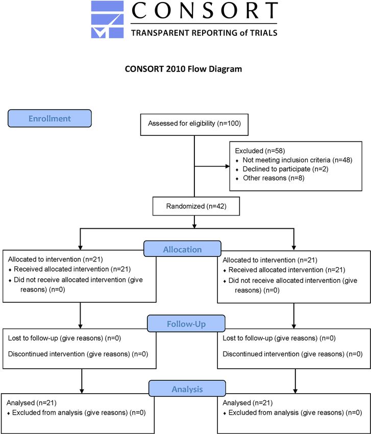
There was not any loss of patients during follow-up (Figure 1). Demographic data, preoperative and post-operative pain
levels, and pain levels on percussion are displayed in the Table 2.
Mean age of patients was 28.24 and 30.52 years for the group 1 and group 2, respectively. There were no significant
differences in the age, gender, tooth number between the groups (P > 0.05). The preoperative pain levels and those on
percussion were around the moderate level (85 mm) in both groups, and no significant differences were found in
preoperative pain levels and these on percussion between two groups (P > 0.05). The postoperative pain levels were
significantly reduced by half at 6 hours for both groups (greater than 50 mm); however, no significant differences were
found in postoperative pain levels between two groups at all considered times (P > 0.05). The postoperative pain levels on
percussion were reduced on day 7 in both groups, and there was no significant difference in this variable between two
groups. There was only one patient who needed to use analgesics postoperatively in the instrumentation with uncon-
nected EAL group (group 1). There was no patient who needed to use analgesics in the instrumentation with connected
EAL group (group 2). There were not any signs of swelling, sinus tract, or palpation pain in the patients, and there were
not any unscheduled appointments for the patients in the study.
Discussion
The result of the present study showed that postoperative pain levels of patients were lower than the “weak” level.
Because the root canal length is changed during root canal preparation, therefore, it must be measured, controlled and
maintained continuously.2 This makes the operator fatigued and exhausted. Using a common, separate EAL, the operator
must use a reference from the remaining tooth structure on the occlusal surface, incisal edge or even the root surface in
teeth missing structure. This reference can be lost during treatment, especially in multiple-appointment treatment. In
addition, using the rubber stop on the shaft of the instrument is improper in certain circumstances. This rubber stop can be
displaced or worse than that, the rubber stop can be overpassed because of its elastic property without notice from the
operator if the instrument is sucked into the canal (screw-in tendency). Instrumentation with a connected EAL device will
overcome these shortcomings of previous methods in WL determination, control, and maintenance. Although there are
many in vitro studies on the WL determination of EALs,1,8-10 there are few studies on endodontic motors with built-in or
connected EAL up to now and therefore, there is not much data on the effect of an endodontic motor on root canal
Page 5 of 10F1000Research 2021, 10:868 Last updated: 27 AUG 2021
Figure 1. CONSORT flow diagram.
treatment.5,11 EAL connected to the endodontic motor in the present study has been investigated in a previous study,
which shows that accuracy was at the highest level when compared with other EAL and cone beam computed
tomography.1 In the present study, we showed that EAL’s accuracy is still at a high level when using with the endodontic
motor.
The PTN is made of M-Wire with special offset design and unique dimensions in order to reduce operator’s fatigue
and exhaustion, and procedure complexity and enhance effectiveness.4,12 In the two previous studies on causing apical
extruded debris of PTN and other rotary NiTi instruments, the results showed that PTN caused significantly less apical
debris extrusion than ProTaper Universal and PTN caused significantly more apical extruded debris than HyFlex
CM.13,14 These studies of comparison between the continuous rotary NiTi and reciprocating instruments showed that
Page 6 of 10F1000Research 2021, 10:868 Last updated: 27 AUG 2021
Table 2. Demographic and pain data according to the two treatment groups.
Instrumentation with Instrumentation with
unconnected EAL (group 1) connected EAL (group 2)
Age 28.24 10.40a 30.52 12.66a
Gender
Male 9 7
Female 12 14
Tooth number
16 1 1
17 0 4
26 2 1
27 4 2
36 1 2
37 5 3
46 3 1
47 5 7
Pain
Preoperative pain after treatment 96.95 41.63a 97.14 45.08a
Postoperative pain levels at 6 h 44.14 41.51a 43.86 36.89a
Postoperative pain levels at 24 h 29.43 38.37 a
23.48 26.67a
Postoperative pain levels at 48 h 21.33 34.38a 13.62 18.85a
a
Postoperative pain levels at 72 h 14.67 27.84 11.10 18.36a
Postoperative pain levels on day 7 08.00 17.32a 06.11 15.62a
Pain on percussion
Preoperative 84.95 45.29a 82.19 41.30a
Postoperative on day 7 20.48 24.41a 17.00 31.60a
Same superscript letters showed no significant differences on the same row (P > .05).
there were not significant differences in apical extruded debris.13,15 Other authors reported that the continuous rotary NiTi
instruments produced less apical extruded debris than the reciprocating instruments.16 These conflicting results may be
due to differences in study design, setup, or type of teeth.
Root canal preparation techniques cause apical extrusion of dentine debris, pulp tissue, microorganisms, and irrigation
solutions through apical foramen, leading to inflammation, resulting in postoperative pain.17 The instrument’s designs or
modes of movement (continuous or reciprocating rotary) are considerable factors that influence the apical extrusion of
debris and therefore, postoperative pain of the patient.18-21 Instruments with the reciprocating movement induce more
debris through apical foramen when compared with instruments with continuous rotary movement.16,22 This affects the
choice of instruments among a great array of nickel-titanium root canal instruments.
Sodium hypochlorite concentration of 3% was used for the present study, that is similar to that of a previous study.11
A lower concentration of sodium hypochlorite has been considered to reduce the postoperative pain in one study,23
however, that result was different from another study.24 Although the standard concentration of 5.25% was used in
other studies,23,24 there was still one study that used a higher concentration of 8.25% with no significant differences of
postoperative pain among other lower concentration groups.25
Because the first 48 hour after endodontic therapy is the most common period when postoperative pain is felt by patient,
five time-points (right after treatment, after 6, 12, 24, 48 hours) were used for evaluation.23 Longer periods are also
selected to further collect the data after 72 hours and 7 days.23
Page 7 of 10F1000Research 2021, 10:868 Last updated: 27 AUG 2021
Postoperative pain related to rotary NiTi instruments was reported by many previous studies using the randomized
clinical trial design.5,11,21,26 Two of these studies performed single appointment endodontic therapy with separate EAL,
and the results revealed that there was no difference in postoperative pain between the reciprocating and continuous
instruments.21,26 These studies enrolled the asymptomatic patients and evaluated only the postoperative pain levels with
different VAS instruments. However, postoperative pain in the single-visit endodontic treatments with NiTi instruments
was at low levels in these two studies.21,26 Another study also recruited asymptomatic patients with postoperative pain
evaluation but used multiple-visit treatment and WL determination during the root canal preparation.11 There were no
differences in postoperative pain among the experimental NiTi instruments with both modes of movement in the above
studies. The remaining study used the same concept as the present study with the different modalities.5 The result of the
previous study revealed that postoperative pain levels on only day 1 of the group using the traditional endodontic motor
with separate WL determination were significantly higher than that of the other groups.5 On the other days of the study,
the postoperative pain levels were not significantly different between two groups using different modalities.5 This result
agreed with that of the present study.
There are many various scales used to record the pain levels for evaluation of the effectiveness of many endodontic
treatments.5,6,27-29 Although the 4- and 5-point rating scales of pain are used commonly and successfully in a clinical
setting because of easy instructions for use, these scales did not have enough sensitivity to record the pain experience of
patients.6 The Numerical Rating Scale for pain evaluation has low sensitivity when compared with the VAS, and in some
previous studies, the VAS proved the high sensitivity and positive correlation with treatment effectiveness. The most
important advantage of the VAS among the other pain scales was the difference in pain intensity at the two different
times showed the actual difference in pain level.30-32 The Heft-Parker VAS is a line with the dimension of 170 mm with
different distances on the scales to describe the pain of the patient.6 The VAS is used commonly in oral-facial pain studies;
however, this scale confuses patients in selection of right position on the scale because there are no guides for ratings other
than the two extremities.6 Using category word designations on the line of a VAS, the graphic rating scale of Heft and
Parker offers more sensitivity than a category scale and is easier to use than a VAS.6
The result of the present study revealed that although the postoperative pain of patients in the instrumentation with
connected EAL group was lower than that of the instrumentation with unconnected EAL group, there was no significant
difference in postoperative pain levels at all points in time between the two experimental groups. Therefore, the null
hypothesis was accepted.
The limitations of the present study were the small sample size, that the operator was not blinded to the applications of the
two different modalities.
Within the limitations of the present study, the result revealed that the preoperative pain levels of the subjects in both
groups were above the “moderate” level and those reduced after single-visit endodontic treatment with the postoperative
pain levels less than the “faint” level. There was just one patient that used anti-inflammation drugs after treatment. Along
with many other advantages of single-visit endodontic treatment such as reduction of appointments, exclusion of leakage
through temporary restorations and removal risk of additional missing of tooth structure in previously severe structure
missing tooth, this mode of treatment is tolerated and preferred better by patients and becomes common practice in many
situations.26 Within the limitations of the present study, the endodontic therapy with the single-visit treatment brings the
benefit to the patients without increasing the pain.
Conclusions
Within the limitations of the present study, endodontic therapy with the single-visit treatment brings the benefit to the
patients without increasing the pain. Both groups using the endodontic motors with unconnected or connected electronic
apex locator have a similar effect on reduction of the postoperative pain for endodontic patients.
Data availability
Underlying data
Mendeley Data: Khoa Cuong PO Pain, https://doi.org/10.17632/48ytd79w39.1.33
Reporting guidelines
Mendeley Data: CONSORT checklist for ‘Comparison of the effect on postoperative pain between instrumentation with
and without connected electronic apex locator: a randomized clinical trial’, https://doi.org/10.17632/48ytd79w39.1.33
Data are available under the terms of the Creative Commons Attribution 4.0 International license (CC-BY 4.0).
Page 8 of 10F1000Research 2021, 10:868 Last updated: 27 AUG 2021
References
1. Pham KV, Khuc NK: The Accuracy of Endodontic Length 18. Pasqualini D, Corbella S, Alovisi M, et al.: Postoperative quality of
Measurement Using Cone-beam Computed Tomography in life following single-visit root canal treatment performed by
Comparison with Electronic Apex Locators. Iran Endod J. 2020; rotary or reciprocating instrumentation: a randomized clinical
15(1): 12–17. trial. Int Endod J. 2016; 49(11): 1030–1039.
Publisher Full Text PubMed Abstract|Publisher Full Text
2. Vasconcelos BC, Bastos LM, Oliveira AS, et al.: Changes in Root 19. Nekoofar MH, Sheykhrezae MS, Meraji N, et al.: Comparison of the
Canal Length Determined during Mechanical Preparation Effect of Root Canal Preparation by Using WaveOne and
Stages and Their Relationship with the Accuracy of Root ZX II. ProTaper on Postoperative Pain: A Randomized Clinical Trial.
J Endod. 2016; 42(11): 1683–1686. eng. J Endod. 2015; 41(5): 575–578.
PubMed Abstract|Publisher Full Text PubMed Abstract|Publisher Full Text
3. Pham K, Nguyen N: Cutting efficiency and dentinal defects using 20. Neelakantan P, Sharma S: Pain after single-visit root canal
two single-file continuous rotary nickel-titanium instruments treatment with two single-file systems based on different
[Original Article]. Saudi Endod J. 2020; 10(1): 56–60. kinematics—a prospective randomized multicenter clinical
Publisher Full Text study. Clin Oral Investig. 2015; 19(9): 2211–2217.
4. Pham K, Phan T: Evaluation of root canal preparation using PubMed Abstract|Publisher Full Text
two nickel-titanium instrument systems via cone-beam 21. Kherlakian D, Cunha RS, Ehrhardt IC, et al.: Comparison of the
computed tomography [Original Article]. Saudi Endod J. 2019; 9(3): Incidence of Postoperative Pain after Using 2 Reciprocating
210–215. Systems and a Continuous Rotary System: A Prospective
Publisher Full Text Randomized Clinical Trial. J Endod. 2016; 42(2): 171–176.
5. Arslan H, Güven Y, Karataş E, et al. : Effect of the Simultaneous PubMed Abstract|Publisher Full Text
Working Length Control during Root Canal Preparation on 22. Caviedes-Bucheli J, Castellanos F, Vasquez N, et al.: The influence of
Postoperative Pain. J Endod. 2017; 43(9): 1422–1427. eng. two reciprocating single-file and two rotary-file systems on the
PubMed Abstract|Publisher Full Text apical extrusion of debris and its biological relationship with
6. Heft MW, Parker SR: An experimental basis for symptomatic apical periodontitis. A systematic review and
revising the graphic rating scale for pain. Pain. 1984; meta-analysis. Int Endod J. 2016; 49(3): 255–270.
19(2): 153–161. PubMed Abstract|Publisher Full Text
PubMed Abstract|Publisher Full Text 23. Mostafa M, El-Shrief YAI, Anous WIO, et al. : Postoperative pain
7. Nusstein J, Steinkruger G, Reader A, et al.: The effects of a 2-stage following endodontic irrigation using 1.3% versus 5.25% sodium
injection technique on inferior alveolar nerve block injection hypochlorite in mandibular molars with necrotic pulps: a
pain. Anesth Prog. 2006; 53(4): 126–130. eng. randomized double-blind clinical trial. Int Endod J. 2020 Feb; 53(2):
PubMed Abstract|Publisher Full Text|Free Full Text 154–166. eng.
PubMed Abstract|Publisher Full Text
8. Van Pham K: Endodontic length measurements using 3D Endo,
cone-beam computed tomography, and electronic apex locator. 24. Farzaneh S, Parirokh M, Nakhaee N, et al. : Effect of two different
BMC Oral Health. 2021; 21(1): 271. concentrations of sodium hypochlorite on postoperative pain
PubMed Abstract|Publisher Full Text|Free Full Text following single-visit root canal treatment: a triple-blind
randomized clinical trial. Int Endod J. 2018; 51(S1): e2–e11.
9. Van Pham K: Endodontic length measurements using PubMed Abstract|Publisher Full Text
cone beam computed tomography with dedicated or
conventional software at different voxel sizes. Sci Rep. 2021; 25. Demenech LS, de Freitas JV, Tomazinho FSF, et al.: Postoperative
11(1): 9432. Pain after Endodontic Treatment under Irrigation with 8.25%
PubMed Abstract|Publisher Full Text|Free Full Text Sodium Hypochlorite and Other Solutions: A Randomized
Clinical Trial. J Endod. 2021 May; 47(5): 696–704. eng.
10. Nguyen P, Pham K: Endodontic length measurements using PubMed Abstract|Publisher Full Text
different modalities: An in vitro study [Original Article]. J Int Soc
Prev Community Dent. 2020; 10(6): 752–758. 26. Çiçek E, Koçak MM, Koçak S, et al.: Postoperative pain intensity
PubMed Abstract|Publisher Full Text|Free Full Text after using different instrumentation techniques: a
randomized clinical study [Original Article]. J Appl Oral Sci. 2017;
11. Oliveira PS, da Costa KNB, Carvalho CN, et al.: Impact of root canal 25(1): 20–26.
preparation performed by ProTaper Next or Reciproc on the PubMed Abstract|Publisher Full Text|Free Full Text
quality of life of patients: a randomized clinical trial. Int Endod J.
2019; 52(2): 139–148. eng. 27. Ramamoorthi S, Nivedhitha MS, Divyanand MJ: Comparative
PubMed Abstract|Publisher Full Text evaluation of postoperative pain after using endodontic needle
and EndoActivator during root canal irrigation: A randomised
12. Pham KV: A Comparison of Cone Beam Computed Tomography controlled trial. Aust Endod J. 2015; 41(2): 78–87.
and Periapical Digital Radiography for Evaluation of Root Canal PubMed Abstract|Publisher Full Text
Preparation. Applied Sciences. 2021; 11(14).
Publisher Full Text 28. Pasqualini D, Mollo L, Scotti N, et al.: Postoperative Pain after
Manual and Mechanical Glide Path: A Randomized Clinical Trial.
13. Ozsu D, Karatas E, Arslan H, et al. : Quantitative evaluation of J Endod. 2012; 38(1): 32–36.
apically extruded debris during root canal instrumentation PubMed Abstract|Publisher Full Text
with ProTaper Universal, ProTaper Next, WaveOne, and self-
adjusting file systems. Eur J Dent. 2014; 8(4): 504–508. eng. 29. ElMubarak AHH, Abu-bakr NH, Ibrahim YE: Postoperative Pain in
PubMed Abstract|Publisher Full Text|Free Full Text Multiple-visit and Single-visit Root Canal Treatment. J Endod.
2010 2010/01/01/; 36(1): 36–39.
14. Koçak MM, Çiçek E, Koçak S, et al.: Comparison of ProTaper Next PubMed Abstract|Publisher Full Text
and HyFlex instruments on apical debris extrusion in curved
canals. Int Endod J. 2016; 49(10): 996–1000. eng. 30. Price DD, Bush FM, Long S, et al.: A comparison of
PubMed Abstract|Publisher Full Text pain measurement characteristics of mechanical visual
analogue and simple numerical rating scales. Pain. 1994; 56(2):
15. Silva EJNL, Carapiá MF, Lopes RM, et al.: Comparison of apically 217–226.
extruded debris after large apical preparations by full-sequence PubMed Abstract|Publisher Full Text
rotary and single-file reciprocating systems. Int Endod J. 2016;
49(7): 700–705. eng. 31. Kremer E, Atkinson HJ, Ignelzi RJ: Measurement of pain: Patient
PubMed Abstract|Publisher Full Text preference does not confound pain measurement. Pain. 1981;
10(2): 241–248.
16. Bürklein S, Schäfer E: Apically extruded debris with reciprocating PubMed Abstract|Publisher Full Text
single-file and full-sequence rotary instrumentation systems.
J Endod. 2012; 38(6): 850–852. eng. 32. Jensen MP, Karoly P, Braver S: The measurement of clinical pain
PubMed Abstract|Publisher Full Text intensity: a comparison of six methods. Pain. 1986; 27(1):
117–126.
17. Relvas JBF, Bastos MMB, Marques AAF, et al. : Assessment of PubMed Abstract|Publisher Full Text
postoperative pain after reciprocating or rotary NiTi
instrumentation of root canals: a randomized, controlled 33. Pham K: Khoa Cuong PO Pain and Consort Checklist. Mendeley
clinical trial. Clin Oral Investig. 2016; 20(8): 1987–1993. Data, V1. 2021.
PubMed Abstract|Publisher Full Text Publisher Full Text
Page 9 of 10F1000Research 2021, 10:868 Last updated: 27 AUG 2021
The benefits of publishing with F1000Research:
• Your article is published within days, with no editorial bias
• You can publish traditional articles, null/negative results, case reports, data notes and more
• The peer review process is transparent and collaborative
• Your article is indexed in PubMed after passing peer review
• Dedicated customer support at every stage
For pre-submission enquiries, contact research@f1000.com
Page 10 of 10You can also read