Diagnosis and Management of Fungal Neglected Tropical Diseases In Community Settings-Mycetoma and Sporotrichosis - MDPI
←
→
Page content transcription
If your browser does not render page correctly, please read the page content below
Tropical Medicine and
Infectious Disease
Article
Diagnosis and Management of Fungal Neglected
Tropical Diseases In Community Settings—Mycetoma
and Sporotrichosis
Roberto Estrada-Castañón 1 , Guadalupe Estrada-Chávez 2 and
María de Guadalupe Chávez-López 3, *
1 Community Dermatology Mexico C.A.; Health Secretary Guerrero, 39355 Acapulco, Guerrero, Mexico;
restrada_13@hotmail.com
2 Department of Dermatology and Dermato-Oncology, Instituto Estatal de Cancerología “Dr. Arturo Beltrán
Ortega”, Health Secretary Guerrero, Faculty of Medicine, Universidad Autónoma de Guerrero Mexico,
Community Dermatology Mexico C.A., 39850 Acapulco, Guerrero, Mexico; estradaguadalupe@hotmail.com
3 Department of Dermatology and Mycology Acapulco General Hospital, Health Secretary Guerrero,
Community Dermatology Mexico C.A., 39355 Acapulco, Guerrero, Mexico
* Correspondence: chavezg13@live.com.mx; Tel.: +52-744-4463882
Received: 19 March 2019; Accepted: 26 April 2019; Published: 16 May 2019
Abstract: Background: This is a retrospective, analytic observational study where we describe cases of
sporotrichosis and mycetoma from Acapulco General Hospital and Community Dermatology Mexico
C.A. over 25 years. Analysis of environmental features that favour the development of such diseases
has been made, as well as the limitations in the study and treatment of such diseases in resource poor
settings. Methods: We reviewed the information on 76 sporotrichosis and 113 mycetoma patients out
of a total of 14,000 consultations at Acapulco General Hospital and from Community Dermatology
clinics. We analysed the epidemiological and mycological characteristics and the investigations used
for diagnosis such as direct examination, culture, intradermal test reactions, and biopsy. Results: In
total 91 confirmed cases of actinomycetoma, 22 of eumycetoma and 76 of sporotrichosis have been
identified including diagnostic studies for both diseases and their treatment. Discussion: The results
obtained have been analysed and interpreted in patients with mycetoma and sporotrichosis in the
state of Guerrero, México, along with limitations in their management in areas with limited economic
and logistical resources. The prevalence of mycetoma in our setting is compared with other centres
where patients from all over the country are seen. The possible causes for variations in prevalence in
specific areas has been looked for, in one of the poorest states of the Mexican Republic.
Keywords: subcutaneous mycosis; actinomycetoma; eumycetoma; sporotrichosis Community
dermatology
1. Introduction
Mycetoma and sporotrichosis are two of the most widely distributed implantation (subcutaneous)
mycoses worldwide. Mycetoma is a chronic infectious disease, which can be caused by different species
of fungus (eumycetoma) or by aerobic filamentous bacteria (actinomycetoma). Clinical characteristics
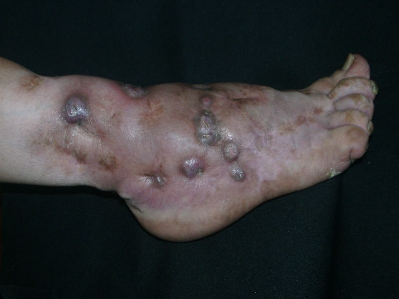
include, local swelling, and draining sinuses with serosanguinous or purulent exudates (Figure 1),
which contains the infective forms known as “grains” (Figure 2) Climatic and geographic conditions
have been described previously and the main endemic zone identified as between the Tropic of Cancer
at lattitudes 15º south and 30º north; this has been referred to as the “Mycetoma belt” by the World
Health Organisation (WHO). It includes countries like Chad, Ethiopia, India, Mauritania, Senegal,
Somalia, Sudan, Yemen, and in the Americas: Mexico and Venezuela [1].
Trop. Med. Infect. Dis. 2019, 4, 81; doi:10.3390/tropicalmed4020081 www.mdpi.com/journal/tropicalmed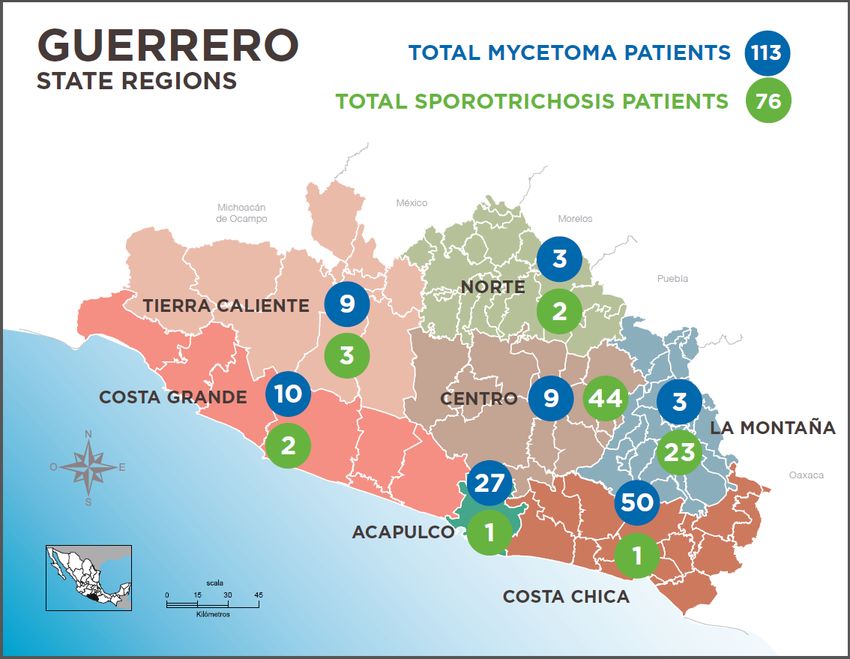
Trop. Med. Infect. Dis. 2019, 4, 81                                                                    2 of 9
 Trop. Med. Infect. Dis. 2018, 3, x FOR PEER REVIEW                                                     3 of 9

                                          Figure1.1.Eumycetoma
                                         Figure      Eumycetomaofofthe
                                                                    thefoot.
                                                                        foot.

      Mexico has been identified as the country with the highest incidence of mycetoma in Latin
America [2] and one of the most endemic countries around the world [3]. Mycetoma has been
recognised by the WHO, as a neglected tropical disease (NTD) in 2016 in a WHA 69.21 resolution [4].
      Even though sporotrichosis does not have the same morbidity patterns, except in disseminated
or systemic forms, as mycetoma, because of its capacity for dissemination, its severity and neglect; it
accounts for significant disability, morbidity, and reduction in the quality of life of affected individuals,
who are mostly peasants; these are mainly farmers living in remote areas where diagnosis and treatment
is frequently delayed due to lack of experience of local health personal working in the communities.
Sporotrichosis is a chronic granulomatous implantation mycosis, caused by a group of dimorphic fungi
belonging to the genus, Sporothrix . It affects either humans or some animal species [5] It is considered,
in some cases, to be an occupational disease, not only in farmers, but also in florists, carpenters, and
workers using hay for packing or building. Latin America has reported a high incidence of the infection,
particularly in Brazil and Mexico [6], although in the former most cases, in the most recent outbreak,
are caused by the zoonotic species Sporothrix brasiliensis, spread by cats, unlike the disease in Mexico.

                    Figure 2. Grain histopathology (actinomycetoma) Haematoxylin and Eosin 100X.
Trop. Med. Infect. Dis. 2019, 4, 81 3 of 9
Figure 1. Eumycetoma of the foot.
Figure2.2.Grain
Figure Grainhistopathology
histopathology(actinomycetoma)
(actinomycetoma)Haematoxylin
Haematoxylinand
andEosin
Eosin100X.
100X.
Guerrero State in Mexico, is located at the south west of the Mexican Republic, on the coast of the
Pacific Ocean between the coordinates 17◦ 360 47”N 99◦ 570 00”O with a territory of 63,794 km2 ; to the
West it is crossed by the mountain chain, the Sierra Madre del Sur, which provides very wide climatic
and environmental diversity, with valleys and hills suitable for the development of different deep and
subcutaneous mycoses. It is divided into seven natural regions, which determined its political division
(Figure 3): The North and Mountain areas “Norte” and “Montaña” are arid and with difficult terrain,
rich in mining and caves that are frequently visited by cave explorers and tourists, in whom cases
of histoplasmosis have been reported [7], the “Tierra Caliente” (warm earth) area, has a wide valley
crossed by the Balsas river and very high daily temperatures which provides the name to the region,
cases of coccidioidomycosis [8] have been reported from there. A mountain chain divides the central
region “Centro” into the other two regions, which are the small and big coastal areas, “Costa chica”
and “Costa Grande”, these are rich in vegetation, with coffee and palm plantations, where cases of
paracoccidioidomycosis [9] and chromoblastomycosis [10] have been reported. Nevertheless, as in
other areas of the world, mycetoma and sporotrichosis are the most frequent subcutaneous mycoses in
Mexico [3] and in Guerrero State [11], making it the country with second or third highest prevalence of
these disesases [1,2].Trop. Med. Infect. Dis. 2019, 4, 81 4 of 9
Trop. Med. Infect. Dis. 2018, 3, x FOR PEER REVIEW 4 of 9
Figure3.3.Map
Figure Mapof
ofGuerrero
GuerreroState
Stateshowing
showingcases
casesofofmycetoma
mycetomaand
andsporotrichosis.
sporotrichosis.
2. Methods
Mycetoma is more frequent in the Coastal and Tierra Caliente areas. Sporotrichosis is more
frequent in the Mountain and Central areas (Figure 3). Affected farmers live in conditions of poverty
We report an analytical, retrospective, observational study in which we reviewed 14,000
that can be severe in isolated regions.
consultations at the Acapulco General Hospital and records of the Community Dermatology program
Community Dermatology Mexico (CDM) is a 28 year old programme whose goal is to assist
over the last 20 years. The information obtained in the communities was the result of visits to the
people in need or without access to specialized dermatological care, by teaching basic dermatology
areas with a high marginality index amongst the seven regions of Guerrero state, with the support of
to health workers working in remote areas where there is less opportunity for continuous medical
the State health programme called Desarollo Integral de la Familia or DIF and the Secretary of Health.
education and to identify, refer, and/or treat patients with both simple and complicated dermatological
Of the mycetoma and sporotrichosis cases that were identified clinically, all were referred to our
problems and to carry out research [12,13], the reason for this communication.
Dermatology Department at the Acapulco General Hospital for further studies and treatment.
Due to limitations associated with field work and the logistics needed for transportation of basic
2. Methods
material and equipment, the studies, that were feasible, were restricted to mycological scrapings for
Weexamination,
direct report an analytical, retrospective,
punch biopsies, observational
and occasionally study in ofwhich
the inoculation samples weofreviewed 14,000
lesional exudate
consultations
onto media for at the Acapulcoculture,
subsequent Generalwhich
Hospitalwere and records of the
subsequently Community
completed Dermatology
in the mycology program
facility in
over the last 20 years. The information obtained in the communities was
our institution. All organisms that were speciated in this study were identified by culture. the result of visits to the
areas In
with a high
order to marginality
facilitate andindex
assistamongst the seven regions
the management of Guerrero
of affected patients,state, with the support
we provided financial
ofsupport
the State health programme called Desarollo Integral de la Familia or
for transportation and food during their trip to Acapulco city, where after having anyDIF and the Secretary of
Health. Of the mycetoma and sporotrichosis cases that were identified clinically,
ancillary studies such as imaging including Computer Assisted Tomography (CAT) scans in specific all were referred to
our Dermatology
cases, culture, or Department at the Acapulco
further mycological studies, General Hospitalfor
free treatment fortheir
further studies
specific andconditions
health treatment.was
Due to limitations associated with field work and the logistics needed
provided after evaluation; this included: Trimethoprim sulfamethoxazole and dapsone (DDS) for transportation of basic
for
material
patients with actinomycetoma, itraconazole (100–200mg daily), and terbinafine (250 mg daily)for
and equipment, the studies, that were feasible, were restricted to mycological scrapings for
direct examination,
eumycetoma punch
patients and biopsies,
potassium and occasionally
iodide the inoculation
for sporotrichosis; the use ofofsamples of medication
the latter lesional exudate
is due
onto media for subsequent
to cost constraints. culture, which were subsequently completed in the mycology facility in our
institution.
Despite Allthe
organisms that were
support given speciated
to the patients,in38%
this did
studynotwere identified
attend by culture.
subsequent consultations because
of economic, language, personal reasons or sometimes because family members or friends, raised theTrop. Med. Infect. Dis. 2019, 4, 81 5 of 9
In order to facilitate and assist the management of affected patients, we provided financial
support for transportation and food during their trip to Acapulco city, where after having any
ancillary studies such as imaging including Computer Assisted Tomography (CAT) scans in specific
cases, culture, or further mycological studies, free treatment for their specific health conditions was
provided after evaluation; this included: Trimethoprim sulfamethoxazole and dapsone (DDS) for
patients with actinomycetoma, itraconazole (100–200mg daily), and terbinafine (250 mg daily) for
eumycetoma patients and potassium iodide for sporotrichosis; the use of the latter medication is due
to cost constraints.
Despite the support given to the patients, 38% did not attend subsequent consultations because of
economic, language, personal reasons or sometimes because family members or friends, raised the
spectre of unnecessary amputation. Distance and poverty were constant barriers to consent, study,
and treatment.
In order to review cases we developed a basic recording format including, patient information, in
Excel and graphics were made with statistics analysis SPSS.9
3. Results
Table 1 shows the results from 113 confirmed mycetomas studied. For logistical reasons not all
mycological studies could be carried out on all patients. There were more actinomycetomas than
eumycetomas with scattered anatomical distributions
Table 1. Mycetoma Patients.
VARIABLE VALUES
Male = 85 (75.2%)
GENDER
Female = 28 (24.8%)
Adults > 18a = 104 (92.0%)
AGE
Children < 18a = 9 (8.0%)
Farmers = 53 (48.6%)
OCCUPATION Housewives = 23 (21.1%)
Others = 37 (30.3%)
Feet = 34 (30.1%)
Legs = 11 (9.7%)
Upper limbs = 22 (19.5%)
AFFECTED AREA Pelvic area = 19 (16.8%)
Abdomen = 6 (5.3%)
Trunk = 17 (15.1%)
Cervical column = 4 (3.5%)
Cultures (+) = 82
Direct Exam (+) = 101
MYCOLOGY Biopsy = 79
X ray = 75
CT =7
Actinomycetoma = 81 (78.6%)
TYPE OF MYCETOMA
Eumycetoma = 22 (21.4%)
Nocardia brasiliensis = 64 (70.4%)
Nocardia spp = 19 (20.9%)
ACTINOMYCETES
Actinomadura madurae = 6 ( 6.6%)
N. otitidis caviarum = 2 (2.1%)
Madurella mycetomatis = 16 (72.8%)
Trematosphaeria grisea = 3 (13.7%)
FUNGI
Scedosporium boydii = 2 ( 9.0%)
Phomopsis longicola = 1 ( 4.5%)
five years = 29 (25.7%)
Costa Chica = 50 (44.2%)
Acapulco = 27 (23.9%)
Costa Grande = 10 (8.8%)
REGION Tierra Caliente = 9 (8.0%)
Centro = 9 (8.0%)
Norte = 5 (4.4%)
Montaña = 3 (2.7%)Trop. Med. Infect. Dis. 2019, 4, 81 6 of 9
Studies made for confirmation of the diagnosis were: Biopsy in 79 cases, direct examination in 101
and culture in 82 cases. Actinomycetoma cases were caused by Nocardia brasiliensis in 64 cases, Nocardia
sp 19 cases, Actinomadura madurae 6, Nocardia otitidis caviarum 2. The causative agents in eumycetomas
were in most of the cases Madurella mycetomatis 16, Trematosphaeria grisea 3, Scedosporium apiospermum 2,
and Phomopsis longicolla 1 case.
Table 2 shows the 76 cases of sporotrichosis studied, of which 43 were of the lymphangitic clinical
pattern, fixed type 24 and cutaneous disseminated 8.35 were male, 40 female; 39 adults and 36 children.
Most of these patients were farmers; men, women or children work, or are partially involved, in
field activities.
Table 2. Sporotrichosis Patients.
VARIABLE VALUES
Male = 35 (46.0%)
GENDER
Female = 41 (54.0%)
Adults > 18 = 39 (51.3%)
AGE
Children < 18 = 37 (48.7%)
Farmers = 62 (81.6%)
OCCUPATION Students = 2 (2.6%)
Children not working = 12 (15.8%)
Upper limbs = 32 (42.1%)
Lower limbs = 16 (21.1%)
AFFECTED AREA Face = 16 (21.1%)
Trunk = 4 (5.3%)
Other = 8 (10.4%)
Cultures = 52 (+)
MYCOLOGY IDR (Skin Test) 1 = 45 (+)
Biopsy = 31
Lymphangitic = 43 (56.8%)
CLINICAL FORM Fixed = 24 (32.2%)
Disseminated = 8 (11.0%)Trop. Med. Infect. Dis. 2019, 4, 81 7 of 9
4. Discussion
Neglected diseases in developing countries are usually located in remote, isolated areas with
limited access, and communication. This means that health systems are limited, and there is a high level
of ignorance and malnutrition all of which are reasons why these diseases develop without intervention
and reach extremes of severity, leading to severe morbidity and even death amongst affected patients.
One of the main and persistent obstacles to care and further research is the lack of properly trained
health personnel to identify patients or to perform studies [14]. Additionally personal safety of staff is
a further obstacle to investigation and care, imposing a risk in visits to isolated communities. Even
though, encounters with criminal groups have been few and without severe consequences during
work in the communities, the potential risk for personnel, has now forced us to adapt our working
methods to include the use of teledermatology and telemedicine in order to continue the work of
teaching, research, and advice for the health personnel based in remote areas [15].
In 1988, Lavalle and Padilla described in a communication [16] that Guerrero was the second
commonest source in the country of mycetoma cases attending the “Centro Dermatológico Pascua”
(CDP), which is a referral centre for treatment of dermatological problems. In another review of
mycetomas by Bonifaz et al., Guerrero was rated 3rd in incidence (1). In comparing our data with
that of Lavalle and Bonifaz for patients coming from Guerrero, but diagnosed and treated in the
Department of Mycology at the Hospital General de Mexico (HGM), patient parameters were similar:
male predominance in 70%, mostly middle aged, foot involvement, though in our studies uncommon
areas such as the perianal region [17] or the neck, a source of considerable morbidity, have also
been seen to be involved [18]. Causal agents reported in these other studies are also similar with a
predominance of actinomycetes in 97% (Lavalle), 82% (Bonifaz), and 81% (Current study). It is worth
mentioning that both centres in Mexico city are among the main primary mycology referral laboratories
where patients are seen from every part of the country.
Our studies have also found that there are regional variations in disease prevalence for both
mycoses in our environment. Eumycetomas are predominantly found in the Costa Chica area where
they account for a much higher proportion of cases (30%) compared with the 2–3% reported nationally.
Most sporotrichosis cases originate from the Central and Montaña regions (Figure 3). In explanation,
besides the environmental characteristics–in the Costa Chica there is pastureland and cattle farming
in commo—it is possible that there is a higher genetic predisposition, related to a high proportion of
indigenous Mexican groups, for some mycoses. In the Costa Chica there is also a well established
population of individuals of African Mexican origin. It has also been suggested in cases of sporotrichosis,
in patients of indigenous origin in the Central Mountain areas carry alleles of Class II [19].
Madurella mycetomatis is the most important causal agent found in our series which is similar to
studies reported in patients from other areas with high endemicity [20–22]; these isolates have not
been subject to molecular identification, therefore the presence of other Madurella species is possible.
Mycetomas per se have a low mortality, except those cases with neck and dorsal spine involvement [18],
nevertheless they do have severe morbidity, which has a serious impact on the earning capacity of the
affected patients as they are usually farmers with reduced economic resources and depend directly
on their ability to work in the fields. The high cost of the treatment and the long term course of the
disease, have a critical impact on the family’s health and financial well-being [21].
Histopathological studies are a very valuable diagnostic auxiliary method used to differentiate the
actinomycetomas and eumycetomas, for which treatment is completely different. Samples can be easily
obtained even in remote areas and together with direct examination and culture can be accessible in
areas where resources are extremely limited. Molecular diagnostic techniques are usually out of the
reach of decentralized institutions where mycetoma patients are studied and treated [22] and even
though simple imaging studies and ultrasound are accessible in order to identify bone involvement,
other studies like CAT scan or MRI, can be unaffordable for patients of low income, which represent
most of the patients studied in our group [23].Trop. Med. Infect. Dis. 2019, 4, 81 8 of 9
We firmly believe that mycetoma treatment should be as integrated or “holistic” [24]. In our
opinion this should include (1) training of the local health personnel, providing the basic knowledge
to identify and refer mycetoma cases to centres where they can be properly diagnosed, studied and
treated, (2) facilitating the means for patients to attend for consultation, (3) provide appropriate tests
for identification of causal agents, together with estimation of the severity index in order to determine
the best available treatment for each case, (4) provide free medications throughout treatment duration,
and (5) register of every case in order to contribute with information on the epidemiology of mycetoma
worldwide (6) establishing a robust system for early detection, and ultimately, prevention of cases in
collaboration with local health workers. In the case of sporotrichosis further work needs to be carried
out, in order to establish the specific molecular identification of the causative Sporothrix species and the
reasons for small outbreaks in specific areas, although feline sources of infection are not suspected. As
with mycetoma, improving early case recognition is an important goal.
We should point out that even though the number of suspected mycetoma cases diagnosed
in Community Dermatology over the last 28 years of work exceeds 240 patients, the diagnosis in
many cases has been mainly determined on clinical grounds with some laboratory tests, as discussed
previously; in these patients other essential laboratory studies could not be done in order to establish
the causal species. Hence, the clinically diagnosed cases have not been included in the analysis.
Finally it is important to mention that 2019 is the year when the Community Dermatology Centre
“Dr. Ramon Ruiz Maldonado” (CDM) has been opened in Acapulco in order to treat patients of low
and very low incomes from the urban, and especially, rural areas where access to specialized attention
is limited or absent.
Author Contributions: All participated in reviewing patients, planning the study and writing the manuscript.
Funding: This research received no external funding.
Acknowledgments: We would like to mention our sincere appreciation to Alexandro Bonifaz MSc for the data
from the laboratory of mycology regarding patients from Guerrero State. As well we would like to acknowledge
Roderick Hay for the very valuable help, orientation and his participation during the activities of Community
Dermatology Mexico and constant encouragement for this communication. To the Secretary of State Health
and the DIF Guerrero for the support given to the activities of Community Dermatology Mexico through which
fundamental information for this study was obtained. Also to the International Foundation for Dermatology, the
American Academy of Dermatology, Galderma Skin Pact Awards and Vaseline Direct Relief for their contributions
in order to purchase medicaments and for providing resources to mycetoma and sporotrichosis patients for their
study and treatment.
Conflicts of Interest: Authors have no relevant conflicts of interest for the elaboration of this work
References
1. Bonifaz, A.; Tirado Sánchez, A.; Calderón, L.; Saul, A.; Araiza, J.; Hernández, M.; González, G.M.; Ponce, R.M.
Mycetoma: Experience of 482 cases in a single center in Mexico. PLoS Negl. Trop. Dis. 2014, 8. [CrossRef]
[PubMed]
2. Lopez-Mtnez, R.; Mendez-Tovar, L.J.; Bonifaz, A.; Arenas, R.; Mayorga, J.; Welsh, O.; Vera-Cabrera, L.;
Padilla-Desgarennes, M.C.; Contreras Pérez, C.; Chávez, G. Update on the epidemiology of Mycetoma in
Mexico. A review of 3933 cases. Gac. Med. Mex. 2013, 149, 586–592.
3. Van de Sande, W. Global burden of human mycetoma: A systemic review and metanalisis. PLoS Negl. Trop.
Dis. 2013, 7. [CrossRef]
4. WHO. Available online: https://www.who.int/neglected_diseases/mediacentre/WHA_69.21_Eng.pdf?ua=1
(accessed on 2 February 2019).
5. Werner, A.; Werner, B. Sporotrichosis in man and animals. Int. J. Dermatol. 1994, 33, 692–700. [CrossRef]
[PubMed]
6. Conti, A.D. Sporotrichosis in Latin America. Mycopath 1989, 108, 113–116.
7. Corcho-Berdugo, A.; Muñoz Hndez, B.; Palma-Cortes, G.; Ramirez Hndez, A.; Martínez-Rivera, M.; Frías-de
León, M.; Reyes-Montes, M.; Martínez-Valadez, E.; Manjarrez-Zavala, M.; Alfaro-Ramos, L.; et al. Brote
inusual de histoplasmosis en residentes del estado de México. Gaceta Med. Mex. 2011, 5, 377–384.Trop. Med. Infect. Dis. 2019, 4, 81 9 of 9
8. Mayorga, P.; Epinoza, H. Coccidioidomycosis in México and Central America. Mycopathol. Mycol. Appl.
1970, 41, 13–23. [CrossRef] [PubMed]
9. Lopez-Martinez, R.; Hernandez-Hernandez, F.; Mendez-Tovar, LJ.; Manzano-Gayosso, P.; Bonifaz, A.;
Arenas, R.; Padilla-Desgarennes Mdel, C.; Estrada, R.; Chávez, G. Paracoccidioidomycosis en Mexico.
Clinical and epidemiological data from 93 new cases (1972–2012). Mycoses 2014, 57, 525–530. [CrossRef]
10. Chavez, L.G.; Estrada, C.R.; Estrada, C.G.; Moreno, C.G. Cromoblastomicosis y micetoma. Informe de un
caso por presentación simultanea de Fonseca pedrosoi y Nocardia brasiliensis. Dermatol. Cosm. Med. Quir.
2014, 12, 268–271.
11. Estrada, C.R.; Chavez, L.G.; Estrada, C.G.; Bonifaz, A. Report of 73 cases of cutaneous sporotrichosis in
Mexico. An. Bras. Dermatol. 2018, 93, 907–909. [CrossRef]
12. Estrada, C.R.; Chavez-Lopez, M.G.; Estrada-Chavez, G.; Paredes-Solis, S. Specialized dermatological care
for marginalized populations and education at primary care level: Is community dermatology a feasible
proposal? Int. J. Dermatol. 2012, 51, 1345–1350. [CrossRef]
13. Hay, R.J.; Estrada, C.R.; Grossmann, H. Managing skin disease in resource-poor environments- the role of
community oriented training and control programs. Int. J. Dermatol. 2011, 50, 558–563. [CrossRef]
14. El-Safi, S.; Chappuis, F.; Boelaert, M. The Challenges of Conducting Clinical Research on neglected tropical
diseases in remote endemic áreas of Sudan. PLoS Negl. Trop. Dis. 2016, 10. [CrossRef] [PubMed]
15. Chávez, L.G.; Estrada, C.G.; Orozco, F.M.; Solis, R.A.; Solchaga-Rosas, J.; Armendariz-Valle, F.;
Estrada-Castañón, R.A. Teledermatología, un modelo de enseñanza y asistencia en atención primaria
a la salud. Gac. Med. Mex. 2018, 154, 1–5.
16. Lavalle, P.; Padilla, M.C.; Perez, J.; Reynoso, S. Contribución al conocimiento de los micetomas en el estado
de Guerrero, México. Origen disctribución geográfica y evolución de 100 casos de micetomas. Dermatol. Rev.
Mex. 1988, 42, 232–238.
17. Chávez, G.; Estrada, R.; Bonifaz, A. Perianal actinomycetoma, experience of 20 cases. Int. J. Dermatol. 2002,
41, 491–493. [CrossRef] [PubMed]
18. Estrada-Chavez, G.; Estrada, R.; Fernandez, R.; Arenas, R.; Reyes, A.; Guevara, C.; Chávez-López, G. Cervical
and middle actinomycetomas from Guerrero State, Mexico. Int. J. Dermatol. 2017, 56, 1146–1149. [CrossRef]
[PubMed]
19. Estrada-Chavez, G.; Estrada, R.; Chavez, G.; Vega Memije, M.; Guzmán, R.; García-Lechuga, M.; Granados, J.;
Rangel-Gamboa, L. HLA Class II alleles in human sporotrichosis in Mexican Amerindians. Asia-Pacif. J.
Blood Types Genes 2018, 2, 183–190.
20. Ahmed, A.O.; Van-Leeuwen, W.; Fahal, A.; Van de Sande, W. Mycetoma caused by Madurella mycetomatis:
A neglected infectious burden. Lancet Infect. Dis. 2004, 4, 566–574. [CrossRef]
21. Torres, G.E.; Niebla, M.A. Mycetoma. Clinical and Microbiological Monograph; Scholar Press: Brivibas gatve,
Riga, 2015.
22. Mehantappa, H.; Sruthi, P.; Kusuma, V.; Niveditha, S.R.; Kumar, S.A. Cytological diagnosis of actinomycosis.
and eumycetoma. A report of two cases. Diagn. Cytopathol. 2010, 38, 918–920.
23. Fahal, A.H.; Sjeikh, H.E.; Lider, M.A.; Homeida, M.A.; El Arabi, Y.E.; Mahgoub, E.S. Ultrasonic imaging in
mycetoma. Br. J. Surg. 1997, 78, 765–766.
24. Mubarak, B.S.; Fahal, A.H.; Mudawi, M.A.; El Samani, W.M.; Fathelrahman, R.O.; Eiman, S.A.; El Nour, M.;
El Rayah, M.M.; El Sheikh Rahman, M.A.; Suliman, H.S.; et al. A holistic approach to the mycetoma
management. PLoS Negl. Trop. Dis. 2018, 12. [CrossRef]
© 2019 by the authors. Licensee MDPI, Basel, Switzerland. This article is an open access
article distributed under the terms and conditions of the Creative Commons Attribution
(CC BY) license (http://creativecommons.org/licenses/by/4.0/).You can also read