Construction and validation a nomogram to predict overall survival for colorectal signet ring cell carcinoma
←
→
Page content transcription
If your browser does not render page correctly, please read the page content below

www.nature.com/scientificreports
OPEN Construction and validation
a nomogram to predict overall
survival for colorectal signet ring
cell carcinoma
Jian‑dong Diao1, Li‑xia Ma2, Chun‑jiao Wu2, Xian‑hong Liu2, Xiao‑yun Su3, Hong‑yu Bi4,
Bo Bao4, Hao‑wei Yan5, Lei Shi6* & Yong‑jing Yang4*
To construct and validate a nomogram to predict the overall survival (OS) of colorectal signet ring cell
carcinoma (SRCC). The potentially eligible cases were obtained against the SEER database from 2004
to 2015. Log-rank test and Cox analysis were conducted to identify the independent prognostic factors
for predicting OS. The identified prognostic factors were later integrated for the construction of an OS
prediction nomogram. Altogether 2904 eligible cases were identified, and the median survival time
was 18 (range: 0–155) months. As suggested by multivariate analysis, age, primary site, grade, tumor
size, T stage, N stage, M stage, surgery, lymph node dissection and chemotherapy were identified as
the independent factors for predicting OS. Afterwards, the above variables were incorporated into
the nomogram. The C-index indicated better discriminatory ability of the nomogram than AJCC 8th
TNM staging and SEER summary stage systems (both P < 0.001). Calibration plots further showed
good consistency between the nomogram prediction and actual observation. The time independent
area under the curves (tAUCs) for 3-year and 5-year OS in nomogram were larger than AJCC and SEER
summary stage system. The constructed nomogram could potentially predict the survival of colorectal
SRCC individuals.
Colorectal cancer (CRC) is the second most common cause of cancer-associated mortality in the United States,
and it has posed a great threat to global h ealth1. In recent years, colorectal signet ring cell carcinoma (SRCC),
2
one of the CRC s ubtypes , has aroused wide attention. It is extensively reported that, SRCC commonly origi-
nates from the undifferentiated colorectal mucosal stem cells; therefore, fast proliferation, low differentiation
level, metastasis and diffuse infiltration can be frequently d etected3,4. In addition, SRCC is also identified by the
AJCC 7th TNM classification system as the independent factor to predict the adverse p rognosis5. Nonetheless,
there are only small case series and case reports available for colorectal SRCC, while information regarding its
clinicopathological characteristics and prognostic outcomes remains largely unexplored6. In this regard, it is
important to precisely estimate the prognosis of SRCC cases, which may facilitate the development of risk-based
individualized treatment and the best therapeutic strategies.
TNM stage system is a prevalent method to predict the outcomes in tumor patients through assessing tumor
size and location (T), regional lymph node metastasis (N) and distant metastasis (M)7. However, TNM classifi-
cation is not efficient to encompass cancer biology as well as to precisely predict outcomes of colorectal SRCC8.
In addition, other clinicopathological parameters can also affect prognosis in SRCC patients, including tumor
grade, tumor site, race, age and therapy4,9. Hence, it is urgently demanded to establish a novel stage classification
involving tumor features and patient status.
As a simple, user-friendly statistical method, nomogram has been uncovered to harbor comparative or even
superior predictive capacity over conventional TNM stage systems in various types of malignancies10–12. To be
specific, the successful establishment of nomogram should not only consider the prognostic weight of every
parameter to calculate the possibility of an outcome but incorporate several independent indicators for optimal
1
Department of Oncology and Hematology, China-Japan Union Hospital of Jilin University, Changchun,
Jilin 130033, China. 2Departments of Oncology, Jilin Cancer Hospital, Changchun, Jilin 130012, China. 3School of
Pharmaceutical Sciences, Jilin University, Changchun, Jilin 130012, China. 4Department of Radiation Oncology,
Jilin Cancer Hospital, Changchun, Jilin 130012, China. 5Department of Regeneratve Medicine, School of Pharmacy,
Jilin University, Changchun, Jilin 130012, China. 6Prevention and Health Care Department, Jilin Cancer Hospital,
Changchun, Jilin 130012, China. *email: 1256267702@qq.com; yangyongjing1975@163.com
Scientific Reports | (2021) 11:3382 | https://doi.org/10.1038/s41598-021-82978-8 1
Vol.:(0123456789)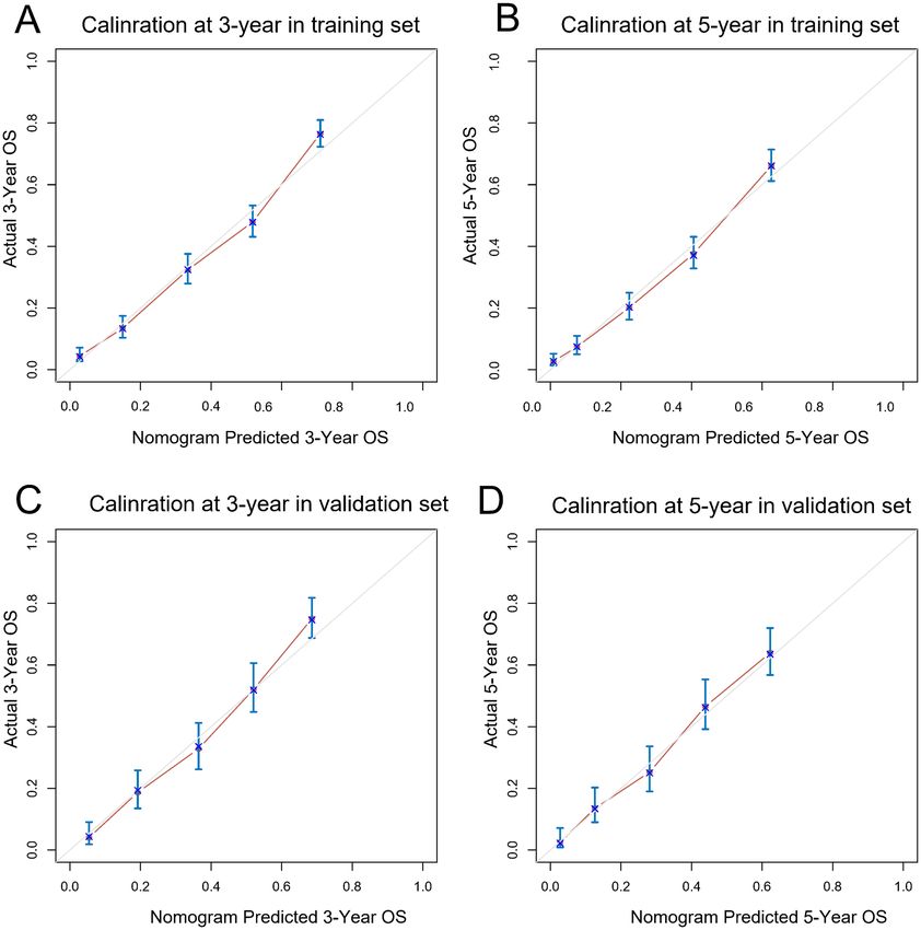
www.nature.com/scientificreports/
conclusion. Of note, nomograms are capable of accurately estimating survival for individuals via the assessment
of vital prognostic factors than TNM stage system13,14. However, as far as we know, there is no such popula-
tion-based nomogram specifically for colorectal SRCC. To this end, we aimed at constructing and verifying a
nomogram for OS prediction in colorectal SRCC based on Surveillance, Epidemiology and End Results (SEER)
database.
Materials and methods
Ethics statement. SEER is the greatest cancer database with the highest authoritativeness in North
merica15, which includes cancer data through covering nearly 30% of the US populations across different geo-
A
graphic regions that can stand for population diversity16. To collect related information from this database, the
SEER Research Data Agreement (No. 19817-Nov2018) was signed in this study, and data were searched against
this database in line with those approved guidelines. The extracted data were publicly accessible and de-iden-
tified, and the data analysis was considered as non-human subjects by Office for Human Research Protection,
thus, no approval was demanded from institutional review board.
Study population. The eligible cases were screened using the SEER*State v8.3.6 approach (released on
August 8th, 2019). In the present work, we included 18 SEER regions between 2004 and 2015. Patients conform-
ing to the following criteria were enrolled: (1) those with primary colorectal SRCC; and (2) those with SRCC
diagnosed according to the third version of the International Classification of Disease for Oncology (ICD-O-3;
coded as 8490/3). At the same time, patients conforming to any one of the following conditions were excluded
from this study: (1) patients with more than one primary tumor; (2) those with clinical diagnosis, or those
diagnosed based on autopsy or the death certificate; (3) those with insufficient data like the mode of surgery
and AJCC stage; (4) those whose tumor location was not mentioned; (5) those with unavailable information on
prognosis. The remaining participants were included into the initial SEER cohort. For establishing and validat-
ing the nomogram, the enrolled cases were randomized into the training or validation set.
Covariates and endpoint. The following patient characteristics were analyzed, including gender, age,race,
marital status, insurance status, primary site, year of diagnosis, tumor size, grade,T, N, M stage, surgery, lymph
node dissection, radiotherapy and chemotherapy. In this study, the single (never married), widowed (having a
domestic partner), separated and divorced cases were classified into unmarried category17. With regard to the
primary tumor site, it was divided into cecum–transverse colon (such as appendix, cecum, ascending colon,
transverse colon, hepatic flexure), descending colon–sigmoid (like descending colon, sigmoid colon, splenic
flexure), multiple, rectum and u nknown18. As for the year of diagnosis, it was classified as 2004–2007, 2008–
2011, 2012–2015, in line with previous s tudies19,20. At the same time, tumor size and age were grouped according
to previous articles as well21–24. The cancer stage was classified according to the AJCC 6th classification system
that was adapted to SEER-derived patients diagnosed from 2004 to 2015. Meanwhile, the qualified patients were
further regrouped in line with the AJCC 8th TNM classification system. In this study, the endpoint was set as
overall survival (OS), which referred to the duration between diagnosis and death due to all c auses25.
Statistical analysis. Nomogram construction. Categorical variables were compared by Fisher’s exact test
or chi-square test and expressed in the manner of proportions and frequencies. Univariate analysis was con-
ducted to predict prognosis using the Kaplan–Meier (K–M) approach as well as log-rank test. Upon univariate
analysis, variables with P-value ≤ 0.1 were screened for multivariate backward stepwise Cox proportional hazard
analysis to determine each possible independent risk factor. Additionally, multicollinearity diagnostics in statis-
tical modeling were conducted by evaluating the correlations, variance inflation factors, and eigenvalues. Then,
we established a nomogram model (based on identified prognostic factors) to predict 3-year, 5-year OS in the
training and validation cohort using the rms package of R. We determined the total nomogram score for every
case based on every variable score in the contour diagram for modeling group.
Nomogram validation. The nomogram was validated by determining its discrimination and calibration abili-
ties using the internal (training) as well as external (validation) set, respectively. In addition, we used the con-
cordance index (C-index) for evaluating our model discrimination performance and assessing the difference in
the predicting ability between predicted and observed values26. As a result, the higher C-index value indicated
the better patient discrimination ability among different prognostic outcomes. Also, we used the Rcorrp.cens
package of Hmisc in R software for comparing the different results from the existing 8th TNM classification sys-
tem and the SEER summary stage system, and utilized C-index to determine them. Using the marginal estimate
versus model, plots presenting the calibration of predicted with measured survival outcomes were made, where
the 45-degree plot was the optimal model with marked consistency in terms of the outcomes. The Receiver
Operating Characteristic (ROC) curves were also plotted to validate the nomogram score. In this study, the
bootstrapping re-sampling approach (1000 repetitions) was applied in obtaining the comparatively unbiased
estimates and checking interval validation. Statistical analysis was conducted using SPSS19.0 (SPSS Inc., Chi-
cago, USA) and R (version 3.51, www.r-project.org). A difference of P < 0.05 (two-tailed) was deemed to be
statistically significant.
Scientific Reports | (2021) 11:3382 | https://doi.org/10.1038/s41598-021-82978-8 2
Vol:.(1234567890)www.nature.com/scientificreports/
Figure 1. Flow chart for patients selection.
Results
Patient characteristics. In total, 2904 qualified subjects who were diagnosed with colorectal SRCC from
2004 to 2015 were enrolled in this research. In addition, 2032 and 872 subjects were assigned into the train-
ing and validation cohorts, respectively. The flow chart of data selection was displayed in Fig. 1. Among all the
subjects, 50.69% were males, with a median age was 63 years (range: 12–103 years). Most subjects were married
(53.99%) and white (81.61%). Cecum–transverse colon (61.95%) was the most prevalent tumor site, followed by
rectum (19.42%), descending colon–sigmoid (15.87%) and multiple (1.48%). Tumor size ˃ 5 cm (42.84%) was the
most common. Most cases of colorectal SRCC had advanced clinical stage (T3: 44.42%; T4:45.83%; N2:46.83%)
and advanced pathological grade (grade III/IV: 80.65%).Operations were performed on 2568 (88.43%) patients,
of which 1728 (59.50%) were total colectomy/ proctectomy. Most patients (79.72%) had more than four lymph
nodes removed. More than half of the patients received chemotherapy (58.13%) and only 13.83% patients
received radiotherapy. The median survival time was 18.0 months (0–155 months). The 3- and 5-year OS rates
were 35.6%, and 28.1%. The demographic and clinicopathological features were listed in Table 1, indicating no
significant difference between two groups.
Nomogram construction. Univariate analysis revealed 11 indicators could affect OS (shown in Table 2).
Among them, marital status was included as an adjusted variable in the step-wise modeling. Consequently, mul-
tivariate analysis showed that age, primary site, grade, tumor size, T stage, N stage, M stage, surgery, lymph node
dissection and chemotherapy were independent predictive indicators of OS (all P < 0.05). Multicollinearity diag-
nostic tests including pair-wise correlations, variance inflation factors plot and eigenvalues plot suggested that
severe multicollinearity issues would not exist (Supplemental Figs. 1 and 2). A nomogram for 3- and 5-year OS
prediction was constructed according to these independent factors (Fig. 2). Nomogram uncovered that AJCC
stage made the greatest contribution to prognosis, followed by surgery, chemotherapy, number of lymph node
dissection and age. By adding the scores of each selected variable, the likelihood of survival of the individual
patient can be easily calculated.
Nomogram validation. The nomogram was validated internally and externally. In the training and valida-
tion cohorts, namely internal validation and external validation cohorts, the OS prediction C-indexes in nomo-
gram were respectively 0.743 (95% CI, 0.730–0.755), 0.730 (95% CI, 0.710–0.751). Furthermore, a comparison
was made between the discrimination ability of nomogram with the ability of SEER summary stage and TNM
8th staging classification, indicating that in the training as well as validation set (P < 0.001), the nomogram is
superior to SEER and TNM 8th staging classification, as shown in Table 3. At last, as shown in Fig. 3, both the
internal calibration plot and external calibration plot of the nomogram exhibited good consistency between the
predictions and practical results based on the nomogram. Figure 4 showed the relevant ROC of the training
and validation cohort. In the training cohort, the time independent area under the curves (tAUCs) of 3– and
5– years OS were 0.830 (0.810–0.850) and 0.840 (0.818–0.862). In the validation cohort, the tAUCs of OS for
3- and 5- years were 0.823 (95% CI: 0.793–0.853) and 0.810 (95% CI: 0.775–0.844), respectively, which were
all greater than AJCC and SEER summary stage system. Bootstrapping with 1000 resamples in the training set
Scientific Reports | (2021) 11:3382 | https://doi.org/10.1038/s41598-021-82978-8 3
Vol.:(0123456789)www.nature.com/scientificreports/
Variables All patients N (%) Validation set N (%) Training set N (%) P value
Year at diagnosis 0.479
2004–2007 984 (33.88) 292 (33.49) 692 (34.06)
2008–2011 954 (32.85) 300 (34.40) 654 (32.19)
2012–2015 966 (33.26) 280 (32.11) 686 (33.76)
Insured status 0.736
Uninsured/unknown 903 (31.10) 275 (31.54) 628 (30.91)
Any medicaid/insured 2001 (68.90) 597 (68.46) 1404 (69.09)
Age 0.528
˂65 1516 (52.20) 463 (53.10) 1053 (51.82)
≥ 65 1388 (47.80) 409 (46.90) 979 (48.18)
Marital status 0.924
Unmarried 1336 (46.01) 400 (45.87) 936 (46.06)
Married 1568 (53.99) 472 (54.13) 1096 (53.94)
Gender 0.373
Female 1432 (49.31) 441 (50.57) 991 (48.77)
Male 1472 (50.69) 431 (49.43) 1041 (51.23)
Race 0.327
Black 275 (9.47) 79 (9.06) 196 (9.65)
White 2370 (81.61) 705 (80.85) 1665 (81.94)
Other 259 (8.92) 88 (10.09) 171 (8.42)
Primary site 0.510
Cecum–transverse colon 1799 (61.95) 527 (60.44) 1272 (62.60)
Descending colon–sigmoid 461 (15.87) 145 (16.63) 316 (15.55)
Multiple 43 (1.48) 10 (1.15) 33 (1.62)
Rectum 564 (19.42) 176 (20.18) 388 (19.09)
Unknown 37 (1.27) 14 (1.61) 23 (1.13)
Grade 0.155
Grade I/II 173 (5.96) 63 (7.22) 110 (5.41)
Grade III/IV 2342 (80.65) 697 (79.93) 1645 (80.95)
Unknown 389 (13.40) 112 (12.84) 277 (13.63)
Tumor size 0.882
≤ 5 cm 1179 (40.60) 360 (41.28) 819 (40.31)
˃5 cm 1244 (42.84) 370 (42.43) 874 (43.01)
Unknown 481 (16.56) 142 (16.28) 339 (16.68)
T stage 0.079
T0-Tis 16 (0.55) 4 (0.46) 12 (0.59)
T1 167 (5.75) 61 (7.00) 106 (5.22)
T2 100 (3.44) 35 (4.01) 65 (3.20)
T3 1290 (44.42) 401 (45.99) 889 (43.75)
T4 1331 (45.83) 371 (42.55) 960 (47.24)
N stage 0.269
N0 854 (29.41) 274 (31.42) 580 (28.54)
N1 690 (23.76) 197 (22.59) 493 (24.26)
N2 1360 (46.83) 401 (45.99) 959 (47.19)
M stage 0.713
M0 1827 (62.91) 553 (63.42) 1274 (62.70)
M1 1077 (37.09) 319 (36.58) 758 (37.30)
Surgery 0.841
No surgery 336 (11.57) 104 (11.93) 232 (11.42)
Local tumor excision/partial colectomy 840 (28.93) 256 (29.36) 584 (28.74)
Total colectomy 1728 (59.50) 512 (58.72) 1216 (59.84)
Lymph node dissection 0.717
None or biopsy 507 (17.46) 148 (16.97) 359 (17.67)
1 to 3 82 (2.82) 22 (2.52) 60 (2.95)
≥4 2315 (79.72) 702 (80.50) 1613 (79.38)
Chemotherapy 0.418
Continued
Scientific Reports | (2021) 11:3382 | https://doi.org/10.1038/s41598-021-82978-8 4
Vol:.(1234567890)www.nature.com/scientificreports/
Variables All patients N (%) Validation set N (%) Training set N (%) P value
No/unknown 1216 (41.87) 375 (43.00) 841 (41.39)
Yes 1688 (58.13) 497 (57.00) 1191 (58.61)
Radiotherapy 0.751
No/unknown 2502 (86.16) 754 (86.47) 1748 (86.02)
Yes 402 (13.84) 118 (13.53) 284 (13.98)
Table 1. Patient demographics and pathological characteristics.
yielded similar discrimination.The 3–year and 5– year tAUCs of the prognostic model in the training set were
0.829(0.812–0.850) and 0.839 (0.820–0.860), respectively.
Discussion
A prognostic nomogram for 3- and 5-year OS prediction was constructed and validated in our study. We analyzed
2904 colorectal SRCC patients from SEER dataset, followed by constructing a nomogram for 3- and 5-year OS
prediction. In addition, internal and external validation of the nomogram demonstrated favorable calibration as
well as discrimination. Moreover, our established nomogram showed more potent predictive capacity compared
to SEER summary stage or TNM staging systems, which could be readily applied in clinical practice to assist
patient counseling as well as individualized therapy.
Some independent factors for predicting prognosis were incorporated into our constructed nomogram.
Besides, the survival was also analyzed based on the colorectal SRCC stage, which discovered that early stage
patients had better prognosis than those at the advanced stage, and such results conformed to almost every
study27,28. Ishihara and colleagues discovered that primary location might serve as the independent factor for
prognosis prediction29. Typically, both tumor stage and primary location were identified as the prognostic fac-
tors in the present work. Moreover, this study identified tumor size and pathological grade as the independent
prognostic factors for colorectal SRCC.
It is necessary to conduct multidisciplinary treatment of colorectal SRCC, so as to select the best therapeu-
tic strategy, and it should be noted that surgery is significant to treat the localized t umors30. As suggested by a
population-based study that enrolls 1972 colorectal SRCC patients between 1989 and 2010 to evaluate whether
adjuvant chemotherapy is significant, adjuvant chemotherapy can offer survival benefits to stage III colon SRCC
patients31. Tao Shi and coworkers also discovered that chemotherapy was linked with the superior survival of
colorectal SRCC with distant metastasis32. Findings in this work also verified that both chemotherapy and surgery
played important roles in diagnosing colorectal SRCC. Further, the surgical retrieval of at least 4 regional lymph
nodes markedly enhanced patient survival. The above-mentioned factors remarkably impacted colorectal SRCC
prognosis. Using our constructed nomogram, patients suffering from diverse tumor differentiation degrees were
assigned with different scores and then with diverse survival outcomes, even though they might be at the same
TNM stage. Besides, these results explicitly clarified the difference between the prognosis estimated using our
constructed nomogram and that estimated by the TNM classification systems, which might explain the better
ability of our nomogram in predicting OS than the TNM classification systems.
Previous studies have also explored nomograms in colorectal signet ring cell c arcinoma33,34. Wang et al. ret-
rospectively evaluated the patient records of mucinous adenocarcinoma and SRCC patients aged ≤ 40 years34.
A nomogram predicting OS was created for risk quantitation. However, compared with our study, the number
of cases enrolled in the previous study was still too small, and only included patients aged ≤ 40 years. Our study
may be more comprehensive and practical.
As a statistical method, the nomogram is capable of providing survival possibility by formula calculation35,36.
Nomogram has been validated to harbor superior predictive capacity in comparison with TNM stage system
in certain types of malignant tumors, which is considered as an alternative or even a novel s tandard37,38. In
particular, it is proper to use nomogram to handle complicated situations without clinical guidelines. And it is
convenient and simple to utilize nomogram for survival prediction. To begin with, in a nomogram, from each
clinicopathological parameter, a vertical line is drawn to “scores” line, followed by score addition to give rise to
“total scores”. Therefore, certain recommendation could accordingly be offer by clinicians. For instance, operation
is suggested in well-differentiated populations in consideration of satisfactory prognosis. On the contrary, pallia-
tive chemotherapy is preferred in poorly-differentiated populations in consideration of decreased life expectancy.
Thus, the presently established nomogram could help to choose patients with prolonged survival, who might
benefit from palliative resection.
Several advantages exist in our research. The detailed clinicopathological data of colorectal SRCC from SEER
database ensured that we successfully constructed a precise prognostic nomogram. Moreover, superior discrimi-
native capacity for OS prediction is detected in our nomogram compared to SEER summary stage and TNM stage
systems. Additionally, available clinical parameters are used, which is convenient for nomogram application.
Certain limitations should be noted in this population-based study. First of all, selection bias was inevitable
due to the retrospective nature. Secondly, prognostic information, including the microsatellite stability/micro-
satellite instability (MSS/MSI) status, the RAS/BRAF/MSI status, family history, vascular invasion and patient
condition, was not available in the SEER database, and future research should focus on these aspects. Thirdly,
the convincing external verification was lacking in this work. At last, our constructed nomogram, which might
Scientific Reports | (2021) 11:3382 | https://doi.org/10.1038/s41598-021-82978-8 5
Vol.:(0123456789)www.nature.com/scientificreports/
Univariate analysis Multivariate analysis
Variable P value HR (95% CI) P value
Year at diagnosis 0.801 NI
2004–2007
2008–2011
2012–2015
Insured status 0.532 NI
Uninsured/unknown
Any medicaid/insured
Age 0.001
˂65 Reference
≥ 65 1.622 (1.452, 1.813) ˂0.001
Marital statusa < 0.001
Unmarried Reference
Married 0.869 (0.783, 0.964) 0.008
Gender 0.842 NI
Female
Male
Race 0.143 NI
Black
White
Other
Primary site < 0.001
Cecum–transverse colon Reference
Descending colon–sigmoid 1.188 (1.020, 1.385) 0.027
Multiple 1.406 (0.967, 2.044) 0.074
Rectum 1.227 (1.055, 1.434) 0.010
Unknown 1.237 (0.775, 1.974) 0.372
Grade < 0.001
Grade I/II Reference
Grade III/IV 1.364 (1.061, 1.753) 0.015
Unknown 1.407 (1.060, 1.868) 0.018
Tumor size < 0.001
≤ 5 cm Reference
˃5 cm 0.950 (0.845, 1.067) 0.387
Unknown 1.197 (1.016, 1.411) 0.032
T stage < 0.001
T0-Tis Reference
T1 1.134 (0.387, 3.324) 0.819
T2 1.881 (0.679, 5.209) 0.224
T3 2.330 (0.861, 6.304) 0.096
T4 3.306 (1.221, 8.952) 0.019
N stage < 0.001 < 0.001
N0 Reference
N1 1.716 (1.457, 2.021) < 0.001
N2 2.898 (2.479, 3.388) < 0.001
M stage < 0.001
M0 Reference
M1 2.377 (2.104, 2.685) < 0.001
Surgery < 0.001 < 0.001
No surgery Reference
Local tumor excision/partial colectomy 0.468 (0.365, 0.599) < 0.001
Total colectomy 0.552 (0.429, 0.712) < 0.001
Lymph node dissection < 0.001 < 0.001
None or Biopsy Reference
1 to 3 0.782 (0.561, 1.091) 0.148
≥4 0.573 (0.462, 0.710) < 0.001
Continued
Scientific Reports | (2021) 11:3382 | https://doi.org/10.1038/s41598-021-82978-8 6
Vol:.(1234567890)www.nature.com/scientificreports/
Univariate analysis Multivariate analysis
Variable P value HR (95% CI) P value
Chemotherapy 0.002
No/unknown Reference
Yes 0.583 (0.519, 0.654) < 0.001
Radiotherapy 0.280
No/unknown
Yes
Table 2. Univariate and multivariate analyses of overall survival (OS) for patients in training set. NI not
included in the multivariate survival analysis, amarital status was used as an adjusted factor in multivariate
analysis.
Figure 2. Nomogram to predict 3-year (A) and 5-year (B) overall survival (OS) of colorectal SRCC patients.
Scientific Reports | (2021) 11:3382 | https://doi.org/10.1038/s41598-021-82978-8 7
Vol.:(0123456789)www.nature.com/scientificreports/
Training set Validation set
Classification C-index (95% CI) P value* C-index (95% CI) P value*
Nomogram 0.743 (0.730, 0.755) 0.730 (0.710, 0.751)
AJCC 8th stage 0.651(0.637, 0.665) < 0.001 0.650 (0.627, 0.673) < 0.001
SEER summary stage 0.645 (0.631, 0.659) < 0.001 0.639 (0.617, 0.661) < 0.001
Table 3. C-indexes for the nomogram and other stage systems in patients with colorectal signet ring cell
carcinoma. *All are compared with Nomogram, HR hazard ratio, CI confidence interval.
Figure 3. Calibration plots of nomogram to predict 3- and 5-year overall survival (OS) in training (A,B) and
validation cohorts (C,D). The X-axis indicated nomogram-predictive survival; the Y-axis suggested actual CSS.
A plot with 45-degree line was suggestive of a perfect calibration where predictive possibilities were identical to
actual ones. Vertical bars indicated 95% CIs.
Scientific Reports | (2021) 11:3382 | https://doi.org/10.1038/s41598-021-82978-8 8
Vol:.(1234567890)www.nature.com/scientificreports/
Figure 4. Discriminatory accuracy for predicting OS assessed by receiver operator characteristics (ROC)
analysis calculating time independent area under the curves (tAUCs). 3-year (A) and 5-year (B) in the training
cohort; 3-year (C) and 5-year (D) in the validation cohorts.
serve as a user-friendly approach for the decision-making of doctors, did not incorporate each prognostic factors
or always offer accurate prognosis prediction in clinical practice.
Conclusion
In conclusion, for patients with colorectal SRCC, we established and validated a nomogram to predict 3-and
5-year OS based on a large, population-based cohort. The nomogram showed excellent performance and could
be thought of as a practical tool to predict prognosis. Nevertheless, further mining of the uncertain prognostic
parameters for the optimization of nomogram is still needed, which requires in-depth external validation.
Scientific Reports | (2021) 11:3382 | https://doi.org/10.1038/s41598-021-82978-8 9
Vol.:(0123456789)www.nature.com/scientificreports/
Received: 29 August 2020; Accepted: 27 January 2021
References
1. El-Shami, K. et al. American cancer society colorectal cancer survivorship care guidelines. CA A Cancer J. Clin. 65, 428–455. https
://doi.org/10.3322/caac.21286(2015).
2. Korphaisarn, K. et al. Signet ring cell colorectal cancer: genomic insights into a rare subpopulation of colorectal adenocarcinoma.
Br. J. Cancer 121, 505–510. https://doi.org/10.1038/s41416-019-0548-9 (2019).
3. Nitsche, U. et al. Mucinous and signet-ring cell colorectal cancers differ from classical adenocarcinomas in tumor biology and
prognosis. Ann. Surg. 258, 775–782. https://doi.org/10.1097/SLA.0b013e3182a69f7e (2013).
4. Mizushima, T. et al. Primary colorectal signet-ring cell carcinoma: clinicopathological features and postoperative survival. Surg.
Today 40, 234–238. https://doi.org/10.1007/s00595-009-4057-y (2010).
5. Meng, Z. W., Pan, W., Hong, H. J., Chen, J. Z. & Chen, Y. L. Modified staging classification for intrahepatic cholangiocarcinoma
based on the sixth and seventh editions of the AJCC/UICC TNM staging systems. Medicine 96, e7891. https://doi.org/10.1097/
md.0000000000007891 (2017).
6. Arifi, S., Elmesbahi, O. & Amarti Riffi, A. Primary signet ring cell carcinoma of the colon and rectum. Bull. Cancer 102, 880–888.
https://doi.org/10.1016/j.bulcan.2015.07.005 (2015).
7. Burke, H. B. Outcome prediction and the future of the TNM staging system. J. Natl. Cancer Inst. 96, 1408–1409. https://doi.
org/10.1093/jnci/djh293 (2004).
8. Belli, S. et al. Outcomes of surgical treatment of primary signet ring cell carcinoma of the colon and rectum: 22 cases reviewed
with literature. Int. Surg. 99, 691–698. https://doi.org/10.9738/intsurg-d-14-00067.1 (2014).
9. Bademci, R., Bollo, J., Martinez, M. C., Hernadez, M. P. & Targarona, E. M. Colorectal cancer prognosis: the impact of signet ring
cell. Gastrointest. Tumors 6, 57–63. https://doi.org/10.1159/000501454 (2019).
10. Fang, C. et al. Nomogram individually predicts the overall survival of patients with gastroenteropancreatic neuroendocrine neo-
plasms. Br. J. Cancer 117, 1544–1550. https://doi.org/10.1038/bjc.2017.315 (2017).
11. Song, W. et al. Development and validation of a nomogram for predicting survival in patients with gastrointestinal stromal tumours.
Eur. J. Surg. Oncol. J. Eur. Soc. Surg. Oncol. Br. Assoc. Surg. Oncol. 44, 1657–1665. https://doi.org/10.1016/j.ejso.2018.07.004 (2018).
12. Dai, D., Jin, H. & Wang, X. Nomogram for predicting survival in triple-negative breast cancer patients with histology of infiltrating
duct carcinoma: a population-based study. Am. J. Cancer Res. 8, 1576–1585 (2018).
13. Huang, J. et al. Nomograms to predict the long-time prognosis in patients with alpha-fetoprotein negative hepatocellular carcinoma
following radical resection. Cancer Med. https://doi.org/10.1002/cam4.2944 (2020).
14. Zhou, Y. et al. Prognostic nomograms and Aggtrmmns scoring system for predicting overall survival and cancer-specific survival
of patients with kidney cancer. Cancer Med. https://doi.org/10.1002/cam4.2916 (2020).
15. Yu, J. B., Gross, C. P., Wilson, L. D. & Smith, B. D. NCI SEER public-use data: applications and limitations in oncology research.
Oncol. (Williston Park, NY) 23, 288–295 (2009).
16. Cahill, K. S. & Claus, E. B. Treatment and survival of patients with nonmalignant intracranial meningioma: results from the
Surveillance, epidemiology, and end results program of the national cancer institute. Clinical article. J. Neurosurg. 115, 259–267.
https://doi.org/10.3171/2011.3.jns101748 (2011).
17. Liu, Y. L. et al. Marital status is an independent prognostic factor in inflammatory breast cancer patients: an analysis of the surveil-
lance, epidemiology, and end results database. Breast Cancer Res. Treat. 178, 379–388. https://doi.org/10.1007/s10549-019-05385
-8 (2019).
18. Nitsche, U. et al. Prognosis of mucinous and signet-ring cell colorectal cancer in a population-based cohort. J. Cancer Res. Clin.
Oncol. 142, 2357–2366. https://doi.org/10.1007/s00432-016-2224-2 (2016).
19. Wu, S. G. et al. Survival in signet ring cell carcinoma varies based on primary tumor location: a surveillance, epidemiology, and
end results database analysis. Expert Rev. Gastroenterol. Hepatol. 12, 209–214. https://doi.org/10.1080/17474124.2018.1416291
(2018).
20. Lv, X. et al. A nomogram for predicting bowel obstruction in preoperative colorectal cancer patients with clinical characteristics.
World J. Surg. Oncol. 17, 21. https://doi.org/10.1186/s12957-019-1562-3 (2019).
21. Shi, T. et al. Chemotherapy is associated with increased survival from colorectal signet ring cell carcinoma with distant metastasis:
a surveillance, epidemiology, and end results database analysis. Cancer Med. 8, 1930–1940. https://doi.org/10.1002/cam4.2054
(2019).
22. Wei, F. & Lyu, H. Postoperative radiotherapy improves survival in gastric signet-ring cell carcinoma: a SEER database analysis. J.
Gastric Cancer 19, 393–407. https://doi.org/10.5230/jgc.2019.19.e36 (2019).
23. Kim, S. E. et al. Sex- and gender-specific disparities in colorectal cancer risk. World J. Gastroenterol. 21, 5167–5175. https://doi.
org/10.3748/wjg.v21.i17.5167 (2015).
24. Yang, L. L., Wang, M. & He, P. Clinicopathological characteristics and survival in colorectal signet ring cell carcinoma: a population-
based study. Sci. Rep. 10, 10460. https://doi.org/10.1038/s41598-020-67388-6 (2020).
25. Diao, J. D. et al. Construction and validation of a nomogram to predict overall survival in patients with inflammatory breast cancer.
Cancer Med. 8, 5600–5608. https://doi.org/10.1002/cam4.2470 (2019).
26. Wolbers, M., Koller, M. T., Witteman, J. C. & Steyerberg, E. W. Prognostic models with competing risks: methods and application
to coronary risk prediction. Epidemiol. (Cambridge, Mass) 20, 555–561. https://doi.org/10.1097/EDE.0b013e3181a39056 (2009).
27. Thota, R., Fang, X. & Subbiah, S. Clinicopathological features and survival outcomes of primary signet ring cell and mucinous
adenocarcinoma of colon: retrospective analysis of VACCR database. J. Gastrointest. Oncol. 5, 18–24. https://doi.org/10.3978/j.
issn.2078-6891.2013.051 (2014).
28. Hyngstrom, J. R. et al. Clinicopathology and outcomes for mucinous and signet ring colorectal adenocarcinoma: analysis from
the national cancer data base. Ann. Surg. Oncol. 19, 2814–2821. https://doi.org/10.1245/s10434-012-2321-7 (2012).
29. Ishihara, S. et al. Tumor location is a prognostic factor in poorly differentiated adenocarcinoma, mucinous adenocarcinoma, and
signet-ring cell carcinoma of the colon. Int. J. Colorectal Dis. 27, 371–379. https://doi.org/10.1007/s00384-011-1343-0 (2012).
30. Ling, C. R., Wang, R., Wang, M. J., Ping, J. & Zhuang, W. Prognosis and value of preoperative radiotherapy in locally advanced
rectal signet-ring cell carcinoma. Sci. Rep. 7, 45334. https://doi.org/10.1038/srep45334 (2017).
31. Hugen, N. et al. Colorectal signet-ring cell carcinoma: benefit from adjuvant chemotherapy but a poor prognostic factor. Int. J.
Cancer 136, 333–339. https://doi.org/10.1002/ijc.28981 (2015).
32. Shi, T. et al. Chemotherapy is associated with increased survival from colorectal signet ring cell carcinoma with distant metastasis:
a surveillance epidemiology, and end results database analysis. Cancer Med. 8, 1930–1940. https://doi.org/10.1002/cam4.2054
(2019).
33. Simkens, G. A. et al. Development of a prognostic nomogram for patients with peritoneally metastasized colorectal cancer treated
with cytoreductive surgery and HIPEC. Ann. Surg. Oncol. 23, 4214–4221. https://doi.org/10.1245/s10434-016-5211-6 (2016).
34. Wang, B. & Zeng, J. Using nomograms to predict prognostic factors in young colorectal mucinous and signet-ring cell adenocar-
cinoma patients. Biosci. Rep. https://doi.org/10.1042/bsr20181863 (2019).
Scientific Reports | (2021) 11:3382 | https://doi.org/10.1038/s41598-021-82978-8 10
Vol:.(1234567890)www.nature.com/scientificreports/
35. Balachandran, V. P., Gonen, M., Smith, J. J. & DeMatteo, R. P. Nomograms in oncology: more than meets the eye. Lancet Oncol.
16, e173-180. https://doi.org/10.1016/s1470-2045(14)71116-7 (2015).
36. Bochner, B. H., Kattan, M. W. & Vora, K. C. Postoperative nomogram predicting risk of recurrence after radical cystectomy for
bladder cancer. J. Clin. Oncol. Off. J. Am. Soc. Clin. Oncol. 24, 3967–3972. https://doi.org/10.1200/jco.2005.05.3884 (2006).
37. Wang, Y. et al. Prognostic nomogram for intrahepatic cholangiocarcinoma after partial hepatectomy. J. Clin. Oncol. Off. J. Am. Soc.
Clin. Oncol. 31, 1188–1195. https://doi.org/10.1200/jco.2012.41.5984 (2013).
38. Cao, J. et al. Clinical nomogram for predicting survival of esophageal cancer patients after esophagectomy. Sci. Rep. 6, 26684. https
://doi.org/10.1038/srep26684 (2016).
Author contributions
Y.Y. conceived the study. J.D. searched the database and literature. L.M, C.W. and X.L. discussed and analyzed
the data. J.D. and L.S. wrote the manuscript. X.S., H.B., Y.Y., B.B. and H.Y. revised the manuscript. All authors
approved the final version.
Competing interests
The authors declare no competing interests.
Additional information
Supplementary Information The online version contains supplementary material available at (https://doi.
org/10.1038/s41598-021-82978-8).
Correspondence and requests for materials should be addressed to L.S. or Y.Y.
Reprints and permissions information is available at www.nature.com/reprints.
Publisher’s note Springer Nature remains neutral with regard to jurisdictional claims in published maps and
institutional affiliations.
Open Access This article is licensed under a Creative Commons Attribution 4.0 International
License, which permits use, sharing, adaptation, distribution and reproduction in any medium or
format, as long as you give appropriate credit to the original author(s) and the source, provide a link to the
Creative Commons licence, and indicate if changes were made. The images or other third party material in this
article are included in the article’s Creative Commons licence, unless indicated otherwise in a credit line to the
material. If material is not included in the article’s Creative Commons licence and your intended use is not
permitted by statutory regulation or exceeds the permitted use, you will need to obtain permission directly from
the copyright holder. To view a copy of this licence, visit http://creativecommons.org/licenses/by/4.0/.
© The Author(s) 2021
Scientific Reports | (2021) 11:3382 | https://doi.org/10.1038/s41598-021-82978-8 11
Vol.:(0123456789)You can also read