Coagulation Profiles and Viscoelastic Testing in Multisystem Inflammatory Syndrome
←
→
Page content transcription
If your browser does not render page correctly, please read the page content below
Posted on Authorea 8 May 2021 — The copyright holder is the author/funder. All rights reserved. No reuse without permission. — https://doi.org/10.22541/au.162049169.98519152/v1 — This a preprint and has not been peer reviewed. Data may be preliminary.

                                                                                                                                                                                                                                                              Coagulation Profiles and Viscoelastic Testing in Multisystem
                                                                                                                                                                                                                                                              Inflammatory Syndrome
                                                                                                                                                                                                                                                              Ashish Ankola1 , Victoria Bradford1 , Jane Newburger1 , Sirisha Emani1 , Audrey Dionne1 ,
                                                                                                                                                                                                                                                              Kevin Friedman1 , Mary Beth Son1 , Lauren Henderson1 , Pui Lee1 , Amy Hellinger1 , Beth
                                                                                                                                                                                                                                                              Hawkins1 , Courtney Ventresco1 , Paul Esteso1 , and Christina VanderPluym1
                                                                                                                                                                                                                                                              1
                                                                                                                                                                                                                                                                  Boston Children’s Hospital

                                                                                                                                                                                                                                                              May 8, 2021

                                                                                                                                                                                                                                                              Abstract
                                                                                                                                                                                                                                                              Objective: To characterize viscoelastic testing profiles of children with multisystem inflammatory syndrome in children (MIS-
                                                                                                                                                                                                                                                              C). Methods: This single-center retrospective review included 30 patients diagnosed with MIS-C from January 1 to September
                                                                                                                                                                                                                                                              1, 2020. Thromboelastography (TEG) with platelet mapping was performed in 19 (63%) patients and compared to age- and
                                                                                                                                                                                                                                                              gender- matched controls via Student’s t-test and Wilcoxon rank sum test. Pearson’s and Spearman correlation were used to
                                                                                                                                                                                                                                                              assess relationships between TEG parameters and inflammatory markers. Results: Patients with MIS-C had abnormal TEG
                                                                                                                                                                                                                                                              results compared to controls, including decreased K time (1.1 vs. 1.7 min, P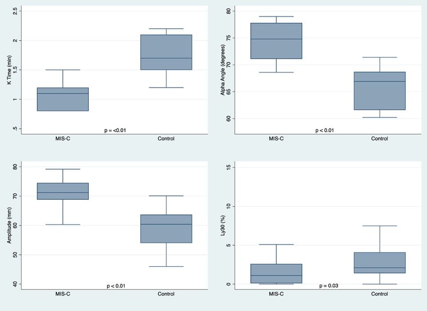
in vitro clot strength on TEG is associated with increased risk of thromboembolism in critically ill adult
Posted on Authorea 8 May 2021 — The copyright holder is the author/funder. All rights reserved. No reuse without permission. — https://doi.org/10.22541/au.162049169.98519152/v1 — This a preprint and has not been peer reviewed. Data may be preliminary.
patients.19 TEG has shown promise in pediatric care, including guiding management of product resuscitation
and thrombosis risk.20–22 We sought to describe the coagulation and viscoelastic testing profiles of children
with MIS-C at our institution, as well as our approach to thromboprophylaxis in these patients.
Methods
Population: This retrospective case series included patients who were diagnosed with MIS-C and admitted
to Boston Children’s Hospital from February 1, 2020 through September 1, 2020. This study was approved
by our institutional review board.
Data collection and definitions: The electronic medical record was reviewed to obtain patients’ demographic,
clinical, and laboratory data. Patients were diagnosed with MIS-C per the Centers for Disease Control and
Prevention’s case definition: age < 21 years presenting with fever, laboratory evidence of inflammation, and
clinically severe illness requiring hospitalization; with multisystem ([?]2) organ involvement; no alternative
plausible diagnosis; and positive for current or recent SARS-CoV-2 infection by reverse transcriptase poly-
merase chain reaction (RT-PCR), serology or antigen test, or COVID-19 exposure within the past 4 weeks.23
All MIS-C cases were adjudicated by rheumatologists and cardiologists.
Demographic data including age and gender were collected. Clinical data were collected including baseline
comorbidities, hospital course, and any bleeding or thrombosis events. Bleeding was defined per the Inter-
national Society on Thrombosis and Haemostasis (ISTH): major bleeding (fatal or in critical area or organ),
clinically relevant non-major bleeding (requiring intervention by a medical provider, hospitalization/increased
level of care, or further evaluation) or minor bleeding (all other events).24,25 Thrombosis was described as
any progressive or new thrombosis that is clinically symptomatic or diagnosed on imaging, including deep
vein thrombosis, sinus venous thrombosis, stroke and pulmonary embolism. Ventricular dysfunction was
defined as a left ventricular ejection fractionResults
Posted on Authorea 8 May 2021 — The copyright holder is the author/funder. All rights reserved. No reuse without permission. — https://doi.org/10.22541/au.162049169.98519152/v1 — This a preprint and has not been peer reviewed. Data may be preliminary.
Cohort Characteristics: Thirty patients were diagnosed with MIS-C during the study period. Median age
was 9.5 ± 5.1 years and 14 (47%) were male. Twenty-four (80%) of patients had significant comorbidities,
with the most common being obesity/overweight (n=17, 56%), followed by asthma (n=7, 23%). All patients
had either positive SARS-CoV-2 RT-PCR (n=16, 53%) and/or positive antibodies (n=22, 73%), 8 (27%)
had both. Eight patients (27%) had an additional documented COVID-19 exposure. Eleven (37%) patients
required admission to an ICU, with 6 (20%) requiring vasopressor support, 5 (17%) requiring positive pressure
ventilation, and 1 (3%) requiring mechanical ventilation. Median ICU length of stay was 6 days (interquartile
range [IQR] 3-9). On echocardiogram, 7 (23%) had coronary artery dilation/aneurysm and 17 (57%) had
ventricular dysfunction (mean ejection fraction 54%, standard deviation [SD] 9.2 %).
Laboratory Characteristics: Initial and peak laboratory data are described in Table 1. Markers of inflamma-
tion, including ferritin, erythrocyte sedimentation rate (ESR), and C-reactive protein (CRP), were elevated
on initial presentation. Peak white blood cell count occurred on median day 0 (IQR 0-12) of illness in those
not started on steroids, and on median day 8 (IQR 4-12) of illness in those on steroids. Patients did not have
significant anemia and were initially mildly thrombocytopenic with a subsequent increase in platelet count
later in illness. Liver and renal function was overall normal. Cardiac biomarkers were notable for a mildly
elevated median peak b-type natriuretic peptide and a normal median troponin, although 5 (17%) patients
had an elevated troponin over the course of illness.
Coagulation profiles are summarized in Table 1. Coagulation profiles were obtained on a median day 0 (IQR 0-
1) of illness, prior to initiation of antiplatelet and anticoagulation medication in 21/24 (88%) patients started
on those therapies. Three patients had coagulation profiles sent 1-2 days after initiating therapies; aspirin in
3/3 and treatment dose low-molecular weight heparin (LMWH) in 2/3, and had no significant prolongation
in their coagulation profiles. Baseline values for activated thromboplastin time (aPTT) and prothrombin
time (PT) were slightly prolonged, though international normalized ratio (INR) was within normal limits.
Peak values were notable for an elevated mean aPTT and PT, with mean INR slightly increased. D-dimer
and fibrinogen were elevated on initial presentation and increased later in illness.
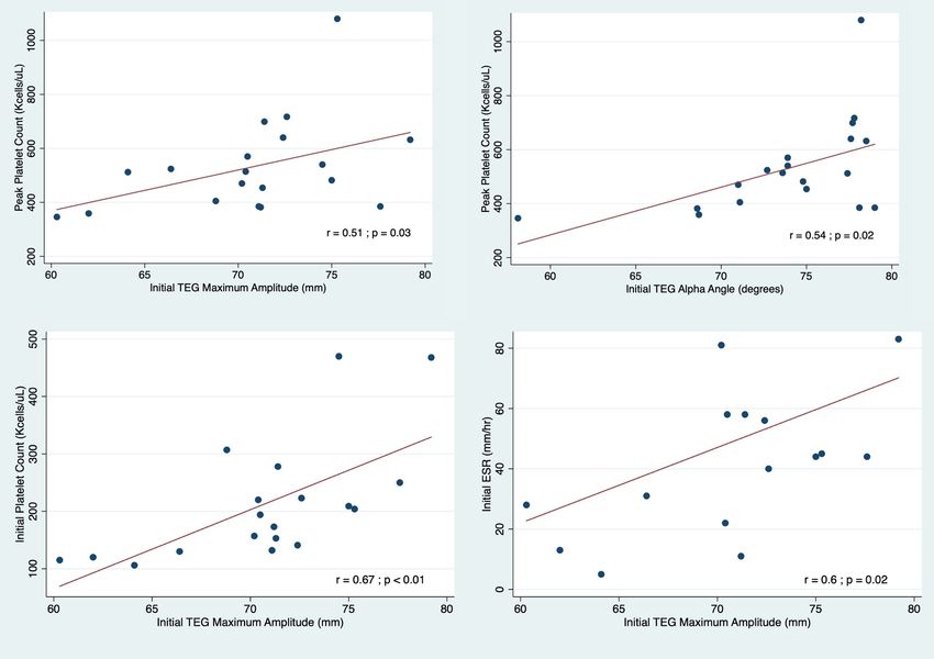
TEG Profiles: Table 2 contains serial TEG data over the course of MIS-C illness with patients’ corresponding
thromboprophylaxis regimens. The initial TEG profile was obtained on day 4 (IQR 3 – 8) of illness in 19
(63%) patients. At time of presentation, patients with MIS-C had abnormal TEG profiles compared with age-
and gender- matched controls (Figure 1), including decreased K time (1.1 vs. 1.7 min,Pis described for all 30 patients with MIS-C, not solely those who had TEG performed. Twenty-two (73%)
Posted on Authorea 8 May 2021 — The copyright holder is the author/funder. All rights reserved. No reuse without permission. — https://doi.org/10.22541/au.162049169.98519152/v1 — This a preprint and has not been peer reviewed. Data may be preliminary.
patients received aspirin (ASA), 7 (23%) received prophylactic LMWH, 11 (37%) received treatment-dose
LMWH, 2 (8%) received unfractionated heparin, and 12 (40%) patients received apixaban. Seventeen (57%)
patients were on dual therapy at one point during their treatment: 14/17 (82%) were on ASA and LMWH (9
on treatment-dose LMWH, 5 on prophylactic LMWH) and 3/17 (18%) were on ASA and apixaban (2 were
briefly bridged on a heparin drip to apixaban). Only 5 (17%) patients received no thromboprophylaxis. In 4/5
of these cases, the patients had short hospitalizations and normal echocardiograms, and thromboprophylaxis
was not provided based on individual provider preference. One patient had significant thrombocytopenia
precluding thromboprophylaxis.
No thrombotic events were documented in this population during their hospitalization or clinical follow-up
at our institution. Six (20%) patients had a total of 7 bleeding events, of which 2 (7%) were clinically relevant
non-major (including bloody stools, epistaxis, or gum bleeding requiring further evaluation or intervention)
and 5 (17%) were minor (including epistaxis, gum bleeding, or bleeding from LMWH sites or sites of trauma).
All of the bleeding events occurred when the patient was on some form of anticoagulation or antiplatelet
agent: 6/7 events were on ASA, 3/7 were on apixaban, and 2/7 were on LMWH (1 treatment-dose, 1
prophylactic). 4/7 events happened while on dual therapy, and 2/4 of these events resulted in discontinuation
of the anticoagulant (apixaban) with continuation of ASA. 2/7 events happened while just on ASA, which
was stopped as a result. Only one bleeding event occurred on prophylactic LMWH in a patient with bloody
stools and a diagnosis of inflammatory bowel disease, and was therefore unlikely to be exclusively attributed
to thromboprophylaxis.
Discussion
This study describes viscoelastic testing profiles in children with MIS-C and demonstrates the presence of
a pro-thrombotic state characterized by increased clot formation rate and strength, and slower fibrinolysis.
Increased ESR and platelet count were both associated with higher clot strength. The majority of patients
received treatment with ASA or anticoagulation, which was well tolerated with 20% incidence of minor or
clinically relevant non-major bleeding events and with no subsequent development of thrombosis.
Our findings build on prior reports in patients with MIS-C that demonstrated laboratory evidence of si-
gnificant inflammation and coagulopathy.6–10 As in our series, children with MIS-C have been reported to
have elevations in multiple biomarkers of inflammation, including ESR, CRP, and ferritin.6–8 Prior work has
demonstrated that baseline platelet count is significantly lower in patients with MIS-C compared to Ka-
wasaki disease.8 Although the MIS-C patients in our cohort had thrombocytopenia early in illness, peak
platelet counts later in illness were just above the normal range, consistent with prior literature showing
a trend towards higher platelet counts upon recovery from MIS-C.9 Mild prolongation of PT and aPTT
are rarely reported in the MIS-C literature,7 but are much more prevalent in adult patients with COVID-19
infection12,27 While prolonged PT and aPTT standardly denote increased bleeding risk, in critically ill adults
with COVID-19 pneumonia, they have been shown to be associated with increased incidence of thrombo-
tic complications.16 In the MIS-C population, elevations in fibrinogen and D-dimer have been extensively
described;6–10 however, it is unclear whether these laboratory changes reflect an underlying inflammatory
state or are indicative of increased risk for thrombosis.10,28
Our finding that patients with MIS-C have TEG profiles consistent with faster clot formation, increased
clot strength and slower fibrinolysis is novel and provides a potential unique biomarker to distinguish risk
for thrombosis in pediatric patients. The MIS-C TEG profiles described in our study are consistent with
multiple prior TEG studies in adult COVID-19 patients, which also demonstrate varying degrees of decreased
K time, higher alpha angle, higher maximum amplitude, and low Ly30.12–14 One study in pediatric patients
with acute SARS-CoV-2 infection (not meeting criteria for MIS-C) found evidence of increased maximal
clot firmness on rotational thromboelastometry, another form of viscoelastic testing.29 In adults, abnormal
TEG profiles predict risk for thrombosis in COVID-19. Indeed, Wright et al showed that patients with a
complete lack of clot lysis at 30 minutes and elevated D dimer had a venous thromboembolism rate of 50%
compared to patients with neither risk factor.13 In adult patients with COVID-19 infection, TEG with platelet
4mapping, used according to an algorithm to guide anti-thrombotic therapy, can reduce mortality risk.30 Our
Posted on Authorea 8 May 2021 — The copyright holder is the author/funder. All rights reserved. No reuse without permission. — https://doi.org/10.22541/au.162049169.98519152/v1 — This a preprint and has not been peer reviewed. Data may be preliminary.
study was not sufficiently powered to detect thromboembolism, particularly as rates of thromboembolism
in the pediatric MIS-C population have been reported at 3-6.5%,6,10,11 far lower than in adults with severe
COVID-19.15–17 Future studies should explore the role of abnormal TEG profiles in patients with MIS-C as
a surrogate for potential thrombosis risk to help guide thromboprophylaxis management.
Proposed mechanisms of thrombosis in adult patients with acute COVID-19 infection include all three com-
ponents of Virchow’s triad.12,31,32 Rates of thromboembolism may be lower in pediatric patients with MIS-C
due to more robust anti-coagulant pathways associated with developmental hemostasis and baseline healthier
endothelial linings.33 Our finding that increased platelet counts (both initial and peak) are associated with
increased maximal amplitude is consistent with the fact that clot strength is primarily derived from interacti-
ons between fibrin and platelets.34 Adult COVID-19 literature supports a potential role for increased platelet
count and activation in mediating thrombosis risk, although further research is needed in children.35–37 The
association between degree of ESR elevation and increased clot strength in patients with MIS-C supports
a role for inflammation as a potential driver of thromboembolism risk. In adult patients with COVID-19;
increased ESR, increased CRP, and increased D-dimer were all predictive of thromboembolic complications.38
Although guidelines for management of thromboprophylaxis in patients with MIS-C have emerged, the
recommendations are primarily based on evidence in analogous conditions such as Kawasaki disease and
adult COVID-19 infection.39–41 Low-dose ASA, which was administered to the majority of patients with
MIS-C in our study, could be considered in all patients with MIS-C requiring hospitalization, particularly
those with features of Kawasaki disease, coronary artery aneurysm, and thrombocytosis.39,40 More than
half of the patients with MIS-C in our study were on anticoagulation prophylaxis or treatment, with 57%
on dual platelet and anticoagulation therapy. Current guidelines suggest that prophylactic anticoagulation
(as well as mechanical thromboprophylaxis) be considered in patients with higher baseline risk for venous
thromboembolism (such as patients> 12 years old, with altered mobility, obesity, known thrombophilia or
history of thrombus, or critical presentation).40,41 Some guidelines suggest using D-dimer to risk stratify
pediatric patients hospitalized for COVID-19 in order to guide initiation of prophylactic anticoagulation,41
although this is controversial due to the non-specific nature of an elevated D-dimer.28,40 Our institution’s
algorithm utilizes D-dimer and additionally incorporates TEG clot strength to guide thromboprophylaxis,
as described in Supplemental Figure S1. Per current guidelines recommended by the American College of
Rheumatology and International Kawasaki Disease Registry, therapeutic anticoagulation should be initiated
in patients with MIS-C with giant coronary artery aneurysm (Z-score >10), moderate or severe ventricular
dysfunction, or concern for thrombosis.39,40 The lack of major bleeding complications in our cohort suggests
that antiplatelet and anticoagulation therapies in the MIS-C patient population are relatively safe.
Limitations of our study include the single center retrospective design, which limits potential generalizability.
The small sample size of patients with MIS-C was underpowered to detect the outcome of thrombosis. Due
to variations in clinical practice and management of these patients, there were incomplete data particularly
with regard to TEG profiles, and there may have been selection bias regarding which patients had TEG
profiles obtained. Moreover, we were unable to draw conclusions about the causal relationship between
anticoagulation/antiplatelet regimens and outcomes in our cohort due to our retrospective design, small
sample size and variable clinical practice. There was no formal control group for our study. TEG data are
not routinely obtained in other hospitalized patient cohorts with similar degrees of inflammation; however,
they are often obtained prior to cardiac surgery, the patient group from which we derived matched controls.
In conclusion, patients with MIS-C have evidence of hypercoagulability on TEG, particularly in patients
with elevated ESR and platelet counts. This study supports the safety and utility of antiplatelet and anti-
coagulation medication in the management of patients with MIS-C. Further longitudinal multi-center study
is required to determine the impact of these therapies on the rate of thrombosis and outcomes in patients
with MIS-C.
Conflict of Interest Disclosure: The authors have no conflicts of interest relevant to this article to disclose.
5Funding/Support : Dr. VanderPluym received funding from the Georgia Claire Bowen (GCB) Foundation
Posted on Authorea 8 May 2021 — The copyright holder is the author/funder. All rights reserved. No reuse without permission. — https://doi.org/10.22541/au.162049169.98519152/v1 — This a preprint and has not been peer reviewed. Data may be preliminary.
IMPACT (Imagine More Possibilities for Advanced Cardiac Therapies) innovation lab.
Role of Funder/Sponsor: The funder/sponsor did not participate in the work.
References
1. Health Department-Reported Cases of Multisystem Inflammatory Syndrome in Children (MIS-C) in the
United States | CDC. Accessed March 1, 2021. https://www.cdc.gov/mis-c/cases/index.html
2. Belhadjer Z, Meot M, Bajolle F, et al. Acute heart failure in multisystem inflammatory syndrome in
children (MIS-C) in the context of global SARS-CoV-2 pandemic. Circulation . Published online 2020.
doi:10.1161/circulationaha.120.048360
3. Verdoni L, Mazza A, Gervasoni A, et al. An outbreak of severe Kawasaki-like disease at the Italian
epicentre of the SARS-CoV-2 epidemic: an observational cohort study. Lancet . Published online 2020.
doi:10.1016/S0140-6736(20)31103-X
4. Riphagen S, Gomez X, Gonzalez-Martinez C, Wilkinson N, Theocharis P. Hyperinflammatory
shock in children during COVID-19 pandemic.Lancet . 2020;395(10237):1607-1608. doi:10.1016/S0140-
6736(20)31094-1
5. Toubiana J, Poirault C, Corsia A, et al. Kawasaki-like multisystem inflammatory syndrome in children
during the covid-19 pandemic in Paris, France: prospective observational study. BMJ . Published online
2020. doi:10.1136/bmj.m2094
6. Feldstein LR, Rose EB, Horwitz SM, et al. Multisystem Inflammatory Syndrome in U.S. Children and
Adolescents. N Engl J Med . Published online 2020. doi:10.1056/nejmoa2021680
7. Lee PY, Day-Lewis M, Henderson LA, et al. Distinct clinical and immunological features of SARS–
CoV-2–induced multisystem inflammatory syndrome in children. J Clin Invest . Published online 2020.
doi:10.1172/JCI141113
8. Whittaker E, Bamford A, Kenny J, et al. Clinical Characteristics of 58 Children With a Pediatric
Inflammatory Multisystem Syndrome Temporally Associated With SARS-CoV-2. JAMA - J Am Med Assoc
. Published online 2020. doi:10.1001/jama.2020.10369
9. Del Borrello G, Giraudo I, Bondone C, et al. SARS-COV-2-associated coagulopathy and thromboem-
bolism prophylaxis in children: A single-center observational study. J Thromb Haemost . Published online
2020. doi:10.1111/jth.15216
10. Aronoff SC, Hall A, Del Vecchio MT. The Natural History of SARS-Cov-2 Related Multisystem Inflam-
matory Syndrome in Children (MIS-C): A Systematic Review. J Pediatr Infect Dis Soc . Published online
2020. doi:10.1093/jpids/piaa112
11. Whitworth HB, Sartain SE, Kumar R, et al. Rate of thrombosis in children and adolescents hospitalized
with COVID-19 or MIS-C.Blood . Published online 2021. doi:10.1182/blood.2020010218
12. Panigada M, Bottino N, Tagliabue P, et al. Hypercoagulability of COVID-19 patients in Intensive Care
Unit. A Report of Thromboelastography Findings and other Parameters of Hemostasis. J Thromb Haemost
. 2020;(April):1738-1742. doi:10.1111/jth.14850
13. Wright FL, Vogler TO, Moore EE, et al. Fibrinolysis Shutdown Correlation with
Thromboembolic Events in Severe COVID-19 Infection.J Am Coll Surg . 2020;231(2):193-203.e1.
doi:10.1016/j.jamcollsurg.2020.05.007
14. Salem N, Atallah B, El Nekidy WS, Sadik ZG, Park WM, Mallat J. Thromboelastography findings in
critically ill COVID-19 patients.J Thromb Thrombolysis . Published online 2020. doi:10.1007/s11239-020-
02300-7
615. Bilaloglu S, Aphinyanaphongs Y, Jones S, Iturrate E, Hochman J, Berger JS. Thrombosis in Hospitalized
Posted on Authorea 8 May 2021 — The copyright holder is the author/funder. All rights reserved. No reuse without permission. — https://doi.org/10.22541/au.162049169.98519152/v1 — This a preprint and has not been peer reviewed. Data may be preliminary.
Patients with COVID-19 in a New York City Health System. JAMA - J Am Med Assoc . Published online
2020. doi:10.1001/jama.2020.13372
16. Klok FA, Kruip MJHA, van der Meer NJM, et al. Incidence of thrombotic complications in critically ill
ICU patients with COVID-19.Thromb Res . Published online 2020. doi:10.1016/j.thromres.2020.04.013
17. Helms J, Tacquard C, Severac F, et al. High risk of thrombosis in patients with severe SARS-
CoV-2 infection: a multicenter prospective cohort study. Intensive Care Med . Published online 2020.
doi:10.1007/s00134-020-06062-x
18. Hartmann J, Mason D, Achneck H. Thromboelastography (TEG) Point-of-Care Diagnostic for Hemosta-
sis Management. Point Care . Published online 2018. doi:10.1097/POC.0000000000000156
19. Harahsheh Y, Duff OC, Ho KM. Thromboelastography predicts thromboembolism in critically ill coag-
ulopathic patients. Crit Care Med . Published online 2019. doi:10.1097/CCM.0000000000003730
20. Russell RT, Maizlin II, Vogel AM. Viscoelastic monitoring in pediatric trauma: a survey of pediatric
trauma society members. J Surg Res . Published online 2017. doi:10.1016/j.jss.2017.03.016
21. Emani S, Sleeper LA, Faraoni D, et al. Thromboelastography Is Associated With Surro-
gates for Bleeding After Pediatric Cardiac Operations. Ann Thorac Surg . Published online 2018.
doi:10.1016/j.athoracsur.2018.04.023
22. Gong C, Yu K, Zhang N, Huang J. Predictive value of thromboelastography for postoperative lower
extremity deep venous thrombosis in gastric cancer complicated with portal hypertension patients. Clin Exp
Hypertens . Published online 2020. doi:10.1080/10641963.2020.1836194
23. Information for Healthcare Providers about Multisystem Inflammatory Syndrome in Children (MIS-C)
| CDC. Accessed March 1, 2021. https://www.cdc.gov/mis-c/hcp/
24. Schulman S, Kearon C, Subcommittee on Control of Anticoagulation of the Scientific and Standardization
Committee of the International Society on Thrombosis and Haemostasis. Definition of major bleeding in
clinical investigations of antihemostatic medicinal products in non-surgical patients. J Thromb Haemost .
2005;3(4):692-694. doi:10.1111/j.1538-7836.2005.01204.x
25. Kaatz S, Ahmad D, Spyropoulos AC, Schulman S. Definition of clinically relevant non-major bleed-
ing in studies of anticoagulants in atrial fibrillation and venous thromboembolic disease in non-surgical
patients: Communication from the SSC of the ISTH. J Thromb Haemost . Published online 2015.
doi:10.1111/jth.13140
26. McCrindle BW, Rowley AH, Newburger JW, et al. Diagnosis, treatment, and long-term manage-
ment of Kawasaki disease: A scientific statement for health professionals from the American Heart Associa-
tion.Circulation . Published online 2017. doi:10.1161/CIR.0000000000000484
27. Tang N, Li D, Wang X, Sun Z. Abnormal coagulation parameters are associated with poor prognosis in pa-
tients with novel coronavirus pneumonia. J Thromb Haemost . Published online 2020. doi:10.1111/jth.14768
28. Biss TT, Brandao LR, Kahr WHA, Chan AKC, Williams S. Clinical probability score and D-dimer
estimation lack utility in the diagnosis of childhood pulmonary embolism. J Thromb Haemost . Published
online 2009. doi:10.1111/j.1538-7836.2009.03572.x
29. Al-Ghafry M, Aygun B, Appiah-Kubi A, et al. Are children with SARS-CoV-2 infection at high risk for
thrombosis? Viscoelastic testing and coagulation profiles in a case series of pediatric patients.Pediatr Blood
Cancer . Published online 2020. doi:10.1002/pbc.28737
30. Hranjec T, Estreicher M, Rogers B, et al. Integral Use of Thromboelastography With
Platelet Mapping to Guide Appropriate Treatment, Avoid Complications, and Improve Survival of Pa-
7tients With Coronavirus Disease 2019–Related Coagulopathy. Crit Care Explor . 2020;2(12):e0287.
Posted on Authorea 8 May 2021 — The copyright holder is the author/funder. All rights reserved. No reuse without permission. — https://doi.org/10.22541/au.162049169.98519152/v1 — This a preprint and has not been peer reviewed. Data may be preliminary.
doi:10.1097/cce.0000000000000287
31. The Lancet Haematology. COVID-19 coagulopathy: an evolving story.Lancet Haematol . Published
online 2020. doi:10.1016/S2352-3026(20)30151-4
32. Pons S, Fodil S, Azoulay E, Zafrani L. The vascular endothelium: The cornerstone of organ dysfunction
in severe SARS-CoV-2 infection.Crit Care . Published online 2020. doi:10.1186/s13054-020-03062-7
33. Kuhle S, Male C, Mitchell L. Developmental hemostasis: Pro- and anticoagulant systems during child-
hood. Semin Thromb Hemost . Published online 2003. doi:10.1055/s-2003-42584
34. Lam WA, Chaudhuri O, Crow A, et al. Mechanics and contraction dynamics of single platelets and
implications for clot stiffening.Nat Mater . Published online 2011. doi:10.1038/nmat2903
35. Hottz ED, Azevedo-Quintanilha IG, Palhinha L, et al. Platelet activation and platelet-monocyte aggre-
gate formation trigger tissue factor expression in patients with severe COVID-19. Blood . Published online
2020. doi:10.1182/blood.2020007252
36. Allegra A, Innao V, Allegra AG, Musolino C. Coagulopathy and thromboembolic events in patients with
SARS-CoV-2 infection: pathogenesis and management strategies. Ann Hematol . Published online 2020.
doi:10.1007/s00277-020-04182-4
37. Manne BK, Denorme F, Middleton EA, et al. Platelet gene expression and function in patients with
COVID-19. Blood . Published online 2020. doi:10.1182/blood.2020007214
38. Al-Samkari H, Karp Leaf RS, Dzik WH, et al. COVID-19 and coagulation: Bleeding and thrombotic
manifestations of SARS-CoV-2 infection. Blood . Published online 2020. doi:10.1182/BLOOD.2020006520
39. Henderson LA, Canna SW, Friedman KG, et al. American College of Rheumatology Clinical Guidance
for Multisystem Inflammatory Syndrome in Children Associated With SARS–CoV-2 and Hyperinflammation
in Pediatric COVID-19: Version 1. Arthritis Rheumatol . Published online 2020. doi:10.1002/art.41454
40. Elias MD, McCrindle BW, Larios G, et al. Management of Multisystem Inflammatory Syndrome in
Children Associated With COVID-19: A Survey From the International Kawasaki Disease Registry. CJC
Open . Published online 2020. doi:10.1016/j.cjco.2020.09.004
41. Goldenberg NA, Sochet A, Albisetti M, et al. Consensus-based clinical recommendations and research
priorities for anticoagulant thromboprophylaxis in children hospitalized for COVID-19–related illness. J
Thromb Haemost . Published online 2020. doi:10.1111/jth.15073
FIGURE 1 Comparison of TEG parameters in patients with MIS-C vs. control patients
Data presented as box and whisker blots of median (interquartile range). Statistical significance was deter-
mined via Wilcoxon rank sum test.
FIGURE 2 Sample TEG tracing in a patient with MIS-C vs. a control patient
R = reaction time or latency until clot formation begins, K = kinetic clot time or time until clot reaches
a fixed strength, angle = rate of clot formation, maximum amplitude = maximal clot strength, Ly30 = clot
lysis in 30 minutes after maximum amplitude is reached.
FIGURE 3 Association of TEG parameters with inflammatory markers
Data presented as scatter plots, with statistical significance determined by Pearson or Spearman
correlation.
SUPPLEMENTAL FIGURE S1 MIS-C Antithrombosis Management Guide
8This figure describes the algorithm used by our institution’s Cardiac Antithrombosis Management Program.
Posted on Authorea 8 May 2021 — The copyright holder is the author/funder. All rights reserved. No reuse without permission. — https://doi.org/10.22541/au.162049169.98519152/v1 — This a preprint and has not been peer reviewed. Data may be preliminary.
Additional abbreviations include AC = anticoagulation, ULN = upper limits of normal, UFH = unfraction-
ated heparin, ECG = electrocardiogram, VTE = venous thromboembolism, DOAC = direct oral anticoagulant.
Hosted file
Table 1.pdf available at https://authorea.com/users/412632/articles/521257-coagulation-
profiles-and-viscoelastic-testing-in-multisystem-inflammatory-syndrome
Hosted file
Table 2.pdf available at https://authorea.com/users/412632/articles/521257-coagulation-
profiles-and-viscoelastic-testing-in-multisystem-inflammatory-syndrome
Hosted file
Table 3.pdf available at https://authorea.com/users/412632/articles/521257-coagulation-
profiles-and-viscoelastic-testing-in-multisystem-inflammatory-syndrome
9Posted on Authorea 8 May 2021 — The copyright holder is the author/funder. All rights reserved. No reuse without permission. — https://doi.org/10.22541/au.162049169.98519152/v1 — This a preprint and has not been peer reviewed. Data may be preliminary. 10
Posted on Authorea 8 May 2021 — The copyright holder is the author/funder. All rights reserved. No reuse without permission. — https://doi.org/10.22541/au.162049169.98519152/v1 — This a preprint and has not been peer reviewed. Data may be preliminary.
Patient < 21 years of age admitted with MIS-C
-Therapeutic AC as
Yes clinically indicated
-Consider additional
Confirmed thrombosis OR pre-existing ASA based on labs
(platelet count), echo
anticoagulation for other indication?
and risk of bleeding
No
Platelet count >100,000 and fibrinogen >100 mg/dl;
Start ASA (3-5 mg/kg, max 81 mg daily)
-Moderate to severe
ventricular dysfunction
Normal Echocardiogram -Coronary dilation,
aneurysm (z-score ≥10)
-Intracardiac thrombosis
-Mild ventricular dysfunction
-Mild-moderate coronary
dilation (z-score 2.5-10)
-Therapeutic AC
(UFH or LMWH)
-Re-assess need
for therapeutic
D-dimer D-dimer >10x ULN AC with serial
echo and labs as
clinically
indicated at the
discretion of the
-D-dimer 5-10x ULN or
D-dimer care team
-TEG clot strength >80 orYou can also read