XII. THE PATHOLOGICAL HISTOLOGY OF THE SPLEEN AND LIVER IN SPONTANEOUS RAT-PLAGUE, WITH OBSERVATIONS ON THE EXPERIMENTAL INFECTION. BY J. C. G ...
←
→
Page content transcription
If your browser does not render page correctly, please read the page content below
359

                 XII. THE PATHOLOGICAL HISTOLOGY OF THE SPLEEN
                     AND LIVER IN SPONTANEOUS RAT-PLAGUE, WITH
                     OBSERVATIONS ON THE EXPERIMENTAL INFECTION.

                                   BY J. C. G. LEDINGHAM, M.B., B.Sc, M.A.
                                                            Lister Institute.

                                                   (PLATES VIII AND IX.)

                    The pathological histology of plague in man and experimental plague
                in animals has already been the subject of several researches, notably
                those of Aoyama (1896), Yamagiwa (1897), Albrecht and Gohn (1897),
                Diirck (1904) and Hamdi (1904) in the case of human plague, and of
                van der Stricht (1897), Honl (1897), Lustig and Zardo (1897), Babes
                and Livadite (1897), Albrecht and Gohn (1900) and Sata (1900) in the
                case of experimental plague.
                    The guinea-pig and rat have been mainly employed as experimental
                animals owing to their great susceptibility to infection, but in view of
                the natural occurrence of plague in the latter animal and the important
                part it plays in the propagation of human plague, it is highly essential
                that we should have all details regarding the histology of the natural
                rat infection even though the differences between such and the experi-
                mental form may be presumably insignificant.
                    The pathological histology of natural plague infection in rats has
                been referred to only by Albrecht and Gohn (1900) who gave brief
                histological details of the organs of five rats which had died of plague in
                Bombay. Ogata (1897) also contributed a few observations on the
                subject.
                    It is to fill up this lacuna in our knowledge of spontaneous rat
                plague that the following histological examinations have been made
                from material collected in Bombay by the Plague Commission. The
                primary Cause, however, of this inquiry was the elucidation of the
                peculiar characteristic post-mortem appearances of the spleen and liver,

Downloaded from https://www.cambridge.org/core. IP address: 46.4.80.155, on 22 Dec 2020 at 08:08:03, subject to the Cambridge Core terms of use,
available at https://www.cambridge.org/core/terms. https://doi.org/10.1017/S0022172400033362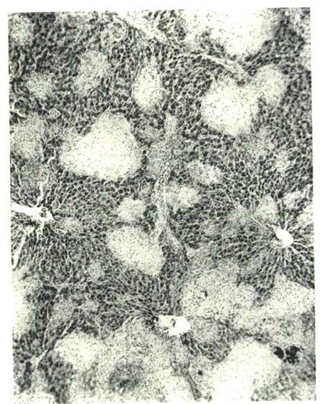
360                              Pathological                Histology
                to which reference has been made in another article in this number.
                For this purpose, therefore, only the spleens and livers from the various
                cases were forwarded.
                    It was felt, moreover, that a more minute investigation of the
                histological changes might throw some light on the conditions leading
                up to the chronic form of rat plague which is associated with the
                presence, in the spleen particularly, of encapsulated abscesses.
                    To add completeness to the work and for purposes of comparison
                the organs of several rats which had been inoculated in Bombay with
                virulent plague, were submitted to detailed examination by similar
                methods. Finally one or two cases of chronic experimental plague in
                vaccinated rats were investigated histologically. The results obtained
                in these experimental cases will be discussed in Part II of this paper.

                                                                 PART I.
                                                    Spontaneous Rat Plague.
                   The very brief histological details recorded by Albrecht and Gohn
                may be here summarised:—
                   Eat I. Spleen :—Numerous haemorrhages were present in the pulp. Necroses
                had also commenced especially at the periphery of the nodes. Bacilli were scarce.
                Liver :—Showed cloudy swelling. Bacilli scarce.
                    Rat II. Spleen:—Pulp was haemorrhagic and infiltrated with polynuclear
                leucocytes. The nodes were surrounded by a fine or coarse network of connective
                tissue. Bacilli were numerous. Liver :—Liver cells showed karyorrhectic nuclei in
                many cases. The capillaries contained enormous masses of bacilli.
                    Rat III. Spleen :—Pulp contained large amounts of nuclear de'bris especially in
                the vicinity of the nodes. Liver :—Showed degeneration of parenchyma. Capil-
                laries were full of bacilli.
                    Rat IV. Spleen :—Pulp presented commencing necroses. Bacilli numerous.
                    Rat V. Spleen :—Numerous haemorrhages and necroses in the pulp. The
                peripheral portions of the nodes and the adjoining pulp-tissue were transformed into
                a coarse network staining intensely with eosin.
                    Rat VI. Spleen :—Showed haemorrhages and nuclear disintegration. Bacilli
                very numerous. Liver:—Liver cells showed fatty degeneration. Bacilli very
                abundant in the capillaries.

                    Ogata (1897), during the epidemic of plague in Formosa, obtained
                six rats which had died of plague. Referring to the condition of the
                organs he merely remarks that the spleen was much swollen and the
                liver congested. Small haemorrhages were also present in the liver and
                bacilli were numerous, as also in the spleen and glands.

Downloaded from https://www.cambridge.org/core. IP address: 46.4.80.155, on 22 Dec 2020 at 08:08:03, subject to the Cambridge Core terms of use,
available at https://www.cambridge.org/core/terms. https://doi.org/10.1017/S0022172400033362
Reports on Plague Investigations in India 361
From the above summary, it will be evident that the grosser lesions
only have been recorded, possibly owing to the difficulty of procuring
material fresh enough for detailed histological examination. The
organs at my disposal were the spleens and livers of thirteen cases of
rat plague. These had been fixed in Orth's fluid and sent to this
country in spirit. The tissues were embedded in paraffin and the stains
chiefly employed were Unna-Pappenheim's methyl-green-pyronin and
Ehrlich's haematoxylin and orange-rubin. The former stain proved
eminently satisfactory for the demonstration of plague bacilli and
plasma cells.
Rat I. Protocol:—Primary bubo—left axillary. Congestion of spleen and
subcutaneous tissues. Bacilli present in bubo, heart blood and spleen.
Spleen :—Capsule: shows no pathological changes.—Nodes: The nodes are few in
number but regular in outline. Karyorrhexis of the lymphoid cells of the node is
a marked feature and a large amount of nuclear detritus is lying either free or
included in large endothelioid phagocytic cells. Some of these particles show their
cytoplasmic origin by taking up the pyronin stain. Large mononuclear cells of endo-
thelioid type are abundant throughout the node, many of them presenting mitotic
figures (see Plate VIII, Fig. 2). The protoplasm of these cells stains a deep red,
the nucleus being rather vesicular. No bacilli were detected within the node though
a specially rich zone of them was present in the perinodal lymph sinus.—Pulp :
Extravasation of red cells was very marked and plague bacilli were fairly uniformly
distributed and in large numbers. No necrotic foci were present. Rows of plasma
cells, many showing mitosis, were arranged in the sheaths of the trabecular vessels.
Liver :—The protoplasm of the liver cells was coarsely vacuolated. Necrotic foci,
which are the outstanding feature of plague livers, were in this case few in number
and of very small size, in some cases only two or three liver cells being involved.
Within these necrotic areas were a few vesicular nuclei and occasionally a small
group of bacilli lying in what remained of the intraacinar capillaries. Enormous
numbers of bacilli were present in the liver capillaries. Many of the liver cell-nuclei
showed karyorrhexis.
Rat II. Protocol:—Primary bubo—left submaxillary. Spleen, lungs, and sub-
cutaneous tissues congested. The surface of the liver had a mottled appearance.
Bacilli in spleen and bubo.
Spleen:—Subcapsular haemorrhage was considerable. — Nodes: These were
greatly diminished in number. Large mononuclear endothelioid cells were numerous,
the small lymphoid cells showing extreme karyorrhexis. In some nodes were small
necrotic foci containing large endothelioid cells and nuclear detritus, similar to those
described in the diphtheritic spleen (see Waschkewitsch Virch. Archiv, Bd. 159,
1900).—Pulp : There was great congestion of the pulp sinuses accompanied with
red cell extravasation. Around the trabecular vessels were many plasma cells
showing extreme karyorrhexis and pyknosis (see Plate VIII, Fig. 5). The cells of
the spleen pulp presented as a whole only a slight degree of karyorrhexis and no
actual necrotic foci were observed. Bacilli occurred in swarms throughout the pulp
especially in the areas of blood extravasation. Liver :—The protoplasm of the
Downloaded from https://www.cambridge.org/core. IP address: 46.4.80.155, on 22 Dec 2020 at 08:08:03, subject to the Cambridge Core terms of use,
available at https://www.cambridge.org/core/terms. https://doi.org/10.1017/S0022172400033362362 Pathological Histology
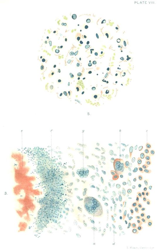
EXPLANATION OF PLATES VIM AND IX.
Plate VIM. Fig. 1. Portion of necrotic focus of liver (natura rat plague). a=Blood in
capillaries. 6 = Remains of liver-acini. c=Liver cell showing vacuolar honeycomb
degeneration. d=Mononuclear cell in capillary.
Fig. 2. Portion of malpighian body (natural rat plague) showing large mononuclear
endothelioid cells, a = Mitosis of the same. A degenerated endothelial cell contain-
ing nuclear detritus is also seen.
Fig. 3. Portion of spleen in chronic experimental rat plague, a=Giant-cell (of tuber-
cular type). g = Commencing agglomeration of nuclei to form giant-cell. 6 = Mega-
karyocyte. d=Granulation spindle cell. c=Bows of plasma cells. / = Leucocytic
debris round bacilli, e = Degenerated bacilli.
Fig. 4. Portion of liver (natural rat plague), a = Megakaryocyte in capillary. 6 = Liver-
acinus.
Fig. 5. Portion of spleen-pulp (natural rat plague) showing bacilli and pyknotic plasma
cells of all types, and effused red cells.
Fig. 6. Giant-cell (of Langhans' type) from periphery of malpighian body (experimental
rat-plague). The drawings are made from five successive sections through the same
cell. Note the grape-like nuclear agglomeration in the 4th drawing.
Plate IX. Photomicrograph of liver necroses (natural rat-plague).
Downloaded from https://www.cambridge.org/core. IP address: 46.4.80.155, on 22 Dec 2020 at 08:08:03, subject to the Cambridge Core terms of use,
available at https://www.cambridge.org/core/terms. https://doi.org/10.1017/S0022172400033362JOURNAL OF. HYGIENE,VOL.VII, PLAGUE NUMBER.
©
O
e
6.
^p
©
a..
Downloaded from https://www.cambridge.org/core. IP address: 46.4.80.155, on 22 Dec 2020 at 08:08:03, subject to the Cambridge Core terms of use,
available at https://www.cambridge.org/core/terms. https://doi.org/10.1017/S0022172400033362PLATE VIII.
c •••
• • - * •
•o- I
5.
3.
O
E.Wilson, Cambridge.
Downloaded from https://www.cambridge.org/core. IP address: 46.4.80.155, on 22 Dec 2020 at 08:08:03, subject to the Cambridge Core terms of use,
available at https://www.cambridge.org/core/terms. https://doi.org/10.1017/S0022172400033362JOURNAL OF HYGIENE (PLAGUE NO.), VOL. VII. NO. 3 PLATE IX Downloaded from https://www.cambridge.org/core. IP address: 46.4.80.155, on 22 Dec 2020 at 08:08:03, subject to the Cambridge Core terms of use, available at https://www.cambridge.org/core/terms. https://doi.org/10.1017/S0022172400033362
Reports on Plague Investigations in India                                                363
                 liver cells was finely vacuolated but not fatty. Acinar necroses were few in number
                 and of very irregular contour. In the capillaries of the necrotic area, bacilli were
                 still present. The portal vessels and intraacinar capillaries were extremely con-
                 gested and contained large numbers of bacilli.
                      Rat III. Protocol:—Primary bubo—right submaxillary. The liver had a
                 granular appearance and presented minute haemorrhages on the serosa. Bacilli
                 present in heart blood, spleen and bubo.
                      Spleen:—The capsule presented no marked changes.—Nodes: These were few
                 and of irregular shape and size and showed the characteristic endothelioid prolifera-
                 tion with numerous mitoses. At the periphery of the node plasma cells were
                 abundant. A marked feature was the presence of a reticular zone encircling each
                 node. The few pulp cells remaining in this zone showed extreme karyolysis and
                 bacilli were specially abundant.—Pulp : The pulp showed great congestion with
                 haemorrhages and polynuclear infiltration. No definite necrotic foci were seen
                 although bacilli occurred in swarms. Plasma cells were numerous in the sheatha
                 of the vessels and there was a very pronounced catarrh of the vascular endothelial
                  cells.    Liver :—Focal liver cell necroses were fairly numerous especially in the
                 subcapsular area. They were apparently of recent origin and contained many
                 extravasated red blood corpuscles with bacilli still lying in the capillaries. The
                 liver cells showed fine vacuolation of their cytoplasm. Bacilli were not quite so
                 numerous in the vascular system of the liver as in the previous cases.
                      Rat IV. Protocol:—Primary bubo—right submaxillary. The serosa of spleen
                 and liver had a granular appearance. Bacilli few in heart blood, spleen and bubo.
                      Spleen :—The capsule was markedly thickened and oedematous, the subcapsular
                 lymph space showing extreme endothelial catarrh.—Nodes: The nodes were
                 greatly diminished in number and in some cases contained rounded areas with
                 vacuolated endothelial cells and nuclear de'bris. Many lymphoid cells had under-
                 gone karyorrhexis.—Pulp : Even with low magnification large clumps of bacilli
                 could be seen throughout the pulp, each surrounded by a zone of karyorrhectic
                 nuclei. The condition was thus one of multiple small abscess formation. Around
                 the nodes and vessel-sheaths plasma cells and vesicular epithelioid cells abounded.
                 The karyorrhectic focus was immediately surrounded by a zone of vesicular epithe-
                 lioid nuclei and this again by a barrier of plasma cells. These epithelioid cells showed
                 a slight tendency to nuclear grouping but no fully formed giant-cells of tubercular
                 type were met with.         Liver :—Liver cell necroses were present in enormous
                 numbers and in fact little healthy liver tissue was left (see Plate VIII, Fig. 1 and
                 Plate IX). Some foci contained only red blood corpuscles, with no traces of bacilli
                 remaining, while others contained a central clump of bacilli surrounded by karyor-
                 rhectic nuclei or proliferating epithelioid cells. The production of some necroses by
                 vascular bacillary emboli was quite patent though in others such a connexion could
                 not be traced. Among the vesicular nuclei surrounding one focus a fully formed
                 giant-cell of tubercular type was detected. In the healthy liver capillaries, bacilli
                 were scarce and evidently degenerated, their remains being taken up by the endo-
                 thelial cells of the vessel wall. A very interesting feature was the presence in the
                 dilated capillaries of great numbers of epithelioid cells some of which were under-
                 going mitosis. An occasional giant-cell of the splenic megakaryocyte type was
                 detected in the capillary lumen. Round the portal vessels in Glisson's capsule
                 small lymphomata were sometimes met with.

Downloaded from https://www.cambridge.org/core. IP address: 46.4.80.155, on 22 Dec 2020 at 08:08:03, subject to the Cambridge Core terms of use,
available at https://www.cambridge.org/core/terms. https://doi.org/10.1017/S0022172400033362
364 Pathological Histology
Rat V. Protocol:—Congestion of subcutaneous tissue, lungs and spleen.—Liver
fatty and granular. No bacilli in heart blood, spleen or liver.
Spleen:—The capsule is very oedematous.—Nodes: There is no diminution in the
number of malpighian bodies, each containing many endothelioid cells with plasma
cells at the periphery. Among the latter, very many mitotic forms were observed.
A perinodal zone of reticular tissue with few nuclei was a constant feature, forming
a species of capsule for the node.—Pulp : The pulp was extremely congested but
only a few bacilli were detected in small groups lying among the extravasated red
blood corpuscles. Many necrotic foci were present. These at times consisted of
karyorrhectic nuclei and detritus or of vesicular epithelioid cells. The plasma cell
reaction was very conspicuous. Pigment cells containing haemosiderin and catarrhal
endothelial cells occurred in fairly large numbers. Liver:—Very numerous sharply
demarcated necroses were pi esent in all stages of development. In some of them,
red blood corpuscles were the predominant elements, while in others the endothelial
cells of the capillaries showed a marked proliferation. A few bacilli could still be
detected in these foci. Elsewhere, the capillaries of the liver contained here and
there small bunches of bacilli which appeared to have undergone degenerative
changes. The phagocytic endothelial cells frequently contained such bacillary
debris.
Rat VI. Protocol :—Double pelvic bubo containing swarms of bacilli. No
bacilli in heart blood or spleen.—Liver coarsely granular and spleen congested.
Spleen :—The capsule was oedematous and the subcapsular area very much
congested, the endothelial cells actively proliferating.—Nodes : These were much
diminished in number but presented only slight pathological changes.—Pulp : The
pulp was greatly congested. There were no definite areas showing karyorrhexis of
the pulp cells but here and there could be seen foci in which the nuclei had
disappeared or had become vesicular. Pyknotic plasma cells were present in
moderate numbers. No bacilli could be demonstrated. Liver :—The liver cells
looked fairly healthy. Focal necroses were moderate in number, a few remains of
bacilli being still apparent in them. The portal vessels were extremely congested.
Free bacilli were never seen in the capillaries but the endothelial cells contained
here and there what appeared to be bacillary remains.
Rat VII. Protocol:—Primary bubo unknown. Liver fatty and granular.
—Spleen granular. Many bacilli in spleen.
Spleen :—The capsule was oedematous and infiltrated with red cells, the super-
ficial endothelium being catarrhal.—Nodes : These showed no important changes.
No bacilli were detected in them.—Pulp : Large irregular necrotic areas were
present showing no very definite boundary and frequently lying alongside a
malpighian body. At the margins of these areas or inside them were wisps of bacilli.
In one case, a vein leading to such a focus was completely blocked by plague bacilli
for a considerable distance. Extraordinary numbers of bacilli were present in the
pulp among the extravasated red cells and polynuclear infiltrates. Karyorrhectic and
pyknotic plasma cells and catarrhal endothelial cells were specially numerous.
Liver :—The liver cells throughout the lobule showed large protoplasmic vacuoles
due to fat. Focal liver cell necroses were very numerous and sharply demarcated
from the healthy liver substance. Some foci contained very few nuclei, while others
contained numerous vesicular nuclei tending at times to coalesce. The contents of
Downloaded from https://www.cambridge.org/core. IP address: 46.4.80.155, on 22 Dec 2020 at 08:08:03, subject to the Cambridge Core terms of use,
available at https://www.cambridge.org/core/terms. https://doi.org/10.1017/S0022172400033362Reports on Plague Investigations in India 365
the capillaries were polynuclear cells, red cells and occasional degenerated bacilli.
A few small lymphoid nodes were observed around the portal vessels in the large
trabeculae. Large mononuclear endothelioid cells showing mitoses could be seen in
these small lymphomata as in the splenic nodes.
Rat VIII. Protocol:—Primary bubo—left axillary. Liver fatty and granular.
— Spleen faintly granular.—No bacilli in heart blood or spleen.
Spleen:—The chief feature was the presence of numerous pulp necroses in which
only a few vesicular nuclei remained. These areas were invariably perinodal. Outside
these areas the pulp cells showed slight karyorrhexis. Pyknotic plasma cells occurred
in enormous numbers. Bacilli were exceedingly scarce and confined to the necrotic
foci. Liver :—Cell necroses were very numerous and contained nuclear debris and
vesicular epithelioid cells. Sometimes a few degenerated bacilli could be seen in
them. Elsewhere, the liver capillaries contained numerous degenerated endothelial
cells and karyorrhectic nuclei. Bacilli were very scarce and only in phagocytic endo-
thelial cells. There was marked congestion throughout.
Rat IX. Protocol :—Primary bubo—left submaxillary. Liver fatty and granu-
lar.—Spleen granular.—No bacilli in heart blood, spleen or bubo.
Spleen:—In this case the areas of pulp degeneration were very numerous and
extensive. The malpighian bodies were encroached upon and very little normal
splenic tissue remained. In the degenerated areas were enormous masses of nuclear
detritus with occasional degenerated bacillary clumps in the centre. Bounding these
areas were rows of plasma cells in active division and catarrhal endothelial cells.
Signs were already present of the replacement of these foci by spindle-shaped granu-
lation cells formed from the actively dividing plasma cells. Liver :—Necroses were
few but of all sizes. Some bacilli still remained in the capillaries of the necrotic area
along with red cells. Throughout the liver substance, the capillaries were greatly
dilated with red cells, desquamated endothelial cells containing nuclear detritus
and large mononuclear cells suggesting a splenic origin. Typical giant cells of mega-
karyocyte type were also met with not infrequently lying in the capillary lumen (see
Plate VIII, Fig. 4). It seems most probable that these cells, along with many of
the types filling up the liver capillaries, have found their way to this organ from
the spleen. Babes and Livadite (1897) noted the presence of similar giant cells in
the guinea-pig's liver (in experimental plague), but apparently assigned to them a
different origin. They write " Sehr bemerkenswerth ist das Auftreten von Riesen-
zellen mit gelappten Kern wohl auf Kosten gewisser Endothelien." Only a very few
bacilli were present in the capillaries.
Rat X. Protocol:—No primary bubo. Liver fatty and granular.—Spleen
congested.—No bacilli in heart blood, spleen or liver.
Spleen :—Not available for histological examination. Liver :—Liver cell
necroses were few and of small size. The subcapsular region was mainly the site of
these foci and there also bacilli were most frequent. The capillaries of the necrotic
area were blocked by bacilli in many cases. Throughout the liver substance the
capillaries were dilated with red cells and splenic elements as in the previous case.
Megakaryocytes were also occasionally detected. Bacilli as a rule were scarce
and many were included in the endothelial phagocytes.
Rat XI. Protocol:—Primary bubo—left axillary. Liver and spleen congested.
Many bacilli in spleen and bubo.
Downloaded from https://www.cambridge.org/core. IP address: 46.4.80.155, on 22 Dec 2020 at 08:08:03, subject to the Cambridge Core terms of use,
available at https://www.cambridge.org/core/terms. https://doi.org/10.1017/S0022172400033362366 Pathological Histology
Spleen:—The lymphoid tissue was greatly diminished. Large mononuclear
endothelioid cells in active division were present in the nodes. Necrotic areas
containing vesicular nuclei and bacillary debris were distributed throughout the
pulp. Megakaryocytes, pyknotic plasma cells and catarrhal endothelial cells were
specially abundant. An interesting feature was the presence of a large number of
eosinophile cells. Liver :—Necroses were few but of all sizes. The capillaries
were dilated as in the previous cases with splenic elements. Plasma cells were also
numerous. A slight degree of perivascular lymphoid infiltration was noted in the
portal spaces.
Rat XII. Protocol :—Liver coarsely granular and fatty, spleen finely granular.—
Subcutaneous and pulmonary haemorrhages.—No bacilli in heart blood, few clumps
in spleen.
Spleen :—The nodes were few in number and badly differentiated from the
surrounding pulp.—Pulp : Small irregular necrotic foci were distributed through-
out the pulp. The centre of each focus was occupied by swarms of bacilli and the
periphery by vesicular nuclei, detritus and blood-pigment-carrying cells. The
subcapsular area was infiltrated with red cells and polynuclear leucocytes. Mega-
karyocytes were exceedingly numerous especially in the neighbourhood of the necrotic
foci. Liver :—Fatty infiltration of the liver cell-protoplasm was far advanced.
Many nuclei also exhibited karyorrhexis. Only a few small necrotic foci were noted.
The intraacinar capillaries were greatly dilated, their lumina being filled with small
and large mononuclear cells and endothelial phagocytes.
Rat XIII. Protocol:—Lungs both consolidated. Gray hepatisation of upper
and middle lobes of right lung.—Liver has a mottled appearance.—Spleen congested.
—Swarms of bacilli in heart blood, spleen and bubo.
Spleen:—Malpighian bodies were numerous and of irregular form and size. Many
karyorrhectic nuclei were present in the nodes along with large endothelial cells
containing nuclear detritus.—Pulp : A few clumps of degenerated bacilli were seen
here and there but no necrotic foci were in evidence. The cell types met with in
the pulp were very varied, small and large mononuclear cells, endothelial cells,
megakaryocytes and nucleated red cells. An interesting feature was the great
abundance of coarsely granular eosinophile cells many of which showed mitotic
figures. They occurred in greatest numbers in the subcapsular area. A few
clumps of mast cells were also noted. . Plasma cells were very scarce. In this
case the pneumonia was evidently the main plague-lesion, while the spleen
presented none of the characteristic changes observed in the foregoing cases.
The splenic picture, in fact, was that associated with an actively functioning
haemopoietic organ. Liver :—Unsuitable for histological examination.
Survey of the above cases.
The cases may be divided into two main groups:—
1. Those in which bacteriaemia of the spleen and liver is at a
maximum and has been of recent development.
2. Those in which bacteriaemia is less prominent or is rapidly
disappearing as a result of reactive tissue changes.
Downloaded from https://www.cambridge.org/core. IP address: 46.4.80.155, on 22 Dec 2020 at 08:08:03, subject to the Cambridge Core terms of use,
available at https://www.cambridge.org/core/terms. https://doi.org/10.1017/S0022172400033362Reports on Plague Investigations in India 367
In the first group, the incursion of bacillary swarms into the spleen
and liver has been accompanied by extensive haemorrhages and conges-
tion of the pulp sinuses and liver capillaries. Definite abscess formation
in the spleen has not had time to develop and focal liver cell-necroses
are few in number, the latter being very largely confined to the sub-
capsular region.
In the second group, definite abscess formation in the spleen is far
more frequent and is accompanied by extensive reactive changes on the
part of the plasma cells. The reduction in the amount of lymphoid
tissue is due in great measure to the perinodal distribution of degenerated
pulp areas. A barricade of plasma cells separates the lymphoid tissue
of the node from the necrotic zone. Proliferation of the large mono-
nuclear endothelioid cells of the node is a noteworthy feature. Diirck
(1904) has described a similar change in the splenic nodes in human
plague.
In the liver, focal necroses may be so numerous that little healthy
liver tissue remains. The demonstration of bacilli in the central
capillaries of these foci was made in nearly every case and frequently
actual bacillary embolism was noted. The formation of giant-cells of
Langhans' type in the neighbourhood of necrotic foci is also of great
importance, although in some of the cases these cells had not reached
their full development. It can readily be conceived how, providing the
animal lives long enough, the reaction of the fixed tissue cells may
proceed to complete encapsulation of abscess areas and so bring about
a more or less chronic condition. So far I have had no opportunity of
examining histologically such cases of chronic spontaneous rat plague,
but it is hoped that suitable material of this kind may soon be available.
The chronic experimental case, which will be described later, gives
however a very clear notion of the later stages in this process of abscess-
encapsulation.
Frequent mention has been made in the protocols of a granular and
mottled appearance of the liver. The spleen has also been described as
granular in some cases. In the case of the liver such changes are
readily accounted for by the distribution of the haemorrhages and the
focal necroses together with the fatty changes in the liver cells. It
must be understood, however, that a peculiar, honeycomb-like vacuolar
degeneration of the liver cell protoplasm was far more frequent than
an actual coarse fatty infiltration. The granular appearance of the
spleen is due partly to endothelial catarrh and partly to subcapsular
changes.
Downloaded from https://www.cambridge.org/core. IP address: 46.4.80.155, on 22 Dec 2020 at 08:08:03, subject to the Cambridge Core terms of use,
available at https://www.cambridge.org/core/terms. https://doi.org/10.1017/S0022172400033362368 Pathological Histology
With regard to the disappearance of bacilli from the intraacinar
capillaries of the liver it appears that phagocytosis by endothelial cells
is largely responsible. Indeed in some cases no free bacilli were
demonstrable. The presence of large giant-cells of megakaryocyte type
in the liver capillaries of some cases is highly interesting: in such
cases the capillaries generally contained so many extraneous cell
elements that one is forced to assign to them a splenic origin.
Finally, with regard to the distribution of plague bacilli in the
spleen, it was exceedingly rare to find the organisms in the interior of
the nodes. Yet, though bacilli were absent, karyorrhexis of the nodal
cells was frequently far advanced. Toxaemia must then be largely
responsible for the alterations in the nodes, as we know that analogous
changes take place in the malpighian bodies after the inoculation, for
instance, of diphtheria toxin.
PART I I .
Experimental Rat Plague.
The following is a brief resume of our knowledge regarding the
histology of the spleen and liver in experimental plague.
Spleen. In the guinea-pig van der Stricht (1897) noted a diminution
in the size of the malpighian bodies with dilatation of the capillaries of
the node, leading to actual rupture. Abscesses occurred in the pulp
but did not affect the nodes. The splenic megakaryocytes were very
numerous and the capsule of the organ was infiltrated with white
corpuscles. Honl (1897), working with the same animal, found an
enlargement of the follicles and extreme congestion of the pulp. Bacilli
occurred in " zoogloeal" groups round which were numerous fragmented
cell elements. Lustig and Zardo (1897) working with rats, mice,
guinea-pigs and rabbits noted that the periphery of the follicle was the
seat par excellence of the necrotic foci. The trabeculae had a hyaline
appearance and the arteries were dilated. Haemorrhage into the pulp
was a constant feature. Plague bacilli were numerous throughout the
pulp and might occur inside the nodes. Babes and Livadite (1897)
recorded in guinea-pigs and mice the presence of large numbers of giant-
cells of megakaryocyte type, especially in the vicinity of the necrotic areas.
Blood-corpuscle-containing cells and pigment cells were scarce in spite
of extensive pulp haemorrhages.
Albrecht and Gohn (1900) found a great similarity in the main
Downloaded from https://www.cambridge.org/core. IP address: 46.4.80.155, on 22 Dec 2020 at 08:08:03, subject to the Cambridge Core terms of use,
available at https://www.cambridge.org/core/terms. https://doi.org/10.1017/S0022172400033362Reports on Plague Investigations in India 369
splenic lesions in experimental rat plague by whatever method the inocu-
lation was performed. The nodes were as a rule much diminished and
necrotic pulp-foci were of constant occurrence.
Sata (1900) gives a fairly detailed description of the histology of
experimental rat plague and lays great stress on the variations met with
as regards the distribution of plague bacilli in the organs. Subcapsular
haemorrhage was a frequent feature in the spleen and bacilli might
be very numerous, very scarce or not demonstrable at all in sections.
Liver. Van der Stricht observed areas of fatty degeneration of
the liver cells and also a fine vacuolated condition of the liver cell
protoplasm. In one animal small necrotic areas due to capillary emboli
were noted.
Honl noted focal necroses surrounded by a zone of leucocytes.
Groups of bacilli arranged in zoogloeal masses were present in these foci.
Babes and Livadite recorded the presence in the liver capillaries of
" Riesenzellen mit gelappten Kern" which have been already alluded to.
Fatty degeneration of the liver cells was noted both by Albrecht and
Gohn and Sata. The latter also demonstrated fibrin in the larger
vessels. The occurrence of bacilli in the liver capillaries was found to
be a very variable factor.
The material of the following six cases was obtained from rats inocu-
lated by the cutaneous method in Bombay. For Rat No. VII of chronic
experimental plague I am indebted to Capt. S. R. Douglas.
Rat I. Spleen:—Serous endothelium swollen, capsule oedematous with effu-
sion of red cells into it. The endothelial cells of serosa contained red cell debris.
There was marked subcapsular haemorrhage.—Nodes : The lymphoid tissue was
increased in amount. At the periphery of each node was a circular zone of
necrotic cells with bacillary clumps in the neighbourhood. Near the margin of one
node was a typical Langhans' giant-cell. The appearance of this cell in five serial
sections is indicated in Plate VIII, Fig. 6. In one cross section the constituent
nuclei are seen to fill practically the whole cell leaving only a faint rim of protoplasm.
The pulp was much congested and numerous zoogloeal bacillary masses were noted.
Plasma cells were abundant. The pulp cells showed a slight degree of karyorrhexis.
No bacilli were seen in the nodes. Liver :—The liver cells were generally healthy
apart from the necrotic areas which were numerous. Each focus contained small
heaps of bacilli and a good deal of haemorrhage was present at the periphery.
Endothelial catarrh of the capillaries was a marked feature. Bacilli were distributed
in clumps and many of them were noted inside phagocytic endothelial cells. Fibrin
was also observed in the capillaries, along with numerous red cells.
Rat II. Spleen :—Marked catarrh of the serous endothelium with subcapsular
haemorrhage. The lymphoid tissue was increased and irregularly distributed.
Numerous actively dividing large mononuclear endothelial cells were observed in the
Downloaded from https://www.cambridge.org/core. IP address: 46.4.80.155, on 22 Dec 2020 at 08:08:03, subject to the Cambridge Core terms of use,
available at https://www.cambridge.org/core/terms. https://doi.org/10.1017/S0022172400033362370 Pathological Histology
nodes. A zone of karyorrhectic pulp cells with detritus forms a sort of capsule to
each node. Bacilli occurred in clumps surrounded occasionally by necrotic pulp cells.
Pyknotic plasma cells were very abundant. In the haemorrhagic areas, fibrin was
present in large amount and blood-corpuscle-containing cells were numerous.
Liver:—Liver cells were not fatty. Marked subcapsular haemorrhage. Necroses
were few and small and most of them contained bacillary clumps, surrounded by
karyorrhectic nuclei. Bacilli were very numerous in the capillaries and the endo-
thelial cells frequently contained ingested bacilli.
Rat III. Spleen :—Capsule swollen and subcapsular haemorrhage. The mal-
pighian bodies were swollen but regular in contour. Round each was a zone of
thickened reticulum containing few cells. For the first time, a node was seen
whose central artery contained a fibrin thrombus with bacilli. In the pulp,
enormous numbers of bacilli were present along with red cells and fibrin. Poly-
nuclear cells, pyknotic plasma cells and endothelial cells were abundant. Haemo-
siderin cells also occurred frequently. Liver :—The liver cells were markedly
fatty. Their nuclei also frequently presented karyorrhexis. Necroses were fairly
numerous and often of large size. Capillaries containing bacillary emboli were noted
in these foci. In the capillaries of the healthy liver substance were enormous
numbers of bacilli generally clinging to the walls of the vessels. Round the portal
vessels were lymphoid and plasma cell infiltration.
Rat IV. Spleen :—Large numbers of bacilli surrounded each node but in only
one case were bacilli seen in the follicular artery. The large mononuclear endo-
thelioid cells were abundant in the node and showed signs of active division. Also
small necrotic areas containing large endothelial phagocytes and detritus sometimes
appeared inside the node. The pulp showed extensive haemorrhages with polynuclear
infiltrates and haemosiderin cells. No necrotic foci were present but here and
there were small areas in which the cell-nuclei were vesicular. Liver :—The liver
cells were fatty. Only one or two minute necroses were seen each containing bacilli
in its central capillary with effusion of red cells at the periphery. The capillaries
throughout the liver substance were quite filled with bacilli and many polynuclear
cells were present. Colonies of plasma cells were seen in the sheaths of the large
portal vessels in the Glisson's space.
Rat V. Spleen :—Capsule thickened, trabeculae increased, and marked subcap-
sular haemorrhage. The nodes were greatly diminished and irregular in contour.
They contained numerous large mononuclear cells of endothelioid type, many of
which were dividing. The spleen pulp was greatly congested and bacilli occurred in
swarms especially in the large trabecular sinuses. Polynuclear cells, haemosiderin
pigment cells and blood-corpuscle-containing cells were abundant in the areas of red
cell extravasation. Plasma cells were very numerous and were in active division. No
actual pulp necroses were observed. Liver :—Only a very few minute necrotic foci
were observed in the subcapsular region, involving individual liver cells or a few
adjacent ones. Bacilli were also most numerous in this region, lying in clumps in
the capillary vessels. The intraacinar capillaries throughout the organ were filled
with red cells, infiltrating cells of polynuclear type, and catarrhal endothelial cells.
Rat VI. Spleen :—Marked subcapsular haemorrhage and great reduction in
the amount of lymphoid tissue. Many of the nodes showed a hyaline thrombosis of
the central artery, the lymphoid cells in the neighbourhood being karyorrhectic.
Downloaded from https://www.cambridge.org/core. IP address: 46.4.80.155, on 22 Dec 2020 at 08:08:03, subject to the Cambridge Core terms of use,
available at https://www.cambridge.org/core/terms. https://doi.org/10.1017/S0022172400033362Reports on Plague Investigations in India 371
Many endothelial phagocytic cells were present, containing nuclear detritus in their
interior. A perinodal zone of karyorrhectic pulp cells was a conspicuous feature.
Vesicular epithelioid cells and plasma cells invariably bounded this zone. The pulp
was greatly congested with extravasated red cells and polynuclear leucocytes.
Bacilli occurred in enormous numbers. Haemosiderin cells and megakaryocytes
were also very abundant. Liver :—Necroses were few in number. Bacilli were
generally to be found in the central capillaries of the focus with effused red
cells at the periphery. The large portal sinuses and the intraacinar capillaries
throughout the organ were extremely congested and contained enormous numbers of
bacilli and polynuclear leucocytes.
Eat VII. Chronic experimental rat plague.
The rats in which these chronic plague lesions were found had been inoculated
with virulent plague ten days after a partial immunisation with plague vaccine.
Those which survived were killed on the eleventh day following the inoculation
with the virulent culture.
Protocol:—Spleen much enlarged and contained large grayish caseous-looking
areas.—Liver also showed a few grayish nodules.
Spleen :—Microscopical examination (Unna-Pappenheim's stain). No differ-
entiation of the splenic tissue into nodes and pulp was possible. In fact the organ
was transformed into a veritable plasma-cell granuloma with abscesses interspersed
here and there. A large clump of degenerated bacilli occupied the centre of each
necrotic area and all around were broken down polynuclear cells. Bounding this
zone of degenerated cells was a band of epithelioid cells, spindle cells and numerous
giant-cells of tubercular type. Megakaryocytes also appeared in this zone.
Enclosing the whole was a barricade of plasma cells in active division, and transition
forms were readily demonstrable between these latter cells and the spindle cells
from which the granulation zone surrounding the abscess was being developed
(see Plate VIII, Fig. 3). The presence of giant-cells of Langhans' type was
especially interesting as confirming the view that the small agglomerations of
nuclei above referred to in some of the cases of spontaneous plague were really
developing giant-cells. Fully formed cells of this type have been already noted in
the liver of Bat IV, Part I. Liver:—A section through one of the small subcapsular
nodules showed that nothing remained of the original abscess. The nodule
consisted solely of spindle cells and fine connective tissue fibres with a boundary
zone of actively proliferating plasma cells.
Survey of the experimental cases.
The changes met with in the experimental cases present far more
points of resemblance with those of group I of the spontaneous cases
than with those of group II.
Extreme bacteriaemia is the rule and the infiltrating cells are found
to belong mainly to the polynuclear type. In the spontaneous cases
the latter feature was not so prominent.
Focal necroses of the liver cells were invariably scanty. The vessels
were all greatly congested and contained large deposits of fibrin.
Journ. of Hyg. VII 25
Downloaded from https://www.cambridge.org/core. IP address: 46.4.80.155, on 22 Dec 2020 at 08:08:03, subject to the Cambridge Core terms of use,
available at https://www.cambridge.org/core/terms. https://doi.org/10.1017/S0022172400033362372 Pathological Histology
In the spleen, a noteworthy feature in two cases was the occurrence
of thrombosis of the follicular artery accompanied by bacillary incursion
and the production of karyorrhectic changes in the nodal cells. Giant-
cell formation (of Langhans' type) had evidently not proceeded to any
extent, but the discovery of a typical cell of this nature at the periphery
of one follicle and bordering a necrotic patch shows that, even in acute
experimental cases, the tendency to giant-cell development is at least
not in entire abeyance.
The importance of Rat VII, as showing the later stages of the
pathological processes already at work in the more acute cases, has been
sufficiently alluded to at the close of the histological summary.
REFERENCES.
ALBBEOHT and GOHN (1900), Ueber die Beulenpest in Bombay im Jahre 1897.
{Oesterr. Pest-Commission.) Theil n. C. p. 694.
AOYAMA (1896), Ueber die Pestepidemie in Hong-Kong im Jahre 1894. Mitteil. a.
d. med. Fakult. d. Kaiserlich. Jap. Unioersitat, Bd. in. H. 2.
BABES and LIVADITE (1897), Ueber einige durch den Pest-bacillus veruraachte
histologische Veranderungen. Virch. Archiv, Bd. 150, p. 343.
DURCK (1904), Beitrage zur pathologischen Anatomie der Pest. Ziegl. Beitr. vi.
Supp. Heft.
HAMDI (1904), Ueber die histologischen Veranderungen bei der Pest des Men-
sohen. Zeitschr. f. Hyg. Bd. 48, p. 337.
HONL (1897), Pestis bubonica. Casopis Uk. cesk. Ma'rz. Ref. Centralbl. f. allg.
Path. Bd. 9, p. 188.
LUSTIG and ZAEDO (1897), Beitrag zum Studium der feineren Gewebeveranderun-
gen bei der experimentellen Beulenpest. Centralbl. f. allg. Path. Bd. 8, p. 389.
OGATA (1897), Ueber die Pestepidemie in Formosa. Centralbl. f. Bakt. Pt I. Bd.
21, p. 769.
SATA (1900), Experimentelle Beitrage zur Aetiologie und pathologischen Anatomie
der Pest. Arch. f. Hyg. Bd. 37, p. 105.
VAN DER STRICHT (1897), Le'sions anatomo-pathologiques produites par le
microbe de la peste. Bull, de I'Acad. roy. de mM. de Belg. Stance du 27
Mars, p. 215.
YAMAGIWA (1897), Ueber die Bubonenpest. Virch. Arch. Bd. 149, Supp. Heft.
Downloaded from https://www.cambridge.org/core. IP address: 46.4.80.155, on 22 Dec 2020 at 08:08:03, subject to the Cambridge Core terms of use,
available at https://www.cambridge.org/core/terms. https://doi.org/10.1017/S0022172400033362You can also read