Nitrogen Starvation and Stationary Phase Lipophagy Have Distinct Molecular Mechanisms - MDPI
←
→
Page content transcription
If your browser does not render page correctly, please read the page content below
International Journal of
            Molecular Sciences

Article
Nitrogen Starvation and Stationary Phase Lipophagy
Have Distinct Molecular Mechanisms
Ravinder Kumar 1,† , Muhammad Arifur Rahman 2                    and Taras Y. Nazarko 1,2, *
 1    Section of Molecular Biology, Division of Biological Sciences, University of California, San Diego,
      La Jolla, CA 92093, USA; fnu.ravinderkumar@ucsf.edu
 2    Department of Biology, Georgia State University, Atlanta, GA 30303, USA; mrahman27@gsu.edu
 *    Correspondence: tnazarko@gsu.edu; Tel.: +1-404-413-5349
 †    Current affiliation: Department of Obstetrics, Gynecology and Reproductive Science,
      University of California, San Francisco, CA 94143, USA.

 Received: 10 November 2020; Accepted: 26 November 2020; Published: 29 November 2020

 Abstract: In yeast, the selective autophagy of intracellular lipid droplets (LDs) or lipophagy can
 be induced by either nitrogen (N) starvation or carbon limitation (e.g., in the stationary (S) phase).
 We developed the yeast, Komagataella phaffii (formerly Pichia pastoris), as a new lipophagy model
 and compared the N-starvation and S-phase lipophagy in over 30 autophagy-related mutants using
 the Erg6-GFP processing assay. Surprisingly, two lipophagy pathways had hardly overlapping
 stringent molecular requirements. While the N-starvation lipophagy strictly depended on the core
 autophagic machinery (Atg1-Atg9, Atg18, and Vps15), vacuole fusion machinery (Vam7 and Ypt7),
 and vacuolar proteolysis (proteinases A and B), only Atg6 and proteinases A and B were essential
 for the S-phase lipophagy. The rest of the proteins were only partially required in the S-phase.
 Moreover, we isolated the prl1 (for the positive regulator of lipophagy 1) mutant affected in the
 S-phase lipophagy, but not N-starvation lipophagy. The prl1 defect was at a stage of delivery of the
 LDs from the cytoplasm to the vacuole, further supporting the mechanistically different nature of the
 two lipophagy pathways. Taken together, our results suggest that N-starvation and S-phase lipophagy
 have distinct molecular mechanisms.

 Keywords: autophagic machinery; autophagy; Komagataella phaffii; lipid droplets; lipophagy;
 Pichia pastoris; Prl1; selective autophagy; vacuole; yeast

1. Introduction
     Autophagy is a highly conserved degradation process in which proteins, protein aggregates,
and even entire organelles can be sequestered from the cytoplasm by the vacuoles/lysosomes either
directly at the vacuolar/lysosomal membrane (microautophagy) or via the double-membrane vesicular
intermediates, autophagosomes (macroautophagy) [1,2]. Autophagy is strongly induced by starvation
for nutrients, such as the sources of nitrogen (N), carbon (C), and other elements. The lack of several
amino acids can also induce autophagy [3,4]. Therefore, this process acts as an internal supply of
building blocks for cells when the external nutrients become unavailable and it allows cells to survive
the prolonged periods of starvation.
     Lipophagy is an important autophagic process, which delivers the intracellular lipid droplets
(LDs) to the vacuoles/lysosomes for degradation and recycling [5]. Lipophagy was initially described
in hepatocytes, which become a major site of excessive lipid accumulation in obesity and metabolic
syndrome [6]. However, the intracellular lipid metabolism in most eukaryotic cells is also regulated by
lipophagy [7], and impaired lipophagy may contribute to the development of many liver and non-liver

Int. J. Mol. Sci. 2020, 21, 9094; doi:10.3390/ijms21239094                              www.mdpi.com/journal/ijms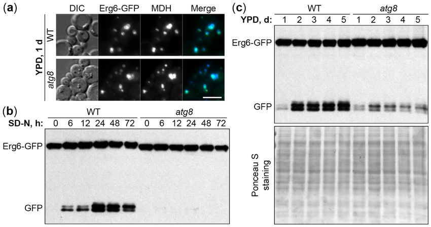
Int. J. Mol. Sci. 2020, 21, 9094 2 of 14
diseases [8,9]. Thus, understanding the mechanisms of lipophagy is very important for the prevention
and treatment of various lipid accumulation diseases.
The budding yeast Saccharomyces cerevisiae was used as a simple lipophagy model by several
groups to study the mechanisms of lipophagy. Precisely, lipophagy was induced by either acute
N-starvation [10,11] or C-limitation (either acute [12] or gradual due to the prolonged incubation
of cells in the same medium and entering them into stationary (S) phase [11,13]). These studies
described the morphological features of lipophagy and tested the requirements of lipophagy for
known autophagy-related (Atg) factors. They suggested that both N-starvation and C-limitation
induce microlipophagy [10–13], the selective microautophagy of LDs, and that this microlipophagy
depends on the same core autophagic factors, which are necessary for the formation of autophagic
double-membrane in other autophagic pathways [10,12,13]. However, such autophagic membrane
was never reported to be associated with LDs in the yeast lipophagy studies questioning the role of the
autophagic machinery in yeast lipophagy.
Here, we developed the yeast, Komagataella phaffii (formerly Pichia pastoris), as a new simple
model to study lipophagy. The K. phaffii has proven to be an excellent model organism for the studies
of autophagy-related (Atg) pathways and contributed a lot of mechanistic insights to the field of
autophagy [14]. Then, we run the entire collection of K. phaffii atg-mutants through the lipophagy
assay under both N-starvation and S-phase conditions. As a result, we found that the core autophagic
machinery is essential only for the N-starvation lipophagy. The only overlapping stringent molecular
requirements for two lipophagy pathways were Atg6 and vacuolar proteinases A and B. In addition,
we isolated a new positive regulator of lipophagy 1 (prl1) mutant that was deficient only in the S-phase
lipophagy. Therefore, we suggest that the N-starvation and S-phase lipophagy pathways have distinct
molecular mechanisms.
2. Results
2.1. K. phaffii Is a Good Model for Both N-Starvation and S-Phase Lipophagy
To develop K. phaffii as a new lipophagy model, we used the established LD marker protein,
Erg6 [10–12], tagged with the green fluorescent protein (GFP) on the integrative plasmid, pRK2.
To confirm the localization of Erg6-GFP to LDs, wild-type (WT) PPY12h cells with pRK2 integrated
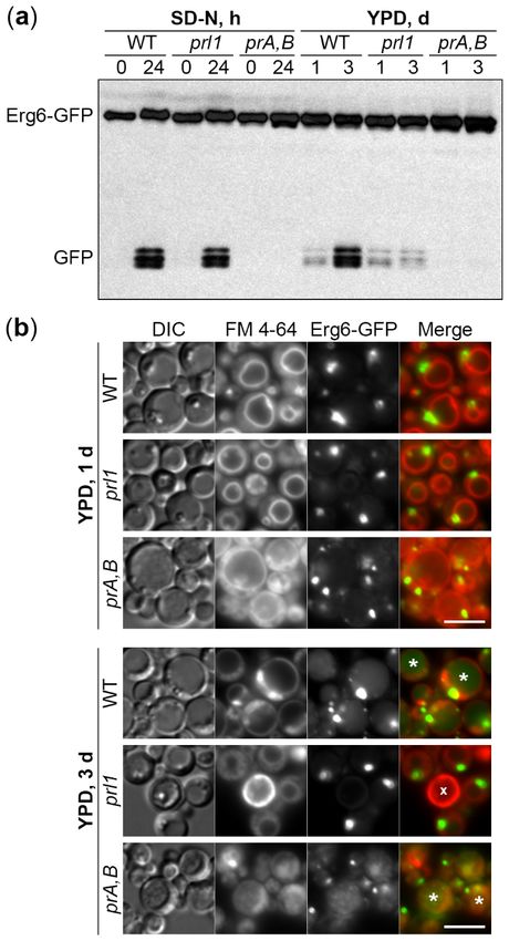
into the HIS4 locus were grown in YPD medium for 1 d to an early S-phase and stained with a blue
LD dye, monodansylpentane (MDH) [15]. The Erg6-GFP displayed a complete co-localization with
MDH (Figure 1a), suggesting that it is a good LD protein marker for K. phaffii under our experimental
conditions. The lack of a key Atg protein, Atg8, did not affect the localization of Erg6-GFP to LDs in
atg8 cells (Figure 1a), making it possible to use the Erg6-GFP as a lipophagy reporter.
Then, we developed two Erg6-GFP processing assays to monitor lipophagy: One after the transfer
of cells from early S-phase in YPD medium to N-starvation in SD-N medium and another one after the
prolonged S-phase in YPD medium. When the LDs with Erg6-GFP are delivered from the cytoplasm
to the vacuole, Erg6, but not GFP moiety, is degraded by vacuolar proteases resulting in free GFP,
which can be detected by Western blot [16]. The processing of Erg6-GFP to GFP in WT (PPY12h)
cells culminated after 24 h of N-starvation (Figure 1b) and after 2-3 days in YPD medium (Figure 1c).
Therefore, we picked 0 and 24 h, and 1 and 3 d time-points for the N-starvation and S-phase lipophagy
assays, respectively.confirm the localization of Erg6-GFP to LDs, wild-type (WT) PPY12h cells with pRK2 integrated into
the HIS4 locus were grown in YPD medium for 1 d to an early S-phase and stained with a blue LD
dye, monodansylpentane (MDH) [15]. The Erg6-GFP displayed a complete co-localization with MDH
(Figure 1a), suggesting that it is a good LD protein marker for K. phaffii under our experimental
conditions.
Int. J. Mol. Sci. The
2020, lack of a key Atg protein, Atg8, did not affect the localization of Erg6-GFP to LDs
21, 9094 in
3 of 14
atg8 cells (Figure 1a), making it possible to use the Erg6-GFP as a lipophagy reporter.
Figure 1. Komagataella phaffii is a good model for both N-starvation and S-phase lipophagy. (a) K. phaffii
Erg6-GFP co-localizes with MDH-stained LDs in both WT and atg8 cells. DIC: Differential interference
contrast. Scale bar, 5 µm. (b) Atg8 is essential for N-starvation lipophagy. Cells were normalized in
SD-N at OD600 1, and an equal volume of culture (1 mL) was processed at all time-points for both
strains to nullify the differential growth (Erg6-GFP dilution) effects in SD-N medium (loading control
is not applicable). (c) Atg8 is only partially required for S-phase lipophagy. Since biomass slightly
decreased during the time-course in S-phase, equal biomass (1 OD600 ) was taken at all time-points for
both strains. Ponceau S staining was used as a loading control for S-phase samples.
Interestingly, while atg8 cells were completely deficient in the Erg6-GFP processing under
N-starvation conditions (Figure 1b), they were only partially compromised in it in S-phase (Figure 1c),
suggesting that N-starvation and S-phase lipophagy pathways might have differences in their molecular
requirements. In summary, both N-starvation and S-phase lipophagy pathways were readily induced
in K. phaffii yeast, making it a good model for comparison of their machinery.
2.2. Molecular Requirements of N-Starvation and S-Phase Lipophagy in K. phaffii
Encouraged by atg8 results under two lipophagy conditions, we introduced Erg6-GFP into the
collection of K. phaffii strains deficient in genes that were previously implicated in various Atg-pathways
in either K. phaffii or other species (Table 1). The collected mutants belong to 4 different WT backgrounds:
GS115, GS200, PPY12h, and PPY12m. Therefore, we grouped mutants by genetic background and
studied them together with the corresponding WT strain, as a control, in both N-starvation and S-phase
lipophagy conditions (Figures 2 and 3, respectively).Int. J. Mol. Sci. 2020, 21, 9094 4 of 14
Table 1. K. phaffii strains used in this study.
Mutant Name Strain Name Background Genotype and Plasmid Source
WT GS115 GS115 his4 [17]
WT GS200 GS200 arg4 his4 [18]
WT PPY12h PPY12h arg4 his4 [19]
WT PPY12m PPY12m arg4 his4 [19]
ape1 SJCF434 PPY12m ∆ape1::GeneticinR arg4 his4 [20]
atg1 R12 GS115 atg1-1::ZeocinR his4 [21]
atg2 WDK011 GS115 ∆atg2::ZeocinR his4 [21]
atg3 gsa20 GS115 atg3::ZeocinR his4 [21]
atg4 PPM408 PPY12h atg4::ZeocinR arg4 his4 [22]
atg5 SJCF2320 GS115 ∆atg5::ZeocinR his4 SL 1
atg6 SRDM006 PPY12m ∆atg6::GeneticinR arg4 his4 [23]
atg7 WDK07 GS200 ∆atg7::ScARG4 arg4 his4 [24]
atg8 SJCF925 PPY12h ∆atg8::GeneticinR arg4 his4 [25]
atg9 R19 GS115 atg9-1::ZeocinR his4 [21]
atg11 R8 GS115 atg11-2::ZeocinR his4 [26]
atg17 SJCF929 PPY12h ∆atg17::GeneticinR arg4 his4 [25]
atg11-2::ZeocinR
atg11 atg17 SJCF948 GS115 [25]
∆atg17::GeneticinR his4
atg18 R2 GS115 atg18-1::ZeocinR his4 [21]
atg20 SRDM020 PPY12m ∆atg20::GeneticinR arg4 his4 SL
atg24 paz16 PPY12h atg24::ZeocinR arg4 his4 [27]
atg25 SJCF1231 PPY12h ∆atg25::GeneticinR arg4 his4 [23]
atg26 ∆pdg3 GS200 ∆atg26::ScARG4 arg4 his4 [28]
atg28 ∆pdg2 GS200 ∆atg28::ScARG4 arg4 his4 [29]
atg30 SJCF936 PPY12h ∆atg30::ZeocinR arg4 his4 [25]
atg32 SJCF1715 PPY12h ∆atg32::GeneticinR arg4 his4 [30]
atg35 SVN1 GS200 ∆atg35::ScARG4 arg4 his4 [31]
atg37 STN96 PPY12h ∆atg37::GeneticinR arg4 his4 [32]
atg40 SRK2 PPY12h ∆atg40::ZeocinR (pRK4) arg4 his4 This study
pep4 prb1 SMD1163 GS115 pep4 prb1 his4 [33]
pex3 SEW1 PPY12h ∆pex3::PpARG4 arg4 his4 [34]
pex19 SKF13 PPY12h ∆pex19::ZeocinR arg4 his4 [35]
prl1 SRK3 PPY12h prl1::ZeocinR (pRK6) arg4 his4 This study
vac8 WDY53 GS200 ∆vac8::ZeocinR arg4 his4 [36]
vam7 SRDM050 PPY12m ∆vam7::ZeocinR arg4 his4 [37]
vps15 OP5 GS200 ∆vps15::ScARG4 arg4 his4 [38]
vps17 SRDM122 PPY12m ∆vps17::GeneticinR arg4 his4 [23]
uvrag SRDM083 PPY12m ∆uvrag::ZeocinR arg4 his4 [23]
ypt7 SRRM197 PPY12h ∆ypt7::GeneticinR arg4 his4 [37]
1 SL: Subramani laboratory.vam7 SRDM050 PPY12m ∆vam7::ZeocinR arg4 his4 [37]
vps15 OP5 GS200 ∆vps15::ScARG4 arg4 his4 [38]
vps17 SRDM122 PPY12m ∆vps17::GeneticinR arg4 his4 [23]
uvrag SRDM083 PPY12m ∆uvrag::ZeocinR arg4 his4 [23]
Int. J. ypt7 SRRM197
Mol. Sci. 2020, 21, 9094 PPY12h ∆ypt7::GeneticinR arg4 his4 [37]5 of 14
1 SL: Subramani laboratory.
Figure 2. N-starvation lipophagy strictly depends on the core autophagic machinery (Atg1-Atg9, Atg18,
and Vps15), vacuole fusion machinery (Vam7 and Ypt7), and vacuolar proteolysis (proteinases A and B).
Cells were normalized in SD-N at OD600 1, and an equal volume of culture (1 mL) was processed at
both time-points for all strains to nullify the differential growth (Erg6-GFP dilution) effects in SD-N
medium (loading control is not applicable). prA,B: Proteinases A and B-deficient mutant pep4 prb1.Figure 2. N-starvation lipophagy strictly depends on the core autophagic machinery (Atg1-Atg9,
Atg18, and Vps15), vacuole fusion machinery (Vam7 and Ypt7), and vacuolar proteolysis (proteinases
A and B). Cells were normalized in SD-N at OD600 1, and an equal volume of culture (1 mL) was
processed at both time-points for all strains to nullify the differential growth (Erg6-GFP dilution)
Int.effects in 2020,
J. Mol. Sci. SD-N21,medium
9094 (loading control is not applicable). prA,B: Proteinases A and B-deficient
6 of 14
mutant pep4 prb1.
Figure 3. 3.S-phase
Figure S-phaselipophagy
lipophagystrictly
strictlydepends
depends only
only on Atg6, Prl1
on Atg6, Prl1(positive
(positiveregulator
regulatorofoflipophagy
lipophagy 1),1),
and
and vacuolar proteolysis (proteinases A and B). Since biomass slightly decreased after three days in in
vacuolar proteolysis (proteinases A and B). Since biomass slightly decreased after three days
S-phase, equal
S-phase, equalbiomass
biomass(1(1OD
OD600 ) was taken at both time-points for all strains. Ponceau S staining was
600 ) was taken at both time-points for all strains. Ponceau S staining was
used asas
used a loading
a loadingcontrol
controland
and displayed
displayed ininAppendix
AppendixAFigure
Figure A1. prA,B: ProteinasesAAand
prA,B: Proteinases and B-deficient
B-deficient
mutant
mutantpep4
pep4prb1.
prb1.
WeWe foundthat
found thatmost
mostofofthe
themutants
mutants werewere either
either fully
fully deficient (atg1-atg9,atg18,
deficient(atg1-atg9, atg18,pep4 prb1,
pep4 vam7,
prb1, vam7,
vps15, and ypt7) or fully proficient (ape1, atg11, atg20, atg24-atg26, atg30, atg32, atg35, atg37, atg40,
vps15, and ypt7) or fully proficient (ape1, atg11, atg20, atg24-atg26, atg30, atg32, atg35, atg37, atg40, pex3, pex3,
pex19,
pex19, prl1,
prl1, vac8,vps17,
vac8, vps17,and uvrag) in
anduvrag) in the
the Erg6-GFP
Erg6-GFP processing
processingunder
underN-starvation
N-starvation conditions.
conditions.Only
Only
three strains (atg17, atg11 atg17, and atg28) had an intermediate phenotype (Figure
three strains (atg17, atg11 atg17, and atg28) had an intermediate phenotype (Figure 2 and Table 2 and Table 2).2).
These results suggested that N-starvation lipophagy strictly depends on the core autophagic machinery
These results suggested that N-starvation lipophagy strictly depends on the core autophagic
(Atg1-Atg9, Atg18, and Vps15), vacuole fusion machinery (Vam7 and Ypt7), and vacuolar proteolysis
machinery (Atg1-Atg9, Atg18, and Vps15), vacuole fusion machinery (Vam7 and Ypt7), and vacuolar
(proteinases A and B).
proteolysis (proteinases A and B).
In contrast, the Erg6-GFP processing in S-phase was fully deficient in only three strains
(atg6, pep4 prb1, and prl1). It was fully proficient in nearly as many strains (ape1, atg11, atg20, atg24,
atg25, atg30, atg32, atg35, atg37, vac8, vps17, and uvrag), as under N-starvation conditions. However,
most of the mutants (atg1-atg5, atg7-atg9, atg17, atg11 atg17, atg18, atg26, atg28, atg40, pex3, pex19,
vam7, vps15, and ypt7) had an intermediate phenotype (Figure 3 and Appendix A Figure A1; Table 2).
Therefore, we concluded that S-phase lipophagy strictly depends only on Atg6, Prl1, and vacuolar
proteolysis (proteinases A and B). Summarizing, the N-starvation and S-phase lipophagy pathways
have different molecular requirements.Int. J. Mol. Sci. 2020, 21, 9094 7 of 14
Table 2. Lipophagy phenotype of K. phaffii (this study) and S. cerevisiae mutants.
Kp (This Study) Sc [10] Sc [13] Sc [12]
Strain
(Kp/Sc) SD-N S-Phase SD-N SD-N S-Phase SD-D (0.4%)
Erg6-GFP Erg6-GFP Erg6-GFP Faa4-GFP BODIPY Erg6-DsRed
WT + + + + + +
ape1 + + ND 1 ND ND ND
atg1 − +/− − − − −
atg2 − +/− ND ND − −
atg3 − +/− − − − −
atg4 − +/− − − − ND
atg5 − +/− − − − −
atg6 − − − − − −
atg7 − +/− − − − −
atg8 − +/− − − − −
atg9 − +/− − − − −
atg11 + + ND +/− + +
atg17 +/− +/− ND − − +/−
atg11 atg17 +/− +/− ND ND ND ND
atg18 − +/− − − − −
atg20 + + ND + + +
atg24 + + ND ND + +
atg25 + + NA 2 NA NA NA
atg26 + +/− ND ND + +
atg28/atg29,31 +/− +/− ND ND −, − +/−, +/−
atg30/atg36 + + ND ND + +
atg32 + + ND ND − +/−
atg35 + + NA NA NA NA
atg37 + + NA NA NA NA
atg40 + +/− ND ND ND ND
pep4 prb1 − − ND ND − ND
pex3 + +/− ND ND ND ND
pex19 + +/− ND ND ND ND
prl1 + − NA NA NA NA
vac8 + + ND − ND ND
vam7 − +/− − − ND ND
vps15 − +/− ND ND ND ND
vps17 + + ND ND ND ND
uvrag/vps38 + + +/− +/− ND ND
ypt7 − +/− − − ND ND
1 ND: Phenotype not determined in Sc; 2 NA: Mutant not available in Sc. “ + ”: Fully proficient in lipophagy;
“ − ”: Fully deficient in lipophagy; “ +/− ”: Intermediate phenotype.
2.3. Prl1 Is Essential for the Delivery of LDs to the Vacuole in S-Phase
To probe further into the differences between N-starvation and S-phase lipophagy machinery,
we took advantage of the prl1 mutant. This mutant was obtained by integrating ZeocinR cassette from
the pRK6 plasmid into the genome of PPY12h WT strain (Table 1). The prl1 mutant displayed a uniqueInt. J. Mol. Sci. 2020, 21, x FOR PEER REVIEW 7 of 13
2.3. Prl1 is Essential for the Delivery of LDs to the Vacuole in S-Phase
Int. J. To
Mol.probe further
Sci. 2020, 21, 9094into
the differences between N-starvation and S-phase lipophagy machinery,
8 of 14
we took advantage of the prl1 mutant. This mutant was obtained by integrating Zeocin cassette from
R
the pRK6 plasmid into the genome of PPY12h WT strain (Table 1). The prl1 mutant displayed a unique
phenotype in the screening above: It was fully proficient in the N-starvation lipophagy, but fully
phenotype in the screening above: It was fully proficient in the N-starvation lipophagy, but fully
deficient in the S-phase lipophagy (Table 2).
deficient in the S-phase lipophagy (Table 2).
To compare the phenotypes of prl1 cells in the same experiment, we split the cultures of WT,
To compare the phenotypes of prl1 cells in the same experiment, we split the cultures of WT,
prl1, and pep4 prb1 cells after 1 d in YPD medium: Small aliquots were transferred to SD-N medium
prl1, and pep4 prb1 cells after 1 d in YPD medium: Small aliquots were transferred to SD-N medium
(for 0 and 24 h time-points), while the rest remained in YPD medium (for 1 and 3 d time-points)
(for 0 and 24 h time-points), while the rest remained in YPD medium (for 1 and 3 d time-points)
(Figure 4a and Appendix A Figure A2). While N-starvation and S-phase lipophagy pathways were
(Figure 4a and Appendix Figure A2). While N-starvation and S-phase lipophagy pathways were
equally well induced in WT cells, they were fully blocked in the proteinases A and B-deficient mutant.
equally well induced in WT cells, they were fully blocked in the proteinases A and B-deficient mutant.
Although prl1 cells were indistinguishable from WT cells under N-starvation conditions, they were
Although prl1 cells were indistinguishable from WT cells under N-starvation conditions, they were
indeed incapable of degrading LDs in S-phase (Figure 4a). Therefore, we concluded that the prl1 mutant
indeed incapable of degrading LDs in S-phase (Figure 4a). Therefore, we concluded that the prl1
is specifically deficient in the S-phase lipophagy.
mutant is specifically deficient in the S-phase lipophagy.
Figure
Figure 4.
4. Prl1
Prl1isisessential
essentialto
todeliver
deliverLDs
LDstotothe
thevacuole
vacuolein inS-phase.
S-phase.(a)
(a)S-phase,
S-phase, but
but not
not N-starvation
N-starvation
lipophagy dependson
lipophagy depends onPrl1.
Prl1.N-starvation
N-starvationandand S-phase
S-phase cells
cells werewere processed
processed as described
as described in Figures
in Figures 2 and 23,
and 3, respectively. Ponceau S staining (Appendix A Figure A2) was used as a loading
respectively. Ponceau S staining (Appendix A Figure A2) was used as a loading control for S-phase control for S-
phase samples. (b) Prl1 is required to deliver LDs to the vacuole in S-phase. Vacuole membranes
samples. (b) Prl1 is required to deliver LDs to the vacuole in S-phase. Vacuole membranes were stained were
red with FM 4-64. DIC: Differential interference contrast; prA,B: Proteinases A and B-deficient mutant
pep4 prb1; *: GFP-containing vacuole; x: Dead cell. Scale bar, 5 µm.Int. J. Mol. Sci. 2020, 21, 9094 9 of 14
To get insight into the step of S-phase lipophagy affected in prl1 cells, we studied S-phase
lipophagy by fluorescence microscopy. The WT, prl1, and pep4 prb1 cells with Erg6-GFP reporter were
incubated in the YPD medium with FM 4-64, the lipophilic dye that stains specifically the vacuolar
membrane. After 1 d, the cells of all strains had LDs outside the vacuole and no GFP fluorescence
inside the vacuolar lumen. However, after 3 d, WT and pep4 prb1 cells developed a diffuse or grainy
GFP fluorescence in the vacuolar lumen, respectively (Figure 4b). Those patterns of luminal GFP
fluorescence were consistent with the disintegration of the LD-containing autophagic bodies in WT
vacuoles and Brownian movement of intact LD-containing autophagic bodies in the proteinases A and
B-deficient vacuoles. The prl1 cells did not gain any GFP fluorescence in the vacuolar lumen after 3 d
in YPD, suggesting that Prl1 is required to deliver LDs to the vacuole in S-phase.
3. Discussion
In this study, we introduced K. phaffii yeast as a new lipophagy model and compared the lipophagy
requirements of K. phaffii cells under two conditions: N-starvation and S-phase, the two most popular
ways to induce Atg-pathways in yeast. Previous screenings in S. cerevisiae done for each of these
conditions separately indicated that both of them induced microlipophagy that strongly depended on
the core autophagic machinery [10,13]. However, by comparing the N-starvation and S-phase conditions
in the same study with K. phaffii, we observed a clear difference in lipophagy requirements (Table 2).
Both the N-starvation and S-phase lipophagy pathways strictly depended on the
proteinases A and B, consistent with the vacuolar degradation of LDs under the two conditions. While
the N-starvation lipophagy strongly relied on the core autophagic machinery represented by Atg1-Atg9,
Atg18, and Vps15, the S-phase lipophagy was fully deficient only without the Atg6 protein. Interestingly,
only Atg6 and not the other components of the phosphatidylinositol 3-kinase complex I (i.e., Atg14,
Atg38, Vps15, and Vps34) stably localized to the vacuolar membrane under both acute and gradual
(S-phase) C-limitation conditions in S. cerevisiae [12]. Atg6 was necessary for the formation of raft-like
domains in the vacuolar membrane [12] that are essential for microlipophagy [13]. Combined, this
and previous studies suggest that Atg6 plays a unique role in the S-phase lipophagy, which is
different from its established function in the biogenesis of autophagic double-membrane under
N-starvation conditions [39].
While a reason for the essential role of the core autophagic machinery in the N-starvation lipophagy
is unclear, there is a plausible explanation for the partial requirement of the core autophagic machinery
in the S-phase lipophagy. In S-phase, the core autophagic machinery is essential for the correct vacuolar
localization of Niemann-Pick type C proteins, Ncr1 and Npc2, which transport sterol from the vacuolar
lumen to the vacuolar membrane for the raft-like domain formation [11]. However, the requirements
of Ncr1 and Npc2 for (1) raft-like domains formation, (2) their internalization as microautophagic
bodies, and (3) S-phase lipophagy are partial [11]. Therefore, the core autophagic machinery has, in the
end, a partial role in the S-phase lipophagy. Interestingly, it is not required for the correct vacuolar
localization of Ncr1 and Npc2 under the N-starvation conditions. Thus, the mechanistic role of the
core autophagic machinery in the N-starvation lipophagy is still unknown.
Recently, it was reported that the vacuolar membrane protein, Vph1, which is normally excluded
from the raft-like domains in S-phase [40], is also degraded, like LDs, by microautophagy [41].
Interestingly, the S-phase microautophagy of Vph1 was independent of the core autophagic machinery,
but relied on the machinery of ESCRT, the endosomal sorting complex required for transport. The same
study also reported that the S-phase microautophagy of LDs was partially independent of the core
autophagic factor, Atg1, but strongly relied on the ESCRT component, Vps27 [41]. Our results are
consistent with these lipophagy observations and extend them to the entire core autophagic machinery
being only partially required specifically in the S-phase. However, it is still unclear how LDs and Vph1
can utilize the same microautophagy pathway in S-phase, since they are associated with different
vacuolar membrane domains, the raft-like liquid-ordered domain and the liquid-disordered domain,
respectively. Since the S-phase microautophagy of Vph1 does not require Atg6 [41], and the S-phaseInt. J. Mol. Sci. 2020, 21, 9094 10 of 14
lipophagy strongly depends on it (Figure 3), we propose that these pathways have both common
(Vps27) and unique (Atg6) requirements.
Our study also suggests a unique molecular requirement of the S-phase lipophagy versus
N-starvation lipophagy, the positive regulator of lipophagy 1 (Prl1). The prl1 mutant isolated in this
study was deficient in lipophagy only in the S-phase. Moreover, we showed that the prl10 s lipophagy
block is at a trafficking step, since LDs were not delivered from the cytoplasm to the vacuole for
degradation in the S-phase. It will be interesting to determine the gene responsible for prl1 phenotype,
since it can help us to further distinguish the molecular mechanisms of these two clearly distinct
lipophagy pathways, the N-starvation and S-phase lipophagy.
4. Materials and Methods
4.1. Strains and Plasmids
The K. phaffii strains that were used in this study are shown in Table 1. These strains were
transformed by electroporation [42] with the EcoNI-linearized (R0521S; New England Biolabs, Ipswich,
MA, USA) pRK2 plasmid. This plasmid contained the Erg6-GFP expression cassette (for lipophagy
studies) and HIS4 marker gene (for integration into his4 mutant allele of the recipient strains and
selection of His+ -transformants). The resulting transformants had the following genotype: his4::pRK2
(PERG6 -ERG6-GFP, HIS4). They were selected on SD+CSM-His plates (1.7 g/L YNB without amino acids
and ammonium sulfate, 20 g/L dextrose, 5 g/L ammonium sulfate, 0.78 g/L CSM-His, and 20 g/L agar)
and screened for expression of Erg6-GFP by Western blot with anti-GFP bodies (11814460001; Roche
Diagnostics, Mannheim, Germany) and for localization of Erg6-GFP to LDs by fluorescence microscopy.
4.2. Fluorescence Microscopy
Cells were grown for 1 and/or 3 d in culture tubes with 1 mL of YPD medium (10 g/L yeast extract,
20 g/L peptone and 20 g/L dextrose; the autoclaved solution of yeast extract and peptone was mixed
with the filter-sterilized 20× solution of dextrose). LDs were stained with 1 µL of 0.1 M MDH solution
(SM1000a; Abcepta, San Diego, CA, USA) during the last 1 h of incubation of cells in YPD medium.
Vacuolar membranes were stained with 1 µL of 1 mg/mL solution of FM 4-64 (T3166; Molecular Probes,
Eugene, OR, USA) in DMSO added at the beginning of incubation of cells in YPD medium. Then, cells
were immobilized on slides using 1% low-melt agarose. For this, the 2 µL drop of cell culture on the
slide was mixed with the 5 µL drop of 1% low-melt agarose (37 ◦ C) on the coverslip. Microscopy was
done at the Axioskop 2 MOT microscope equipped with the Plan-Apochromat 100×/1.40 NA oil DIC
objective and operated by the AxioVision software (Carl Zeiss Microscopy, White Plains, NY, USA).
All the experiments were done at least in duplicate.
4.3. Biochemical Studies
Cells were grown in culture tubes with 1 mL of YPD medium and 1 OD600 of cells was taken at
1 and 3 d time-points for studies of S-phase lipophagy. For studies of N-starvation lipophagy, 3 OD600
of cells were taken at 1 d time-point in YPD medium, washed twice with 1 mL of 1× YNB solution
(1.7 g/L YNB without amino acids and ammonium sulfate), and resuspended in 3 mL of SD-N medium
(1.7 g/L YNB without amino acids and ammonium sulfate, and 20 g/L dextrose). Then, 1 mL of cell
culture was taken at 0 and 24 h time-points in SD-N medium. Both YPD (1 and 3 d) and SD-N (0 and 24 h)
samples were TCA precipitated [43] and analyzed by Western blot with the same anti-GFP bodies,
as above. All the experiments were done at least in duplicate.
Author Contributions: Conceptualization, R.K. and T.Y.N.; methodology, R.K. and T.Y.N.; validation, T.Y.N.;
formal analysis, T.Y.N.; investigation, R.K. and T.Y.N.; resources, T.Y.N.; data curation, R.K. and T.Y.N.;
writing—Original draft preparation, M.A.R. and T.Y.N.; writing—Review and editing, R.K., M.A.R. and T.Y.N.;
visualization, T.Y.N.; supervision, T.Y.N.; project administration, T.Y.N.; funding acquisition, T.Y.N. All authors
have read and agreed to the published version of the manuscript.Int. J. Mol. Sci. 2020, 21, x FOR PEER REVIEW 10 of 13
writing—Original draft preparation, M.A.R. and T.Y.N.; writing—Review and editing, R.K., M.A.R. and T.Y.N.;
visualization, T.Y.N.;
Int. J. Mol. Sci. 2020, 21, supervision,
9094 T.Y.N.; project administration, T.Y.N.; funding acquisition, T.Y.N. All authors
11 of 14
have read and agreed to the published version of the manuscript.
Funding: This research was funded by the NIH grants, DK106344 and GM119571, to T.Y.N. The APC was funded
Funding:
by This research was funded by the NIH grants, DK106344 and GM119571, to T.Y.N. The APC was funded
GM119571.
by GM119571.
Acknowledgments:
Acknowledgments: We thank Jean-Claude
We thank Jean-Claude Farré
Farré and
and Suresh
Suresh Subramani
Subramani for
for strains
strains and
and helpful
helpful discussions.
discussions.
Conflicts of Interest:
Conflicts of Interest: The
The authors
authors declare
declare no
no conflict
conflict of
of interest.
interest. The
The funders
funders had
had no
no role
role in
in the
the design
design of
of the
the
study; in the collection, analyses, or interpretation of data; in the writing of the manuscript, or in the decision to
publish the
publish the results.
results.
Abbreviations
Abbreviations
Atg
Atg autophagy-related
autophagy-related
C carbon
C carbon
DIC differential interference contrast
DIC differential interference contrast
ESCRT endosomal sorting complex required for transport
ESCRT endosomal sorting complex required for transport
GFP green fluorescent protein
GFP green fluorescent protein
LD lipid droplet
LD lipid droplet
MDH monodansylpentane
MDH monodansylpentane
N nitrogen
N
Prl1 nitrogenregulator of lipophagy 1
positive
Prl1
prA,B positive regulator
proteinases A and of
B lipophagy 1
SprA,B proteinases
stationary A and B
S
WT stationary
wild-type
WT wild-type
Appendix A
Appendix A
Figure A1. Supplemental figure for Figure 3. Loading control (see Figure 3 for details).Int. J. Mol. Sci. 2020, 21, x FOR PEER REVIEW 11 of 13
Int. J. Mol. Sci. 2020, 21, 9094 12 of 14
Figure A1. Supplemental figure for Figure 3. Loading control (see Figure 3 for details).
Figure
Figure A2. A2. Supplemental
Supplemental figure
figure forforFigure
Figure 4a.
4a. Loading
Loadingcontrol (see(see
control Figure 4a for 4a
Figure details).
for details).
References
References
1. Klionsky, D.J. Autophagy: From phenomenology to molecular understanding in less than a decade. Nat. Rev.
1. Klionsky,Mol. D.J. Autophagy:
Cell From phenomenology
Biol. 2007, 8, 931–937. [CrossRef] to molecular understanding in less than a decade. Nat.
Rev.2.Mol.Ohsumi,
Cell Biol.Y. Historical of autophagy research. Cell Res. 2014, 24, 9–23. [CrossRef] [PubMed]
landmarksdoi:10.1038/nrm2245.
2007, 8, 931–937,
3. Takeshige, K.; Baba, M.; Tsuboi, S.; Noda, T.; Ohsumi, Y. Autophagy in yeast demonstrated with
2. Ohsumi, Y. Historical landmarks of autophagy research. Cell Res. 2014, 24, 9–23, doi:10.1038/cr.2013.169.
proteinase-deficient mutants and conditions for its induction. J. Cell Biol. 1992, 119, 301–311. [CrossRef]
3. Takeshige,
4. K.;B.M.;
Sutter, Baba,
Wu, M.; Tsuboi,
X.; Laxman, S.; B.P.
S.; Tu, Noda, T.; Ohsumi,
Methionine Y. Autophagy
inhibits autophagy in yeast
and promotes growthdemonstrated
by inducing with
proteinase-deficient
the SAM-responsive mutants and ofconditions
methylation PP2A. Cell 2013, for154,its403–415.
induction. J. Cell Biol. 1992, 119, 301–311,
[CrossRef]
doi:10.1083/jcb.119.2.301.
5. Singh, R.; Kaushik, S.; Wang, Y.; Xiang, Y.; Novak, I.; Komatsu, M.; Tanaka, K.; Cuervo, A.M.; Czaja, M.J.
4. Autophagy
Sutter, B.M.; Wu, X.; regulates
Laxman, lipidS.;
metabolism. Nature 2009, 458,
Tu, B.P. Methionine 1131–1135.
inhibits [CrossRef]
autophagy and promotes growth by inducing
6. Marchesini, G.; Brizi, M.; Bianchi, G.; Tomassetti, S.; Bugianesi, E.; Lenzi, M.; McCullough, A.J.; Natale, S.;
the SAM-responsive methylation of PP2A. Cell 2013, 154, 403–415, doi:10.1016/j.cell.2013.06.041.
Forlani, G.; Melchionda, N. Nonalcoholic fatty liver disease: A feature of the metabolic syndrome. Diabetes
5. Singh, R.;2001,
Kaushik, S.; Wang,
50, 1844–1850. Y.; Xiang,
[CrossRef] [PubMed] Y.; Novak, I.; Komatsu, M.; Tanaka, K.; Cuervo, A.M.; Czaja, M.J.
Autophagy
7. regulates
Singh, R.; Cuervo,lipid
A.M.metabolism.
Autophagy in the Nature 2009,
cellular 458, balance.
energetic 1131–1135, doi:10.1038/nature07976.
Cell Metab. 2011, 13, 495–504. [CrossRef]
6. Marchesini, G.; Brizi, M.; Bianchi, G.; Tomassetti, S.; Bugianesi, E.; Lenzi, M.; F.;
8. Zhang, Z.; Yao, Z.; Chen, Y.; Qian, L.; Jiang, S.; Zhou, J.; Shao, J.; Chen, A.; Zhang, Zheng, S. Lipophagy
McCullough, A.J.; Natale, S.;
and liver disease: New perspectives to better understanding and therapy. Biomed. Pharmacother. 2018, 97,
Forlani, G.; Melchionda, N. Nonalcoholic fatty liver disease: A feature of the metabolic syndrome. Diabetes
339–348. [CrossRef]
2001, 50, 1844–1850, doi:10.2337/diabetes.50.8.1844.
9. Zhou, K.; Yao, P.; He, J.; Zhao, H. Lipophagy in nonliver tissues and some related diseases: Pathogenic and
7. Singh, R.; Cuervo,implications.
therapeutic A.M. Autophagy in 2019,
J. Cell. Physiol. the cellular energetic
234, 7938–7947. balance. Cell Metab. 2011, 13, 495–504,
[CrossRef]
doi:10.1016/j.cmet.2011.04.004.
10. Van Zutphen, T.; Todde, V.; de Boer, R.; Kreim, M.; Hofbauer, H.F.; Wolinski, H.; Veenhuis, M.; van der
8. Zhang, Z.; Klei, I.J.; Kohlwein,
Yao, Z.; Chen,S.D.Y.; Lipid
Qian,droplet autophagy
L.; Jiang, in the J.;
S.; Zhou, yeast Saccharomyces
Shao, J.; Chen,cerevisiae. Mol. F.;
A.; Zhang, Biol.Zheng,
Cell 2014,
S. 25,
Lipophagy
290–301. [CrossRef]
and liver disease: New perspectives to better understanding and therapy. Biomed. Pharmacother. 2018, 97,
11. Tsuji, T.; Fujimoto, M.; Tatematsu, T.; Cheng, J.; Orii, M.; Takatori, S.; Fujimoto, T. Niemann-Pick type C
339–348,proteins
doi:10.1016/j.biopha.2017.07.168.
promote microautophagy by expanding raft-like membrane domains in the yeast vacuole. Elife
9. Zhou, K.;2017,
Yao,6. P.; He, J.; Zhao, H. Lipophagy in nonliver tissues and some related diseases: Pathogenic and
[CrossRef]
therapeutic
12. Seo,implications.
A.Y.; Lau, P.W.; J. Cell. Physiol.D.;
Feliciano, 2019, 234, 7938–7947,
Sengupta, P.; Gros,doi:10.1002/jcp.27988.
M.A.L.; Cinquin, B.; Larabell, C.A.;
10. Lippincott-Schwartz, J. AMPK and vacuole-associated
van Zutphen, T.; Todde, V.; de Boer, R.; Kreim, M.; Hofbauer, H.F.;µ-lipophagy Atg14p orchestrate Wolinski,for H.;energy production
Veenhuis, M.; van der
and long-term survival under glucose starvation. Elife 2017, 6. [CrossRef]
Klei, I.J.; Kohlwein, S.D. Lipid droplet autophagy in the yeast Saccharomyces cerevisiae. Mol. Biol. Cell 2014,
13. Wang, C.W.; Miao, Y.H.; Chang, Y.S. A sterol-enriched vacuolar microdomain mediates stationary phase
25, 290–301, doi:10.1091/mbc.E13-08-0448.
lipophagy in budding yeast. J. Cell Biol. 2014, 206, 357–366. [CrossRef]
11. Tsuji,
14.T.;Farre,
Fujimoto, M.; Tatematsu,
J.C.; Subramani, T.; Cheng,
S. Mechanistic J.; into
insights Orii,selective
M.; Takatori,
autophagy S.;pathways:
Fujimoto,Lessons
T. Niemann-Pick
from yeast. type C
proteins Nat.
promote
Rev. Mol.microautophagy
Cell Biol. 2016, 17, by expanding
537–552. [CrossRef] raft-like membrane domains in the yeast vacuole. Elife
15. Yang, H.J.; Hsu, C.L.;
2017, 6, doi:10.7554/eLife.25960. Yang, J.Y.; Yang, W.Y. Monodansylpentane as a blue-fluorescent lipid-droplet marker
for multi-color live-cell imaging. PLoS ONE 2012, 7, e32693. [CrossRef]
12. Seo, A.Y.; Lau, P.W.; Feliciano, D.; Sengupta, P.; Gros, M.A.L.; Cinquin, B.; Larabell, C.A.; Lippincott-
16. Shintani, T.; Klionsky, D.J. Cargo proteins facilitate the formation of transport vesicles in the cytoplasm to
Schwartz, J. AMPK
vacuole and
targeting vacuole-associated
pathway. J. Biol. Chem. 2004, Atg14p orchestrate
279, 29889–29894. µ-lipophagy for energy production and
[CrossRef]
long-term survival under glucose starvation. Elife 2017, 6, doi:10.7554/eLife.21690.
13. Wang, C.W.; Miao, Y.H.; Chang, Y.S. A sterol-enriched vacuolar microdomain mediates stationary phase
lipophagy in budding yeast. J. Cell Biol. 2014, 206, 357–366, doi:10.1083/jcb.201404115.
14. Farre, J.C.; Subramani, S. Mechanistic insights into selective autophagy pathways: Lessons from yeast. Nat.
Rev. Mol. Cell Biol. 2016, 17, 537–552, doi:10.1038/nrm.2016.74.Int. J. Mol. Sci. 2020, 21, 9094 13 of 14
17. Cregg, J.M.; Barringer, K.J.; Hessler, A.Y.; Madden, K.R. Pichia pastoris as a host system for transformations.
Mol. Cell. Biol. 1985, 5, 3376–3385. [CrossRef]
18. Waterham, H.R.; de Vries, Y.; Russel, K.A.; Xie, W.; Veenhuis, M.; Cregg, J.M. The Pichia pastoris PER6 gene
product is a peroxisomal integral membrane protein essential for peroxisome biogenesis and has sequence
similarity to the Zellweger syndrome protein PAF-1. Mol. Cell. Biol. 1996, 16, 2527–2536. [CrossRef]
19. Gould, S.J.; McCollum, D.; Spong, A.P.; Heyman, J.A.; Subramani, S. Development of the yeast Pichia pastoris
as a model organism for a genetic and molecular analysis of peroxisome assembly. Yeast 1992, 8, 613–628.
[CrossRef]
20. Farre, J.C.; Vidal, J.; Subramani, S. A cytoplasm to vacuole targeting pathway in P. pastoris. Autophagy 2007, 3,
230–234. [CrossRef]
21. Stromhaug, P.E.; Bevan, A.; Dunn, W.A., Jr. GSA11 encodes a unique 208-kDa protein required for pexophagy
and autophagy in Pichia pastoris. J. Biol. Chem. 2001, 276, 42422–42435. [CrossRef]
22. Mukaiyama, H.; Oku, M.; Baba, M.; Samizo, T.; Hammond, A.T.; Glick, B.S.; Kato, N.; Sakai, Y. Paz2 and 13
other PAZ gene products regulate vacuolar engulfment of peroxisomes during micropexophagy. Genes Cells
2002, 7, 75–90. [CrossRef]
23. Farre, J.C.; Mathewson, R.D.; Manjithaya, R.; Subramani, S. Roles of Pichia pastoris Uvrag in vacuolar protein
sorting and the phosphatidylinositol 3-kinase complex in phagophore elongation in autophagy pathways.
Autophagy 2010, 6, 86–99. [CrossRef]
24. Yuan, W.; Stromhaug, P.E.; Dunn, W.A., Jr. Glucose-induced autophagy of peroxisomes in Pichia pastoris
requires a unique E1-like protein. Mol. Biol. Cell 1999, 10, 1353–1366. [CrossRef]
25. Nazarko, T.Y.; Farre, J.C.; Subramani, S. Peroxisome size provides insights into the function of
autophagy-related proteins. Mol. Biol. Cell 2009, 20, 3828–3839. [CrossRef]
26. Kim, J.; Kamada, Y.; Stromhaug, P.E.; Guan, J.; Hefner-Gravink, A.; Baba, M.; Scott, S.V.; Ohsumi, Y.;
Dunn, W.A., Jr.; Klionsky, D.J. Cvt9/Gsa9 functions in sequestering selective cytosolic cargo destined for the
vacuole. J. Cell. Biol. 2001, 153, 381–396. [CrossRef]
27. Ano, Y.; Hattori, T.; Oku, M.; Mukaiyama, H.; Baba, M.; Ohsumi, Y.; Kato, N.; Sakai, Y. A sorting nexin PpAtg24
regulates vacuolar membrane dynamics during pexophagy via binding to phosphatidylinositol-3-phosphate.
Mol. Biol. Cell 2005, 16, 446–457. [CrossRef]
28. Stasyk, O.V.; Nazarko, T.Y.; Stasyk, O.G.; Krasovska, O.S.; Warnecke, D.; Nicaud, J.M.; Cregg, J.M.; Sibirny, A.A.
Sterol glucosyltransferases have different functional roles in Pichia pastoris and Yarrowia lipolytica. Cell Biol.
Int. 2003, 27, 947–952. [CrossRef]
29. Stasyk, O.V.; Stasyk, O.G.; Mathewson, R.D.; Farre, J.C.; Nazarko, V.Y.; Krasovska, O.S.; Subramani, S.;
Cregg, J.M.; Sibirny, A.A. Atg28, a novel coiled-coil protein involved in autophagic degradation of peroxisomes
in the methylotrophic yeast Pichia pastoris. Autophagy 2006, 2, 30–38. [CrossRef]
30. Farre, J.C.; Burkenroad, A.; Burnett, S.F.; Subramani, S. Phosphorylation of mitophagy and pexophagy
receptors coordinates their interaction with Atg8 and Atg11. EMBO Rep. 2013, 14, 441–449. [CrossRef]
31. Nazarko, V.Y.; Nazarko, T.Y.; Farre, J.C.; Stasyk, O.V.; Warnecke, D.; Ulaszewski, S.; Cregg, J.M.; Sibirny, A.A.;
Subramani, S. Atg35, a micropexophagy-specific protein that regulates micropexophagic apparatus formation
in Pichia pastoris. Autophagy 2011, 7, 375–385. [CrossRef]
32. Nazarko, T.Y.; Ozeki, K.; Till, A.; Ramakrishnan, G.; Lotfi, P.; Yan, M.; Subramani, S. Peroxisomal Atg37
binds Atg30 or palmitoyl-CoA to regulate phagophore formation during pexophagy. J. Cell. Biol. 2014, 204,
541–557. [CrossRef]
33. Tuttle, D.L.; Dunn, W.A., Jr. Divergent modes of autophagy in the methylotrophic yeast Pichia pastoris. J. Cell
Sci. 1995, 108 (Pt 1), 25–35.
34. Wiemer, E.A.; Luers, G.H.; Faber, K.N.; Wenzel, T.; Veenhuis, M.; Subramani, S. Isolation and characterization
of Pas2p, a peroxisomal membrane protein essential for peroxisome biogenesis in the methylotrophic yeast
Pichia pastoris. J. Biol. Chem. 1996, 271, 18973–18980. [CrossRef]
35. Snyder, W.B.; Faber, K.N.; Wenzel, T.J.; Koller, A.; Luers, G.H.; Rangell, L.; Keller, G.A.; Subramani, S. Pex19p
interacts with Pex3p and Pex10p and is essential for peroxisome biogenesis in Pichia pastoris. Mol. Biol. Cell
1999, 10, 1745–1761. [CrossRef]
36. Chang, T.; Schroder, L.A.; Thomson, J.M.; Klocman, A.S.; Tomasini, A.J.; Stromhaug, P.E.; Dunn, W.A., Jr.
PpATG9 encodes a novel membrane protein that traffics to vacuolar membranes, which sequester peroxisomes
during pexophagy in Pichia pastoris. Mol. Biol. Cell 2005, 16, 4941–4953. [CrossRef]Int. J. Mol. Sci. 2020, 21, 9094 14 of 14
37. Manjithaya, R.; Anjard, C.; Loomis, W.F.; Subramani, S. Unconventional secretion of Pichia pastoris Acb1 is
dependent on GRASP protein, peroxisomal functions, and autophagosome formation. J. Cell. Biol. 2010, 188,
537–546. [CrossRef]
38. Stasyk, O.V.; van der Klei, I.J.; Bellu, A.R.; Shen, S.; Kiel, J.A.; Cregg, J.M.; Veenhuis, M. A Pichia pastoris
VPS15 homologue is required in selective peroxisome autophagy. Curr. Genet. 1999, 36, 262–269. [CrossRef]
39. Obara, K.; Ohsumi, Y. PtdIns 3-kinase orchestrates autophagosome formation in yeast. J. Lipids 2011, 2011,
498768. [CrossRef]
40. Toulmay, A.; Prinz, W.A. Direct imaging reveals stable, micrometer-scale lipid domains that segregate
proteins in live cells. J. Cell. Biol. 2013, 202, 35–44. [CrossRef]
41. Oku, M.; Maeda, Y.; Kagohashi, Y.; Kondo, T.; Yamada, M.; Fujimoto, T.; Sakai, Y. Evidence for ESCRT-and
clathrin-dependent microautophagy. J. Cell. Biol. 2017, 216, 3263–3274. [CrossRef]
42. Cregg, J.M.; Russell, K.A. Transformation. Methods Mol. Biol. 1998, 103, 27–39. [CrossRef]
43. Baerends, R.J.; Faber, K.N.; Kram, A.M.; Kiel, J.A.; van der Klei, I.J.; Veenhuis, M. A stretch of positively
charged amino acids at the N terminus of Hansenula polymorpha Pex3p is involved in incorporation of the
protein into the peroxisomal membrane. J. Biol. Chem. 2000, 275, 9986–9995. [CrossRef]
Publisher’s Note: MDPI stays neutral with regard to jurisdictional claims in published maps and institutional
affiliations.
© 2020 by the authors. Licensee MDPI, Basel, Switzerland. This article is an open access
article distributed under the terms and conditions of the Creative Commons Attribution
(CC BY) license (http://creativecommons.org/licenses/by/4.0/).You can also read