Sustained Human Hair Follicle Growth Ex Vivo in a Glycosaminoglycan Hydrogel Matrix - MDPI
←
→
Page content transcription
If your browser does not render page correctly, please read the page content below
International Journal of
           Molecular Sciences

Communication
Sustained Human Hair Follicle Growth Ex Vivo in
a Glycosaminoglycan Hydrogel Matrix
Sandra Fernández-Martos 1,† , María Calvo-Sánchez 1,2,† , Karla García-Alonso 3 ,
Begoña Castro 4 , Bita Hashtroody 3 and Jesús Espada 1,5, *
 1    Experimental Dermatology and Skin Biology Group, Ramon y Cajal Institute for Health Research (IRYCIS),
      Ramón y Cajal University Hospital, 28034 Madrid, Spain; sandy.fernandez@hotmail.com (S.F.-M.);
      calvosanchezmaria@gmail.com (M.C.-S.)
 2    Instituto de Investigaciones Biosanitarias, Facultad de Ciencias Experimentales,
      Universidad Francisco de Vitoria, 28223 Pozuelo de Alarcón, Spain
 3    Cantabria Labs, 28043 Madrid, Spain; Karla.garcia@cantabrialabs.es (K.G.-A.);
      bita.Hashtroody@cantabrialabs.es (B.H.)
 4    Histocell, Bizkaia Technologic Park, 48160 Derio, Bizkaia, Spain; Bcastro@histocell.com
 5    Centro Integrativo de Biología y Química Aplicada (CIBQA), Universidad Bernardo O’Higgins,
      8370854 Santiago, Chile
 *    Correspondence: jespada1968@gmail.com
 †    These authors contributed equally to this work.

 Received: 17 January 2019; Accepted: 6 April 2019; Published: 9 April 2019

 Abstract: Glycosaminoglycans (GAGs) and associated proteoglycans have important functions in
 homeostatic maintenance and regenerative processes (e.g., wound repair) of the skin. However, little
 is known about the role of these molecules in the regulation of the hair follicle cycle. Here we report
 that growing human hair follicles ex vivo in a defined GAG hydrogel mimicking the dermal matrix
 strongly promotes sustained cell survival and maintenance of a highly proliferative phenotype in
 the hair bulb and suprabulbar regions. This significant effect is associated with the activation of
 WNT/β-catenin signaling targets (CCDN1, AXIN2) and with the expression of stem cell markers
 (CK15, CD34) and growth factors implicated in the telogen/anagen transition (TGFβ2, FGF10). As
 a whole, these results point to the dermal GAG matrix as an important component in the regulation of
 the human hair follicle growth cycle, and to GAG-based hydrogels as potentially relevant modulators
 of this process both in vitro and in vivo.

 Keywords: glycosaminoglycans; hyaluronic acid; WNT/β-catenin; BMP/SMAD; TGFβ; stem cells;
 human hair follicle

1. Introduction
     Glycosaminoglycans (GAGs) are highly hydrophilic long-chain linear polysaccharides made
up of a repeating disaccharide unit, consisting of an amino sugar (N-acetylglucosamine or
N-acetylgalactosamine) in combination with a uronic sugar (glucuronic acid or iduronic acid) or
galactose [1–4]. GAGs are usually bound covalently to a protein core forming a proteoglycan molecule
and are essential components of the extracellular matrix of most mammalian tissues and of the
surface of several cell types [1,3–5]. There are six major GAG types, namely, dermatan sulphate (DS),
chondroitin sulphate (CS), keratan sulphate (KS), heparin (H), heparan sulphate (HS), and hyaluronic
acid (HA; the only GAG type that does not contain sulphate and does not bind to a protein core).
Besides their roles as scaffold molecules, GAGs and derived proteoglycans are important players in
the control of critical physiological parameters (e.g., the organism water balance) and in the regulation

Int. J. Mol. Sci. 2019, 20, 1741; doi:10.3390/ijms20071741                        www.mdpi.com/journal/ijms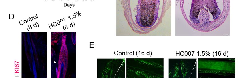
Int. J. Mol. Sci. 2019, 20, 1741                                                                     2 of 8

of key cellular functions such as proliferation, adhesion, and migration, and whose specific disfunction
is associated with different diseases, including cancer [1,2].
      All GAGs are present to some degree in the human skin. The syndecan (containing CS/DS and HS)
and glypican (containing HS) proteoglycan types are extracellularly linked to the plasma membrane of
basal and suprabasal cells in the epidermal layer. HA is the most abundant GAG in the dermal matrix
layer, exerting a basic scaffolding function in combination with the collagen/elastin network [2,3,5].
The dermal matrix also contains different extracellular proteoglycans, including versican (containing
CS), decorin and biglycan (containing CS/DS), fibromodulin, lumican, and keratocan (containing KS),
and perlecan (containing HS) [2,3,5], that complement the GAG setting of the dermis, supposedly
endowing this skin layer with unique functional characteristics. In the human hair follicle, cells in the
epidermal part and outer and inner root sheath basal layers express syndecan-1, whereas the dermal
part, encompassing the dermal papilla and the hair bulb, contain high amounts of HA, HS, CS, DS, KS,
and associated proteoglycans, including perlecan, versican, and biglycan [6].
      Considering the reported roles of extracellular or membrane associated GAGs in the regulation of
key cellular processes, it can be hypothesized that these molecules may have important roles in the skin,
and, particularly, in the hair. Important roles for these molecules have been postulated during skin
regeneration and wound healing and in the process of skin aging [2,3,5]. However, despite different
reports showing a cyclic expression of GAGs and different proteoglycans during the hair follicle
growth cycle [3,5,6], little is actually known at a functional level about the physiological implication
of GAGs in the regulation of this biological oscillator. In this context, our aim in this work was to
investigate potential stimulatory effects of a defined GAG matrix on the hair follicle growth potential
using ex vivo cultured human hair follicles as an experimental system. In this biological model, most
cell types in the tissue rapidly cease to proliferate and enter into an apoptotic cell death program
after serial culturing in basal conditions (e.g., no growth factors added to culture medium), therefore
constituting an adequate system to evaluate the functional effects of GAGs on isolated mini-organs.

2. Results and Discussion
      Here, we used a unique tunable GAG hydrogel mimicking the dermal matrix composition [7,
8], hereafter called HC007, composed of non-crosslinked native GAGs comprising hyaluronic acid
of high and low molecular weight and sulfated GAGs (CS, DS, KS, and HS). HC007 behaves as
a temporary extracellular matrix substitute in damaged tissues by attracting cells and creating an
adequate environment where recruited cells find their innate extracellular matrix to survive, proliferate,
attach, and elicit their natural bioactivity [7,8]. Human follicular units (FUs) grown ex vivo, typically
containing one or two individualized hair follicles in the growing (anagen) phase embedded in
a remnant matrix of fatty and dermal tissues (Figure 1A), were used as the experimental system.
      In our experience, this tissue remnant favors optimal hair follicle survival up to days 7–9 in basal
ex vivo culture conditions. Most hair follicles in basal conditions typically showed a steady decrease
of the hair bulb area, defined here as an area encompassing the hair bulb/dermal papilla and the
most intensely pigmented suprabulbar region (Figure 1A–C; see Materials and Methods), a follicular
area characterized by high cell proliferation and melanogenesis during the anagen phase ([9] and
references therein). Consequently, we found low cell proliferation rates after 6–8 days in ex vivo culture
(Figure 1D), followed by extensive induction of apoptosis in hair bulb and suprabulbar regions around
days 12–16 (Figure 1E). By contrast, growing hair follicles in HC007 1.5% promoted a sustained and
statistically significant enlargement of the hair bulb and suprabulbar regions (Figure 1A–C) associated
with the maintenance of a strong cell proliferation phenotype in the tissue after 8 days in ex vivo
culture (Figure 1D) and with the absence of significant cell death induction by day 16 (Figure 1E) as
compared to control samples. After 16 days in culture, the stimulatory effect of HC007 1.5% appeared
as a significant increase in the number of cells (Figure 1C). Additionally, we were able to spot a clearly
noticeable, although hardly measurable, increase in the length of the hair shaft in a considerable
proportion (about 50%) of hair follicles grown in HC007 1.5% (Figure 1A). These results indicate that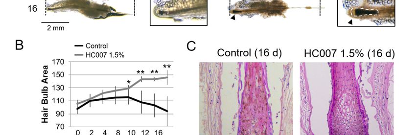
Int. J. Mol. Sci. 2019, 20, 1741 3 of 8
a defined
Int.GAG hydrogel
J. Mol. Sci. 2019, 20, x matrix surrounding an explanted human FU can efficiently support
3 ofsustained
8
cell proliferation and, presumably, hair follicle growth.
Figure 1. Sustained maintenance of human hair follicle growth ex vivo in a defined
Figure 1. Sustained maintenance of human hair follicle growth ex vivo in a defined glycosaminoglycan
glycosaminoglycan hydrogel matrix (HC007). (A) Representative phase-contrast microscopy images
hydrogel matrix (HC007). (A) Representative phase-contrast microscopy images of whole hair follicles
of whole hair follicles showing significant thickening (black arrowheads) of the hair bulb area, defined
showing significant
here as an area thickening
encompassing (black arrowheads)
hair bulb/dermal of the
papilla and hair bulb area,
suprabulbar defined
regions, here as an area
after continuous
encompassing hair bulb/dermal papilla and suprabulbar regions, after continuous
growing in HC007 1.5%. Vertical dotted lines delimitate hair follicle length at time 0. A noticeable growing in HC007
1.5%. Vertical dotted lines
length increase in hairdelimitate hairwas
follicle length follicle length
observed at time
in 50% 0. A
of the noticeable
HC007 length Enlarged
1.5% samples. increase in hair
images depicting
follicle length typical in
was observed hair50%
bulbofareas
the used
HC007 in image
1.5% quantification
samples. Enlargedanalysis images
(white dotted lines). typical
depicting
Bar: 2 mm. (B) Time course quantification of hair bulb area in control and
hair bulb areas used in image quantification analysis (white dotted lines). Bar: 2 mm. (B) Time courseHC007 1.5% samples.
Results are representative of at least 18 hair follicle units per condition. The mean +/− SD of n ≥ 18 for
quantification of hair bulb area in control and HC007 1.5% samples. Results are representative of at
each experimental condition is represented and t-test was used for statistical analysis. *, significant p
least 18 ≤hair follicle
0.1. **, unitspper
significant condition.
≤ 0.05. The mean
(C) Representative +/− SDsections
histological of n ≥ stained
18 for each experimental
with H&E of the hair condition
bulb is
represented and t-test was used for statistical analysis. *, significant p ≤ 0.1. **,
area in control and HC007 1.5% after 16 days in culture. Bar: 50 µm. (D) Confocal microscopy images significant p ≤ 0.05.
(C) Representative histologicalshowing
(maximum projections) sectionsthestained with H&E
localization of theof the
cell hair bulb areamarker
proliferation in control
KI67and
in HC007
morphologically
1.5% after equivalentBar:
16 days in culture. histological
50 µm.sections of hair follicles
(D) Confocal grown ex
microscopy vivo for
images 8 days in control
(maximum projections)
showingbasal
the culture conditions
localization of theor cell
in HC007 1.5%. Bar:marker
proliferation 100 µm.KI67
Enlarged images show nuclear
in morphologically KI67 staining
equivalent histological
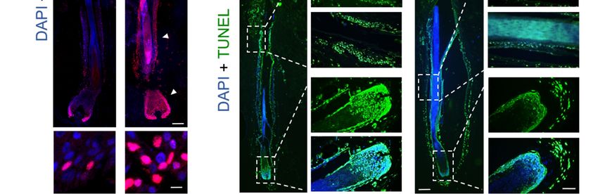
in outer root sheath cells. Bar: 10 µm. (E) Identification of apoptotic cells by the TUNEL assay in
sections of hair follicles grown ex vivo for 8 days in control basal culture conditions or in HC007 1.5%.
morphologically equivalent histological sections of hair follicles grown ex vivo for 16 days in control
Bar: 100 µm. Enlarged images show nuclear KI67 staining in outer root sheath cells. Bar: 10 µm.
(E) Identification of apoptotic cells by the TUNEL assay in morphologically equivalent histological
sections of hair follicles grown ex vivo for 16 days in control basal culture conditions or in HC007
1.5%. Bars: 100 µm. Results shown in (C–E) are representative of at least 10 hair follicles in three
independent experiments.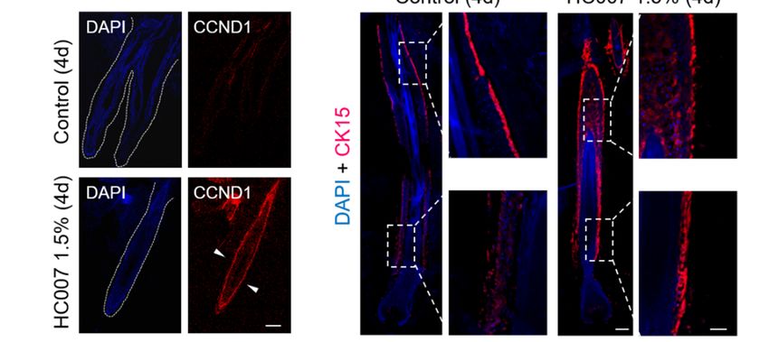
Int. J. Mol. Sci. 2019, 20, x 4 of 8
basal culture conditions or in HC007 1.5%. Bars: 100 µm. Results shown in (C), (D) and, (E) are
Int. J. representative
Mol. Sci. 2019, 20,of1741
at least 10 hair follicles in three independent experiments. 4 of 8
We next quantified the expression of different genes involved in the two major signaling
We next
pathways quantifiedthe
regulating thehair
expression
follicleofgrowth
differentcycle:
genes Transforming
involved in theGrowth
two major signaling
Factor pathways
(TGF)β/Bone
regulating the hair follicle growth cycle: Transforming Growth Factor (TGF)β/Bone
Morphogenetic Protein (BMP)/Mothers Against Decapentaplegic Homolog (SMAD), implicated in Morphogenetic
Protein
the (BMP)/Mothers
maintenance Against Decapentaplegic
of the quiescence state in the hairHomolog (SMAD),
follicle stem cell implicated
niche and thein the maintenance
entrance of
into the
the quiescence state in the hair follicle stem cell niche and the entrance into the resting
resting (telogen) phase, and WNT/β-catenin signaling, which dictates the activation the stem cell (telogen) phase,
and WNT/β-catenin
niche and the entrance signaling,
into thewhich
growingdictates the activation
(anagen) phase [10].theWestemfound
cell niche
that and the entrance
control into
hair follicles
the growing
showed high (anagen)
expressionphase [10].
levels We BMP2
of the found and
that BMP4
controleffectors
hair follicles
and, showed high expression
concomitantly, of the ID1levels
and
of the BMP2 and BMP4 effectors and, concomitantly, of the ID1 and ID2
ID2 BMP/SMAD gene targets in the skin (Figure 2A). Interestingly, this expression pattern BMP/SMAD genewastargets
not
in the skin
affected by (Figure 2A). Interestingly,
HC007 treatments (Figure this
2A).expression
By contrast, pattern was not affected
key WNT/β-catenin bytargets
gene HC007in treatments
the skin,
(FigureD1
cyclin 2A).(CCDN1),
By contrast, key WNT/β-catenin
a general proliferation gene targets
marker, andin the skin, implicated
AXIN2, cyclin D1 (CCDN1), a general
in the metabolic
proliferation marker, and AXIN2, implicated in the metabolic stabilization of
stabilization of β-catenin, were strongly induced by HC007 four days after treatments (Figure β-catenin, were strongly
2A).
induced
The by induction
specific HC007 four days after
of CCDN1 wastreatments (Figure 2A).
further confirmed The specific
by protein induction of CCDN1
immunolocalization was
in the tissue
further confirmed
(Figure 2B). by protein immunolocalization in the tissue (Figure 2B).
Figure 2. Activation of WNT/β-catenin signaling and of stem cell niche proliferation in human hair
Figure 2. Activation of WNT/β-catenin signaling and of stem cell niche proliferation in human hair
follicle growth ex vivo in a defined glycosaminoglycan hydrogel matrix (HC007). (A) Quantitative
follicle growth ex vivo in a defined glycosaminoglycan hydrogel matrix (HC007). (A) Quantitative
expression analysis by qRT-PCR analysis of selected genes. Input mRNA was obtained from three
expression analysis by qRT-PCR analysis of selected genes. Input mRNA was obtained from three or
or four hair follicles per experimental condition, namely, control and HC007 1.5% samples, and the
four hair follicles per experimental condition, namely, control and HC007 1.5% samples, and the mean
mean +/- SD of relative gene expression values, normalized to 18S rRNA, is represented. **, significant
+/-
p ≤SD0.1.of(B,C)
relative gene expression
Confocal microscopy values,
images normalized
(maximumtoprojections)
18S rRNA, of is represented. **, significant
the immunolocalization of pthe
≤
0.1. (B,C) Confocal microscopy images (maximum projections) of the immunolocalization
(B) transcriptional target of WNT signaling CCDN1 (in red; white dotted lines delineate individual of the (B)
transcriptional targetarrowheads
hair follicles; withe of WNT signaling
indicateCCDN1
positive(in red;inwhite
cells dotted
the outer lines
root delineate
sheath) and ofindividual hair
the (C) CK15
follicles; withe arrowheads indicate positive cells in the outer root sheath) and of the
stem cell marker (white dotted boxes are enlarged in right panels) in histological sections of human (C) CK15 stem
cell
hairmarker
follicles(white
grown dotted boxes
ex vivo fourare
daysenlarged in right1.5%
after HC007 panels) in histological
treatments. DAPI sections
was usedofas human hair
chromatin
follicles grown ex vivo four days after HC007 1.5% treatments. DAPI was used as
counterstain. Results shown in (B,C) are representative of at least 10 hair follicles in three independent chromatin
counterstain.
experiments. Results
Bars: 100shown
µm. in (B) and (C) are representative of at least 10 hair follicles in three
independent experiments. Bars: 100 µm.
In addition, we found that the master intracellular inhibitor of WNT/β-catenin signaling,
Glycogen Synthase Kinase- 3 beta (GSK3β), showed high expression levels in control hair follicles
and was significantly repressed by HC007 treatments (Figure 2A). Low levels of the WNT/β-catenin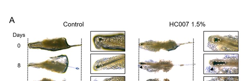
Int. J. Mol. Sci. 2019, 20, 1741                                                                      5 of 8

extracellular antagonist Dickkopf-related protein 1 (DKK1) were found in control hair follicles, in
agreement with previous results in mouse adult hair follicles [11], and this expression pattern was not
affected by HC007 treatments (Figure 2A). Interestingly, we also found a statistically significant and
specific induction of the expression of TGFβ2 and Fibroblast Growth Factor (FGF)10, factors implicated
in the telogen-anagen transition [10], after HC007 treatments, whereas no changes were observed
in the expression of FGF7 or Vascular Endothelial Growth Factor (VEGF) (Figure 2A). Finally, we
analyzed the expression of the stem cell markers cytokeratin 15 (CK15) and CD34, proteins specifically
expressed by skin progenitors after activation of the bulge stem cell niche in the hair follicle and
associated with multipotency and self-renewal potential [12–16]. We found that both stem cell markers
were significantly activated upon ex vivo growing of hair follicles in HC007 1.5% (Figure 2A). We
further confirmed by protein immunolocalization a strong increase in the expression level and in the
number of CK15 positive cells all along the outer root sheath in HC007-treated hair follicles (Figure 2C),
indicating the activation of the bulge stem cell niche.
      The results reported here indicate for the first time that a defined GAG matrix can promote and
support the sustained growth of human hair follicles ex vivo. We have previously shown that HC007
behaves as an effective temporary extracellular matrix substitute in damaged tissues by attracting cells
and creating an adequate environment where recruited cells find their innate extracellular matrix to
survive, proliferate, attach, and elicit their natural bioactivity, also promoting the proliferation and
continuous expansion of primary skin cells [7,8]. Here, we have further shown that the stimulatory
effect of HC007 in the hair follicle occurs in association with an activation of WNT/β-catenin signaling,
bypassing the pervading inhibitory action BMP/SMAD signaling. Interestingly, a specific induction of
TGFβ2 is also observed after HC007 treatments, in agreement with the reported functional interaction
between different GAGs/proteoglycans and member of the TGFβ superfamily [17,18]. Moreover, it has
been reported that WNT/β-catenin signaling during the onset of the anagen phase is accompanied by
the specific expression of TGFβ2, that regulates proliferation, differentiation, and extracellular matrix
production of dermal fibroblasts [19]. The interplay between WNT/β-catenin and TGFβ/BMP/SMAD
to regulate stem cell niche function has been also described in different adult tissues, including the hair
follicle [20]. It can be hypothesized that the GAG matrix produced by dermal fibroblasts in response to
TGFβ/BMP/SMAD signaling is an essential component in the regulation of the hair follicle cycle and,
particularly, in the feedback modulation of WNT/β-catenin signaling. We have also found that the
signaling cascade potentially triggered/modulated by the GAG matrix results finally in the activation
of the hair follicle stem cell niche, showing up as a significant induction of the CK15 and CD34 stem
cell marker and a strong increase in the number of CK15 positive cells. As a whole, these results point
to dermal GAGs as an essential component in the regulation of the human hair follicle growth cycle,
and to the HC007 hydrogel as a potential modulator of this process both in vitro and in vivo, and as
an interesting and useful growth media complement to maintain hair follicle function ex vivo for
longer periods.

3. Materials and Methods
    All methods were performed in accordance with all relevant institutional and European Union
(EU) experimental and ethical guidelines.

3.1. Ex Vivo Culture of Human Hair Follicles and HC007 Treatment
     Hair follicular units (FUs) were obtained from human scalp samples taken from the occipital skin
of volunteer donors during routine hair transplant procedures. Eligible patients provided written
informed consent, and the Ethical Committee of the Ramón y Cajal University Hospital approved
this procedure. Selected follicular units (FUs) typically containing one or two hair follicles and
surrounding fatty and dermal tissue remnants were dissected by expert trichologists at the Ramón
y Cajal Hospital Dermatology Service. Selected FUs encompassed for the most part hair follicles in
the growing (anagen) phase using standard morphological criteria [21]. FUs were grown in Williams
Int. J. Mol. Sci. 2019, 20, 1741 6 of 8
E medium (Sigma-Aldrich, St. Louis, MO, USA), supplemented with 10× penicillin/streptomycin,
10× amphotericin B, and 2 mM L-glutamine (all from Gibco Life Technologies, Carlsbad, CA, USA),
5 µg/mL insulin, 5 µg/mL transferring, 20 pM T3 hormone, 0.083 µg/mL cholera toxin, and 0.4 µg/mL
hydrocortisone at 37 ◦ C in a 5% CO2 humidified atmosphere. Adherent tissue remnants in FUs were
maintained throughout the ex vivo growing process to improve hair follicle viability in basal conditions.
FUs from the same individual and body location were used in each experimental series.
HC007 was used as a sterile solution of non-crosslinked native GAGs comprising hyaluronic
acid of high and low molecular weight and sulfated GAGs (chondroitin sulfate, dermatan sulfate,
keratan sulfate, heparan sulfate), with a purity ≥95%, dissolved in a concentration of 15 mg/mL in
isotonic solution without preservatives. These native GAGs were obtained, through a patented method
(WO 2011/120535), from the Wharton’s jelly of the umbilical cord, a specialized extracellular matrix
that permits the generation of a family of biomaterials with regenerative properties. For treatment
samples, FUs were grown in supplemented Williams E medium containing 1.5% HC007. Working
concentration for HC007 was selected based on previous results in primary cell cultures [7,8]. For
control samples, FUs were grown in supplemented basal Williams E medium. Growth medium was
replenished every two days in all cases.
3.2. Quantification of Hair Bulb Area
For the evaluation of significant morphological changes in vivo in whole hair follicles, high
resolution images of FUs growing in 24-well plates were acquired at time 0 after tseeding in the presence
or absence of HC007 1.5%, and every 24 h onwards for 16 days, depending on experimental settings.
Image acquisition was performed using a Nikon Eclipse Ci LED-fluorescence microscope (Tokio,
Japan) using a 2× objective and a 0.55× Reduction Lens adapter coupled to a Jenoptik PROGRES
GRYPHAX® SUBRA Super HD camera (Jena, Germany) and suited Version 1.1.8.153 image software
pack. We defined the hair bulb area as the hair follicle territory encompassing the hair bulb and dermal
papilla as well as the most intensively pigmented area of the suprabulbar region, including associated
inner and outer root sheets (see Figure 1). This area is characterized by high cell proliferation and
melanogenesis during the anagen phase of the hair follicle cycle [9]. Total hair bulb area in high
resolution images of whole hair follicles was spotted and quantified using suited free FIJI software
packs (https://fiji.sc/). The fold changes with respect to control samples of means +/- SD of hair bulb
measurements in n ≥ 10 samples for each experimental condition was represented and t-test was used
for statistical analysis.
3.3. Protein Immunolocalization
To determine target protein localization and expression patterns in hair follicles, histological
tissues sections were used. At least one FU in each experimental group was fixed in 3.7% aqueous
formaldehyde and embedded in paraffin using standard procedures. Typically, 18–22, 8 µm thick,
longitudinal tissue sections were obtained for each FU. As a rule, the most 6-4 central sections,
encompassing as much as possible the full length of, at least, the suprabulbar fiber and hair bulb regions,
were used for comparative analysis. Antigen retrieval was performed using 10 mM citrate buffer
in hydrated sections following standard procedures. Primary antibodies, including anti-cytokeratin
15 (CK15, clon EPR16Y, Abcam Cambridge, UK), and anti-KI67 (clon SP6, Abcam), and anti-cyclin
D1 (CCDN1, clon EPR2241, Abcam) were incubated overnight at 4 ◦ C in a wet chamber, extensively
washed with PBS, incubated for 1 h at room temperature with appropriate secondary fluorescence-
or HRP-labelled antibodies, and mounted in DAPI (100 ng/mL)-containing Vectashield. Confocal
images were obtained using a Leica TCS SP5 AOBS spectral confocal microscope (Wetzlar, Germany)
and processed using the FIJI software. Bright field images were obtained using a Nikon Eclipse Ci
LED-fluorescence microscope (Tokio, Japan) coupled to a Jenoptik PROGRES GRYPHAX® SUBRA
Super HD camera.Int. J. Mol. Sci. 2019, 20, 1741 7 of 8
3.4. RNA Extraction and Gene Expression Analysis
Total RNA of at least four FUs in each experimental group was extracted four days after
treatments, using an RNeasy micro kit (Qiagen, Venlo, Netherlands). RNA was normalized with
respect to the number of FUs in each experimental condition, and then was converted into cDNA using
a FastGene Scriptase II cDNA Kit (NIPPON Genetics, Tokyo, Japan). qRT-PCR data was analyzed
using a comparative CT method, using 18S ribosomal RNA expression as an internal control. Gene
expression fold changes were represented as the ratio between means of 2−∆Ct values of HC007 FU
and control FU mean values. Primer sequences are available upon request.
Author Contributions: S.F.-M. and M.C.-S. performed all the experiments, prepared figures, and discussed results.
K.G.-A. and B.H. discussed results. B.C. provided materials and discussed results. J.E. performed the experimental
design, discussed results, and wrote the text.
Funding: This work was supported in part by grant PI15/01458 to J.E. from the Ministerio de Economía y
Competitividad, Instituto de Salud Carlos III of Spain.
Conflicts of Interest: K.G.-A., B.C., and B.H. have a financial relationship with companies that have an interest in
the subject of this work.
References
1. Afratis, N.; Gialeli, C.; Nikitovic, D.; Tsegenidis, T.; Karousou, E.; Theocharis, A.D.; Pavão, M.S.;
Tzanakakis, G.N.; Karamanos, N.K. Glycosaminoglycans: Key players in cancer cell biology and treatment.
FEBS J. 2012, 279, 1177–1197. [CrossRef]
2. Salbach, J.; Rachner, T.D.; Rauner, M.; Hempel, U.; Anderegg, U.; Franz, S.; Simon, J.-C.; Hofbauer, L.C.
Regenerative potential of glycosaminoglycans for skin and bone. J. Mol. Med. 2012, 90, 625–635. [CrossRef]
3. Lee, D.H.; Oh, J.-H.; Chung, J.H. Glycosaminoglycan and proteoglycan in skin aging. J. Dermatol. Sci. 2016,
83, 174–181. [CrossRef] [PubMed]
4. Pomin, V.; Mulloy, B. Glycosaminoglycans and Proteoglycans. Pharmaceuticals 2018, 11, 27. [CrossRef]
[PubMed]
5. Smith, M.M.; Melrose, J. Proteoglycans in Normal and Healing Skin. Adv. Wound Care 2015, 4, 152–173.
[CrossRef]
6. Malgouries, S.; Thibaut, S.; Bernard, B.A. Proteoglycan expression patterns in human hair follicle.
Br. J. Dermatol. 2008, 158, 234–242. [CrossRef]
7. Herrero-Mendez, A.; Palomares, T.; Castro, B.; Herrero, J.; Alonso-Varona, A. Generation of tunable
glycosaminoglycan hydrogels to mimic extracellular matrices. J. Tissue Eng. Regen. Med. 2016, 10, 1000–1011.
[CrossRef]
8. Herrero-Mendez, A.; Palomares, T.; Castro, B.; Herrero, J.; Granado, M.H.; Bejar, J.M.; Alonso-Varona, A.
HR007: A family of biomaterials based on glycosaminoglycans for tissue repair. J. Tissue Eng. Regen. Med.
2017, 11, 989–1001. [CrossRef] [PubMed]
9. Slominski, A.; Wortsman, J.; Plonka, P.M.; Schallreuter, K.U.; Paus, R.; Tobin, D.J. Hair follicle pigmentation.
J. Invest. Dermatol. 2005, 124, 13–21. [CrossRef] [PubMed]
10. Hsu, Y.-C.; Li, L.; Fuchs, E. Emerging interactions between skin stem cells and their niches. Nat. Med. 2014,
20, 847–856. [CrossRef]
11. Choi, Y.S.; Zhang, Y.; Xu, M.; Yang, Y.; Ito, M.; Peng, T.; Cui, Z.; Nagy, A.; Hadjantonakis, A.-K.;
Lang, R.A.; et al. Distinct functions for Wnt/β-catenin in hair follicle stem cell proliferation and survival
and interfollicular epidermal homeostasis. Cell Stem Cell 2013, 13, 720–733. [CrossRef]
12. Lyle, S.; Christofidou-Solomidou, M.; Liu, Y.; Elder, D.E.; Albelda, S.; Cotsarelis, G. The C8/144B monoclonal
antibody recognizes cytokeratin 15 and defines the location of human hair follicle stem cells. J. Cell Sci. 1998,
111 Pt 21, 3179–3188.
13. Morris, R.J.; Liu, Y.; Marles, L.; Yang, Z.; Trempus, C.; Li, S.; Lin, J.S.; Sawicki, J.A.; Cotsarelis, G. Capturing
and profiling adult hair follicle stem cells. Nat. Biotechnol. 2004, 22, 411–417. [CrossRef]
14. Cotsarelis, G. Gene expression profiling gets to the root of human hair follicle stem cells. J. Clin. Investig.
2005, 116, 19–22. [CrossRef] [PubMed]Int. J. Mol. Sci. 2019, 20, 1741 8 of 8
15. Ohyama, M.; Terunuma, A.; Tock, C.L.; Radonovich, M.F.; Pise-Masison, C.A.; Hopping, S.B.; Brady, J.N.;
Udey, M.C.; Vogel, J.C. Characterization and isolation of stem cell-enriched human hair follicle bulge cells.
J. Clin. Investig. 2005, 116, 249–260. [CrossRef]
16. Plikus, M.V.; Gay, D.L.; Treffeisen, E.; Wang, A.; Supapannachart, R.J.; Cotsarelis, G. Epithelial stem cells and
implications for wound repair. Semin. Cell Dev. Biol. 2012, 23, 946–953. [CrossRef]
17. Chang, C. Agonists and Antagonists of TGF-β Family Ligands. Cold Spring Harb. Perspect. Biol. 2016, 8,
a021923. [CrossRef]
18. Rider, C.C.; Mulloy, B. Heparin, Heparan Sulphate and the TGF-β Cytokine Superfamily. Molecules 2017,
22, 713. [CrossRef]
19. Lichtenberger, B.M.; Mastrogiannaki, M.; Watt, F.M. Epidermal β-catenin activation remodels the dermis via
paracrine signalling to distinct fibroblast lineages. Nat. Commun. 2016, 7, 10537. [CrossRef]
20. Calvo-Sánchez, M.I.; Fernández-Martos, S.; Carrasco, E.; Moreno-Bueno, G.; Bernabéu, C.; Quintanilla, M.;
Espada, J. A role for the Tgf-β/Bmp co-receptor Endoglin in the molecular oscillator that regulates the hair
follicle cycle. J. Mol. Cell Biol. 2019, 11, 39–52. [CrossRef] [PubMed]
21. Oh, J.W.; Kloepper, J.; Langan, E.A.; Kim, Y.; Yeo, J.; Kim, M.J.; Hsi, T.-C.; Rose, C.; Yoon, G.S.; Lee, S.-J.; et al.
A Guide to Studying Human Hair Follicle Cycling In Vivo. J. Investig. Dermatol. 2016, 136, 34–44. [CrossRef]
© 2019 by the authors. Licensee MDPI, Basel, Switzerland. This article is an open access
article distributed under the terms and conditions of the Creative Commons Attribution
(CC BY) license (http://creativecommons.org/licenses/by/4.0/).You can also read