LACTATE DEHYDROGENASE HAS THE POTENTIAL TO INCREASE MOUSE HAIR FOLLICLE GROWTH BY WNT/ß-CATENIN SIGNALING
←
→
Page content transcription
If your browser does not render page correctly, please read the page content below
Acta Medica Mediterranea, 2020, 36: 685

LACTATE DEHYDROGENASE HAS THE POTENTIAL TO INCREASE MOUSE HAIR FOLLICLE
GROWTH BY WNT/ß-CATENIN SIGNALING

Jun Ye1,2, Qing Zhou3, Yong Miao4, Bojie Lin1,* Guoqian Yin1,*
1
 Department of Plastic and Aesthetic Surgery,The First Affiliated Hospital of Guangxi Medical University, Nanning, Guangxi, 530021,
China - 2Department of emergency surgery, The Affiliated Zhuzhou Hospital Xiangya Medical College CSU, Zhuzhou, Hunan Province,
412007, China - 3Infectious Department, The Affiliated Zhuzhou Hospital Xiangya Medical College CSU, Zhuzhou, Hunan Province,
412007, China - 4Department of Plastic and Reconstructive Surgery, Nanfang Hospital of southern Medical University, Guangzhou,
Guangdong Province, 510515, China

ABSTRACT

       Objective: To investigate whether Lactate dehydrogenase (LDH) promoted the growth of hair follicle stem cells (HFSCs) and
affected the biological characteristics of dermal papilla cells (DPCs) through Wnt /ß-catenin pathway, using vibrissae follicle (VF)
organ culture model.
       Methods: The expression of LDH in DPCs and hair matrix (HM) regions in the anagen phase of the hair cycle was assessed
by immunochemistry. Subsequently, VFs of mice dissected microscopically were treated with different concentrations of LDH (1-10
μg/ml). Then, the proliferation of intracellular keratinocytes in hair follicles was assessed by Ki-67 and TUNEL double immunoflu-
orescence staining. In addition, DPCs were cultured with 5-10 μg/ml LDH and the expression of the associated proteins in the Wnt
/ß-catenin signalling were measured via western blot. The effect of LDH on hair growth was observed in vivo by injecting it into the
mice subcutaneously.
       Results: LDH was mainly expressed in DPCs and HM regions. It significantly promoted hair shaft elongation in cultured VFs.
After treatment with 5-10 μg/ml LDH, considerably stimulation of keratinocyte proliferation was observed in the hair matrix. More-
over, LDH significantly increased expression levels of alkaline phosphatise (ALP) and lymphoid enhancer factor 1 (LEF-1) in DPCs.
The channel proteins of Wnt5a and ß-catenin were also highly expressed in vitro. Finally, LDH noticeably accelerated the transition of
the hair cycle from the telogen to anagen phase in the dorsal skin of mice.
       Conclusion: The results demonstrated that LDH has the potential to stimulate mouse hair growth in vitro, and activate the ini-
tiation of a new hair cycle.

      Keywords: Lactate dehydrogenase, vibrissae follicle, hair follicle, dermal papilla.

      DOI: 10.19193/0393-6384_2020_1_108

Received November 30, 2018; Accepted February 20, 2019

Introduction                                                          follicle stem cells (HFSCs) to form new hair(3). At
                                                                      the beginning of rapid growth, DPCs located in the
      The hair follicle is a mini-organ that is com-                  hair follicle bulb and enveloped by the hair matrix
posed of epithelial and mesenchymal compartments,                     (HM), have an interactive function in the control of
and its growth depends on the signal transfer between                 the hair cycle. Especially, HFSCs receive a series of
epidermal and dermal compartments of the skin(1).                     signals (Wnt /ß-catenin signal) from the dermal pa-
The hair follicle is able to undergo cyclical rounds                  pilla(4-5). Therefore, DPCs emit signals crucial for the
of growth including regeneration (anagen), degen-                     regulation of the proliferation and differentiation in
eration (catagen), and rest (telogen)(2). The dermal                  the hair follicle.
papilla cells (DPCs) play an important role in the                          Some recent studies indicated that LDH plays a
growth and proliferation of the follicle. They could                  vital function   in the activation of not only the HFSCs
promote the proliferation and differentiation of hair                 but also the hair cycle(6-7). During anaerobic glyco-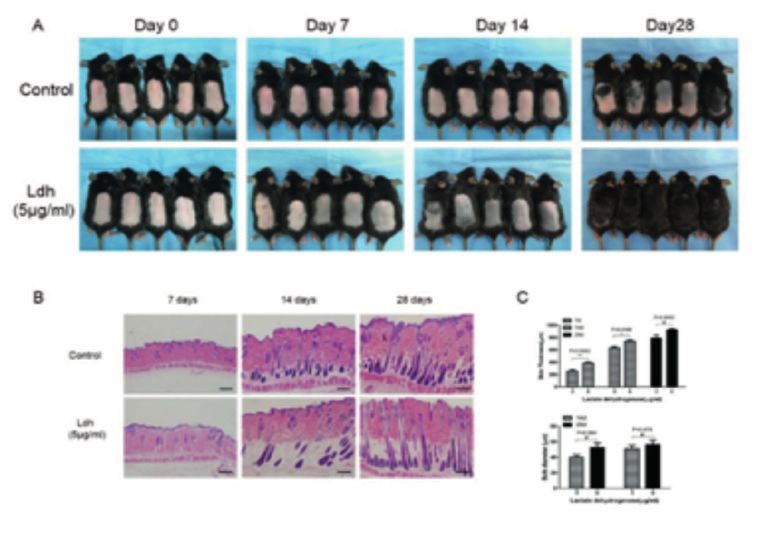
686			                                                                                       Jun Ye, Qing Zhou et Al

lysis, LDH enzyme is able to increasingly convert                 Vibrissae follicle (VF) organ culture and
pyruvate to lactate, which can considerably up-reg-         length measurement of VFs
ulate the activity of HFSCs and stimulate quickly a               VFs in anagen phase were isolated from
series of reactions that initiate next hair cycle(8). Be-   the upper lip pad of 10 four-week-old female
sides its metabolic role, LDH has many other func-          C57BL/6J mice and cultured in Williams E me-
tions, including potential wound healing activity,          dium (Sigma-Aldrich, W1878, USA) supplement-
maintenance of the ability of intestinal stem cells to      ed with 2mmol/L L-glutamine (Gibco, 25030081,
proliferate and differentiate and control of the intrin-    USA), 10mg/ml human insulin (Sigma-Aldrich),
sic mechanisms of stem cell(9-10).                          10ng/ml hydrocortisone (Gibco), 1× penicillin, and
      Several previous studies have shown that elec-        streptomycin solution (Gibco)at 37 °C in a 5% CO2
tron transport chain (ETC) inhibition can be used           atmosphere, as previously described(11-12).
to accelerate the hair cycle(8), however, there is no             After isolation, VFs were incubated individ-
direct evidence that LDH is able to promote DPCs            ually in 24-well plates and were randomized into
proliferation and increase hair follicle growth in vit-     four groups with six VFs in each group. VFs were
ro. Therefore, in this study, we aimed to investigate       cultured in the presence or absence of 1-10μg/ml
whether LDH affects the growth of hair follicle, and        L-Lactic dehydrogenase (Solarbio, Cat, no, L8080;
explore the underlying signalling pathways.                 from rabbit muscle) for 3 days. Length of VFs was
                                                            measured using an inverted microscope (IX71;
Materials and methods                                       Olympus Optical Co. Ltd, Tokyo, Japan). The hair
                                                            shaft growth length was measured on days 0, 1, and
     Animals                                                3 at the same time point.
     A total of 30 Four-week-old female wild type                 The hair cycles were assessed macroscopical-
C57BL/6J mice were obtained from the Experimental           ly, according to the previously described morpho-
Animal Centre for all topical experiments. All animal       logical criteria(13).
experiments were carried out under the approval of
the Institutional Animal Care and Use Committee at                Ki-67 and TUNEL double immunofluores-
Southern Medical University in Guangzhou, China.            cence staining
                                                                  To evaluate the proportion of proliferative and
      Immunohistochemistry                                  apoptotic cells in each intervention group, a Ki-67/
      In order to describe the LDH expression in            TUNEL double staining method was used. VFs were
mouse dorsal skin during anagen to telogen phase of         treated in medium with different concentrations of
the hair follicle. Tissues were isolated from sections      LDH (5 and 10 μg/ml).
of dorsal skin and fixed with 4% paraformaldehyde                 After cultured for 72 h, selected VFs in anagen
overnight. For immunohistochemistry analysis, par-          phase were fixed with 4% paraformaldehyde for 24
affin-embedded tissue slides were prepared with             h. Paraffin embedded tissue sections were obtained
4-μm-thick cuts.                                            with 4-μm-thick cuts.
      The slices were dewaxed in xylene, and rehy-                The sections were dewaxed in xylene, rehy-
drated in a series of graded ethanol rinses. After an-      drated in a series of graded ethanol, and heated in
tigen retrieval in sodium citrate buffer, endogenous        citrate buffer (pH6.0) for 2 min at 100 °C. Subse-
peroxidase activity was blocked with 10% normal             quently, the sections were permeabilized with 0.1%
goat serum 20 min and the sections were incubated           TritonTMX-100 for 10 min, blocked with 10% BSA
with rabbit anti-mouse LDHA antibody (Cell Sig-             in PBST for 30 min, and then incubated with rabbit
naling CST3582; 1:400) at 4 °C overnight. After             anti-mouse Ki-67 primary antibody(Abcam; ab9260;
incubation, the slices were washed 3 times in phos-         1:100) at 4 °C overnight, followed by treatment with
phate buffered saline (PBS) for 3 min and incubated         donkey anti-rabbit lgG secondary antibody (Life-
with goat anti-rabbit secondary antibody (Abcam;            technologies; A21207; 1:200) and TUNEL Kit(Ro-
ab6721; 1:500) for 20 min at room temperature. The          che; Cat. no. 11684817910).
antibody-antigen complexes were visualized with                   Nuclei were stained with 4’, 6-diamidi-
diaminobenzidine.                                           no-2-phenylindole (DAPI) in SlowFade Gold An-
      Slides were then briefly counterstained with          tifade Mountant (Thermo Scientific; S36938). The
haematoxylin and sealed for subsequent visualis-            sections were photographed under upright fluores-
ation using an upright microscope.                          cence microscope (BX51; Olympus, Japan).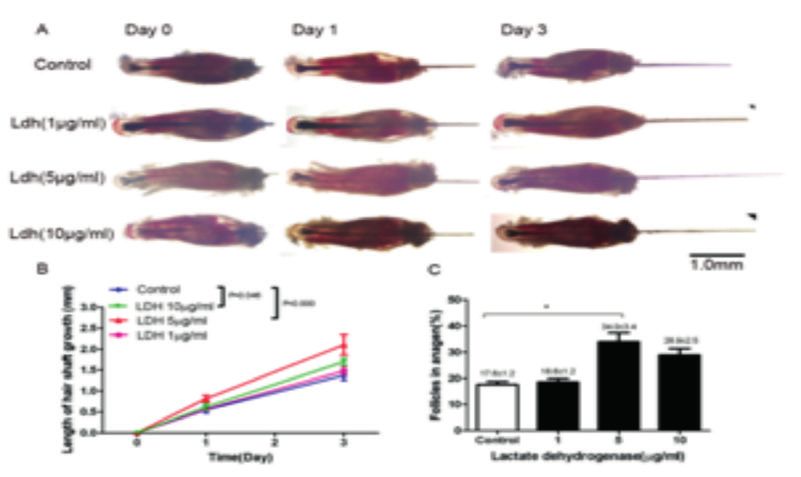
Lactate dehydrogenase has the potential to increase mouse hair follicle growth by WNT/ß-catenin signaling            687

      Isolation and culture of dermal papilla cells                 viously reported(15). 10 four-week-old female
      The method used for isolating and culturing                   C57BL/6J mice were anesthetized and the dor-
DPCs has been previously described(14). Briefly,                    sal skin of all mice was depilated to induce fully
DPCs were dissected from vibrissae follicles of 10                  synchronised anagen stages. After depilation, the
four-week-old female C57BL/6J mice and trans-                       mice were randomly divided into a control and the
ferred onto plastic dishes coated with type IV col-                 LDH-treated group.
lagen (Sigma) for 3 h, then cultured in Dulbecco’s                       For the LDH-treated group (n=5), a total vol-
Modified Eagle’s Medium (DMEM) (Gibco) con-                         ume of 100μl DMEM (containing 5μg/ml LDH)
taining 10,000 μg/ml streptomycin, 10,000 Units/ml                  was injected subcutaneously into the dorsal site,
penicillin, and 10% fetal bovine serum (FBS) (Gib-                  the negative control group (n=5) was injected with
co) at 37 °C in a humidified atmosphere with 5%                     DMEM without LDH.
CO2. Once cell grow to 80% confluence, the cells                         Images were captured 0, 7, 14, 28 days at fol-
were harvested with 0.25% Trypsin-EDTA (Gibco)                      lowing time, and the mice were euthanatized on
in Hank balanced salt solution, and subcultured with                days 7, 14, and 28 at the same time point. Mean-
a split ratio of 1:3.                                               while, the dorsal skin samples of the mice were
      The medium was changed every 3 days.                          harvested, fixed in 4% paraformaldehyde at room
Fourth-passage of confluent DPCs was cultured for                   temperature for 48 h, and paraffin-embedded.
24 h in DMEM, and then treated for 1 day with vari-                      Further, 4 mm thick serial slices from the dor-
ous concentrations of LDH (5-10μg/ml).                              sal skin were stained with haematoxylin and eosin
                                                                    (HE). The sections were observed under an upright
      Western blotting                                              microscope (BX51; Olympus, Japan).
      DPCs at fourth-passage were lysed in RIPA
buffer in the presence of protease inhibitors (Sell-                     Statistical analysis
eck Chemicals, Houston, USA). Protein concentra-                         One-way analysis of variance (ANOVA) was
tions were determined using the Bradford method                     performed for comparison of different groups, using
(Bioss, Beijing, China), and samples were denatured                 SPSS17.0 software.
in protein loading buffer (Bio-RAD) for 5 min at                         All data from three independent experiments
100 °C. Total protein extracts (50 μg) were sub-                    were expressed as mean ± SEM. A p-value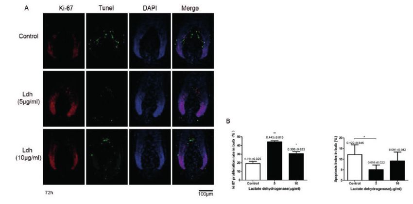
688 Jun Ye, Qing Zhou et Al
LDH promotes the proliferation of hair ma-
trix keratinocytes
To demonstrate that LDH promotes hair folli-
cle growth, Ki-67 and TUNEL double fluorescence
staining experiment was performed after 3 days of
hair follicle organ culture. After treatment with LDH
(5 and 10 μg/ml) for 72 h culture, significant stim-
ulation of keratinocyte proliferation in hair matrix
was detected (by Ki-67 staining) compared with the
control group (Fig. 3A and B).
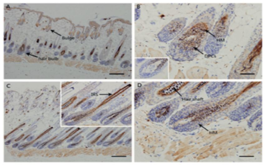
Figure 1: The expression of LDH in dorsal skin.
Macroscopic Immunohistochemistry image of the dorsal
skin in anagen stage.
(A) LDH deposition was found in DPCs and HM. (B) During this
period, the DPCs were surrounded by the hair bulb and became
larger, not fully enclosed by the bulb keratinocytes. The inserted
figure represents the negative control group.(C) Little LDH
immunoreactivity was found in the DPCs during the short anagen
V/VI phase, while some immunoreactivity were found in hair
shaft and IRS.(D) The hair bulb was enlarged and the DPCs were
contracted. DPCs, dermal papilla cells; HM, hair matrix; IRS, inner Figure 3: Effect of LDH on keratinocyte proliferation in
root sheath. Staining with LDHA antibody, original magnification hair matrix.
(A.C) × 40, Scale bars: 400 µm; (B.D) × 400, Scale bars: 100 µm. (A) Compared with the control group, more Ki-67 positive cells
(red) were seen in the hair matrix surrounding the DPCs of VFs
LDH significantly increases hair shaft elon- after treatment with LDH (5 and 10 μg/ml) for 72 h. Limited
immunostaining for TUNEL (green) were observed in the LDH-
gation and the percentage of VFs treated corresponding groups. Original magnification ×400, Scale
VFs were cultured with various concentrations bars: 100 μm. (B) The LDH-treated groups (5 and 10 μg/ml)
of LDH (1-10 μg/ml) for 3 days in vitro. Compared significantly increased the expression of proliferation related genes
with the untreated control group, LDH at concentra- (observed by Ki-67 staining) in bulb compared to the untreated
tions of 5 μg/ml resulted in significantly increased group. By contrast, 5 μg/ml LDH significantly decreased the
expression of apoptosis related genes in the hair bulb area. Data
hair shaft elongation (Fig. 2A and B). In addition, 5 were mean ± SEM of percentages vs. controls (*PLactate dehydrogenase has the potential to increase mouse hair follicle growth by WNT/ß-catenin signaling 689
LDH induced earlier onset of anagen stage
compared with the vehicle-treated group. Histology
showed that LDH-treated hair follicles entered into
the telogen-anagen transition typically after 14 days,
and this was accompanied by typical expansion of
the hypodermis underneath. However, in the back
skin with deletion of LDH, the hypodermis did not
expand (Fig. 5B). In addition, in the back skin treat-
ed with LDH (5 μg/m), the thickness of the skin was
significantly increased compared with that of the
Figure 4: Western blot analysis showing the expression control group at day 7 and 14 (Fig. 5C).
of four proteins (ALP, LEF-1, Wnt5a and ß-catenin) in
DPCs.
(A) Western blotting (WB) was carried out to analyse the Discussion
intrinsic properties of DPCs and their correlation with Wnt/ß-
catenin signalling in three different groups (0, 5 and 10 μg/ml In previous studies, hair follicle stem cells
LDH). (B) The relative protein expressions of ALP and LEF-1 (HFSCs) had been proven to play an important role
in the LDH-treated (5 and 10 μg/ml) group were significantly
up-regulated. Analysis of the expression of Wnts and signal
in maintaining the growth cycle of hair follicles;
transduction cascade in DPCs has shown that Wnt5a and while in the early stage of hair follicle growth, der-
ß-catenin expressions in the LDH-treated (5 and 10 μg/ml) group mal papilla cells (DPCs), as potential mesenchymal
increased significantly (*P690 Jun Ye, Qing Zhou et Al
Next, we further confirmed that LDH Jan 7) 92-105.
can significantly stimulate the proliferation of 4) C.C. Yang, G. Cotsarelis, Review of hair follicle dermal
cells, J Dermatol Sci 57 (2010) 2-11.
intracellular keratinocytes in hair follicle during 5) J. Kishimoto, R.E. Burgeson, B.A. Morgan, Wnt sign-
organ culture in vitro by Ki-67 and TUNEL double aling maintains the hair-inducing activity of the dermal
fluorescence staining. Considering the relationships papilla, Genes Dev 14 (2000) 1181-1185.
between hair matrix keratinocytes and DPCs(24), 6) D. Jelinek, A. Flores, M. Uebelhoer, V. Pasque, K. Plath,
M.L. Iruela-Arispe, H.R. Christofk, W.E. Lowry, H.A.
we speculate that LDH may influence hair growth Coller, Mapping Metabolism: Monitoring Lactate Dehy-
by altering intrinsic characteristics of DPCs. drogenase Activity Directly in Tissue, J Vis Exp (2018).
Previous studies had shown that many signalling 7) A. Flores, J. Schell, A.S. Krall, D. Jelinek, M. Miran-
pathways such as WNT, BMP, and FGF played an da, M. Grigorian, D. Braas, A.C. White, J.L. Zhou, N.A.
important role in the hair follicle morphogenesis and Graham, T. Graeber, P. Seth, D. Evseenko, H.A. Coller,
J. Rutter, H.R. Christofk, W.E. Lowry, Lactate dehydro-
development, of which WNT/ß-catenin signalling genase activity drives hair follicle stem cell activation,
pathway was the most classical one(25-26). Therefore, Nat Cell Biol 19 (2017) 1017-1026.
we assumed that LDH possibly activated the 8) C.H. Miranda M, Jones DL, Lowry WE, Topical Inhibi-
expression of related proteins in the Wnt/ß-catenin tion of the Electron Transport Chain Can Stimulate the
Hair Cycle, J Invest Dermatol 138 (2018 Apr) 968-972.
signalling pathway during the stimulation of DPCs, 9) J.C. Schell, D.R. Wisidagama, C. Bensard, H. Zhao, P.
to induce differentiation of epidermal cells. Wei, J. Tanner, A. Flores, J. Mohlman, L.K. Sorensen,
Furthermore, western blot analysis showed that C.S. Earl, K.A. Olson, R. Miao, T.C. Waller, D. Delker,
LDH stimulated the high expression of ALP and P. Kanth, L. Jiang, R.J. DeBerardinis, M.P. Bronner,
LEF-1, which are both signature proteins in DPCs D.Y. Li, J.E. Cox, H.R. Christofk, W.E. Lowry, C.S.
Thummel, J. Rutter, Control of intestinal stem cell func-
[16.17]. This also suggested that LDH induced hair tion and proliferation by mitochondrial pyruvate metab-
follicle growth and participated in the hair cycle by olism, Nat Cell Biol 19 (2017) 1027-1036.
changing the biological characteristics and functions 10) M. Ito, Y. Liu, Z. Yang, J. Nguyen, F. Liang, R.J. Mor-
of DPCs. Meanwhile, the high expression of the ris, G. Cotsarelis, Stem cells in the hair follicle bulge
contribute to wound repair but not to homeostasis of the
related proteins Wnt5a, ß-catenin, and LEF-1 also epidermis, Nat Med 11 (2005) 1351-1354.
confirmed that LDH might stimulate the activity of 11) D.J. Tobin, Ex vivo organ culture of human hair follicles:
DPCs through Wnt/ ß-catenin signalling pathway, a model epithelial-neuroectodermal-mesenchymal inter-
thus promoting the proliferation of HFSCs and in- action system, Methods Mol Biol 695 (2011) 213-227.
ducing the formation of a new hair cycle. 12) Y. Su, H. Liu, J. Wang, B. Lin, Y. Miao, Z. Hu, Antimi-
crobial peptide lysozyme has the potential to promote
Finally, the in vivo experimental results sup- mouse hair follicle growth in vitro, Acta Histochem 117
port our hypothesis. The new growth cycle of the (2015) 798-802.
hair follicle was found to be advanced when LDH 13) E. Kloepper, K. Sugawara, Y. Al-Nuaimi, E. Gaspar,
was injected into the subcutaneous part of the back N. van Beek, R. Paus, Methods in hair research: how
to objectively distinguish between anagen and catagen
skin of mice. Taken together, LDH, as a rate-limit- in human hair follicle organ culture, Exp Dermatol 19
ing enzyme in glycolytic metabolism, can induce the (2010) 305-312.
intrinsic properties of DPCs and may promote hair 14) A. Osada, T. Iwabuchi, J. Kishimoto, T.S. Hamazaki, H.
follicle growth through WNT signalling pathway. Okochi, Long-term culture of mouse vibrissal dermal
papilla cells and de novo hair follicle induction, Tissue
Eng 13 (2007) 975-982.
15) S. Muller-Rover, B. Handjiski, C. van der Veen, S.
Eichmuller, K. Foitzik, I.A. McKay, K.S. Stenn, R.
Paus, A comprehensive guide for the accurate classifica-
tion of murine hair follicles in distinct hair cycle stages,
J Invest Dermatol 117 (2001) 3-15.
16) K. Yamauchi, A. Kurosaka, Inhibition of glycogen
References synthase kinase-3 enhances the expression of alkaline
phosphatase and insulin-like growth factor-1 in human
1) A.C. Gupta, S. Chawla, A. Hegde, D. Singh, B. Bandy- primary dermal papilla cell culture and maintains mouse
opadhyay, C.C. Lakshmanan, G. Kalsi, S. Ghosh, Es- hair bulbs in organ culture, Arch Dermatol Res 301
tablishment of an in vitro organoid model of dermal pa- (2009) 357-365.
pilla of human hair follicle, J Cell Physiol 233 (2018) 17) Y.L. Wang HD, Yu XJ, He JP, Fan LH, Dong YJ, Dong
9015-9030. CS, Liu TF, Immunolocalization of β-catenin and Lef-1
2) K. Krause, K. Foitzik, Biology of the hair follicle: the during postnatal hair follicle development in mice, Acta
basics, Semin Cutan Med Surg 25 (2006) 2-10. Histochem 114(8) (2012 Dec).
3) P.H. Hsu YC, Fuchs E., Dynamics between stem cells, 18) R.R. Driskell, C. Clavel, M. Rendl, F.M. Watt, Hair
niche, and progeny in the hair follicle, Cell 144 (2011 follicle dermal papilla cells at a glance, J Cell Sci 124Lactate dehydrogenase has the potential to increase mouse hair follicle growth by WNT/ß-catenin signaling 691
(2011) 1179-1182.
19) S.E. Millar, Molecular mechanisms regulating hair folli-
cle development, J Invest Dermatol 118 (2002) 216-225.
20) M.R. Schneider, R. Schmidt-Ullrich, R. Paus, The hair
follicle as a dynamic miniorgan, Curr Biol 19 (2009)
R132-142.
21) Z.Y. Xian, J.M. Liu, Q.K. Chen, H.Z. Chen, C.J. Ye, J.
Xue, H.Q. Yang, J.L. Li, X.F. Liu, S.J. Kuang, Inhibition
of LDHA suppresses tumor progression in prostate can-
cer, Tumour Biol 36 (2015) 8093-8100.
22) J.A. Qiu H, Kilgore JE, Zhong Y, Chan LL, Gehrig PA,
Zhou C, Bae-Jump VL, JQ1 suppresses tumor growth Acknowledgement:
through downregulating LDHA in ovarian cancer, On- Fund Project: The National Natural Science Foundation of Chi-
cotarget 6 (2015 Mar 30) 6915-6930. na (NSFC. 81701938). Zhuzhou Social Public Program in Sci-
23) X. Liu, Z. Yang, Z. Chen, R. Chen, D. Zhao, Y. Zhou, ence and Technology (2018SFK1-013). Natural Science Foun-
L. Qiao, Effects of the suppression of lactate dehydro- dation of Guangxi Province (2018GXNSFBA050025).
genase A on the growth and invasion of human gastric
cancer cells, Oncol Rep 33 (2015) 157-162.
24) V.A. Botchkarev, J. Kishimoto, Molecular control of
epithelial-mesenchymal interactions during hair follicle –––––––––
cycling, J Investig Dermatol Symp Proc 8 (2003) 46-55. Corresponding Author:
25) V. Greco, T. Chen, M. Rendl, M. Schober, H.A. Pasolli, Guoqian Yin, Bojie Lin
N. Stokes, J. Dela Cruz-Racelis, E. Fuchs, A two-step Department of Plastic and Aesthetic Surgery, The First Affiliat-
mechanism for stem cell activation during hair regener- ed Hospital of Guangxi Medical University, Nanning, Guangxi,
ation, Cell Stem Cell 4 (2009) 155-169. 530021, China.
26) M. Rendl, L. Polak, E. Fuchs, BMP signaling in dermal Tel: +86 138 0781 7282; +86 183 7715 3798
papilla cells is required for their hair follicle-inductive Email: yingq61@163.com; linbojie@aliyun.com
properties, Genes Dev 22 (2008) 543-557. (China)You can also read