Original Article Herbacetin treatment remitted LPS induced inhibition of osteoblast differentiation through blocking AKT/NF-κB signaling pathway
←
→
Page content transcription
If your browser does not render page correctly, please read the page content below
Am J Transl Res 2019;11(2):865-874
www.ajtr.org /ISSN:1943-8141/AJTR0086459

Original Article
Herbacetin treatment remitted LPS
induced inhibition of osteoblast differentiation
through blocking AKT/NF-κB signaling pathway
Pengshan Cai, Teng Cai, Xiaobin Li, Lei Fan, Guang Chen, Bofan Yu, Tao Liu

Department of Orthopedics, Henan Provincial People’s Hospital, People’s Hospital of Zhengzhou University,
Zhengzhou 450000, Henan Province, China
Received June 21, 2018; Accepted December 29, 2018; Epub February 15, 2019; Published February 28, 2019

Abstract: Inflammation, a common situation during the process of bone healing, is reported to play a negative
role in bone regeneration. Up to date, therapeutic strategies for inflammation triggered inhibition of osteoblast dif-
ferentiation are still limited. The aim of this study was to explore the potential roles and molecular mechanisms of
Herbacetin in the process of osteoblast differentiation under LPS-mediated inflammatory environment. By using
MC3T3-E1, C2C12 and primary mouse calvarial osteoblast (PMCO) cells as experimental models, we observed that
LPS stimulation suppressed osteoblast differentiation via inhibiting alkaline phosphatase (ALP) activity and the ex-
pression of several osteoblastic genes (osterix, runx2 and osteocalcin). However, the negative role of LPS during os-
teoblast differentiation could be restored by Herbacetin treatment. Mechanistical studies revealed that Herbacetin
treatment suppressed AKT activation and in turn blocked NF-κB signaling pathway. Furthermore, reactivating AKT
by a selective PTEN inhibitor SF1670 suppressed the effect of Herbacetin. These data suggested that Herbacetin
might play a protective role in osteoblast differentiation in MC3T3-E1/C2C12/PMCO cells under LPS stimulation.

Keywords: Herbacetin, LPS, osteoblast differentiation, AKT/NF-κB

Introduction                                                   Herbacetin (3,4,5,7,8-pentahydroxyflavone), an
                                                               active flavonol compound extracted from pl-
The recovery process of bone fracture involves                 ants, exhibited anti-cancer efficacy and strong
in the reprograming of multiple gene expres-                   antioxidant capacity [6-8]. Recently, the anti-
sion as well as the microenvironment [1].                      inflammatory role of Herbacetin was reported
Despite bone formation during fetal skeletal                   in RAW264.7 macrophage cells. In their study,
development and postnatal fracture repair                      Li et al. demonstrated that pretreatment of
share similar processes such as differentia-                   Herbacetin decreased nitric oxide (NO) pro-
tion from mesenchymal stem cells (MSCs) to                     duction in LPS-induced RAW264.7 cells and
osteoblasts, fracture healing is initiated by an               primary macrophages derived from mouse
inflammatory phase during which cellular activi-               bone marrow [9]. However, whether Herbacetin
ties could be regulated by multiple inflammato-                had anti-inflammatory role during osteoblast
ry factors [2, 3]. Lipopolysaccharide (LPS), the               differentiation was still elusive. The aim of this
main pathogenic component of gram-negative                     study was to evaluate the potential roles and
bacteria, is a commonly used stimulator of                     mechanisms of Herbacetin on osteoblast differ-
inflammatory response in cell experiments.                     entiation under LPS-mediated inflammatory
LPS can increase the synthesize and release of                 environment.
IL-6 and other inflammatory factors, activate
pro-inflammatory signaling pathways and thus                   Materials and methods
inhibit osteoblast differentiation [4, 5]. There-
fore, the research based on LPS-mediated inhi-                 Cell culture
bition of osteoblast differentiation might pro-
vide novel opportunity to enhance osteoblastic                 Animal care and the experimental protocol in
differentiation and bone formation.                            this study were approved by the ethics commit-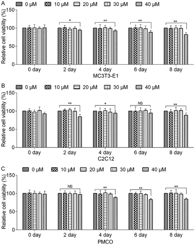
Herbacetin remitted LPS induced inhibition of osteoblast differentiation

Table 1. Primers for osterix, runx2, osteocal-     μM) for 8 days. At the same time point of each
cin, and GAPDH                                     day, 20 μl CCK-8 (Dojindo laboratories) were
Name                    Sequence (5’-3’)           added into each well and OD 450 was mea-
                                                   sured by spectrophotometry (BioTek) 2 h after
Runx2 (Foward)          GCTTGATGACTCTAAACCTA
                                                   incubation.
Runx2 (Reverse)         AAAAAGGGCCCAGTTCTGAA
Osterix (Foward)        AGGCACAAAGAAGCCATAC        Protein extraction and western blot assay
Osterix (Reverse)       AATGAGTGAGGGAAGGGT
Osteocalcin (Foward)    CTCACTCTGCTGGCCCTG         Total cellular protein was lysed using RIPA buf-
Osteocalcin (Reverse)   CCGTAGATGCGTTTGTAGGC       fer (Solarbio, Beijing, China) containing 1%
                                                   PMSF (Solarbio, Beijing, China), protease inhibi-
                                                   tors and phosphatase inhibitors (Selleckchem).
tees of Henan Provincial People’s Hospital.        Nuclear/cytoplasm protein was extracted using
Primary mouse calvarial osteoblasts (PMCO)         the Nuclear and Cytoplasmic Protein Extraction
were obtained from 3-day-old NIH mice calvar-      Kit (Beyotime, Haimen, China). Proteins were
ias following the sequential enzymatic diges-      separated by 10% SDS-polyacrylamide gels
tion method. In brief, skulls were dissected,      and electrotransferred onto polyvinylidene fluo-
and the endosteum and periosteum were              ride (PVDF) membranes (Invitrogen). The mem-
stripped off. Then the bone was cut into approx-   branes were blocked with 5% non-fat milk, in-
imately 1-2 mm2 pieces and subjected to five       cubated with primary antibodies at 4°C over-
sequential digestions at 37°C in 0.1% dispase      night, and then incubated with secondary anti-
and 0.1% collagenase P solution. Finally, the      bodies. Purpose bands were visualized using
Second to Fifth digestions were collected fol-     enhanced chemiluminescence (ECL) system
lowing centrifuge. Preosteoblastic cell line       (Pierce, Thermo Fisher Scientific, Inc.). Primary
MC3T3-E1 and mouse myoblast cell line C2C12        antibodies used in this study were anti-NF-κB
were purchased from the American Type              p65 (8242, Cell Signaling Technology), anti-
Culture Collection (ATCC, Manassas, VA, USA).      Lamin B (13435, Cell Signaling Technology),
MC3T3-E1 and PMCO cells were maintained in         anti-AKT (2920, Cell Signaling Technology),
a-minimum essential medium (a-MEM) contain-        anti-pAKT Thr308 (13038, Cell Signaling Techno-
ing 10% fetal bovine serum (Invitrogen) and 1%     logy), anti-pAKTSer473 (4060, Cell Signaling Tech-
penicillin/streptomycin (Sigma-Aldrich). C2C12     nology) and anti-GAPDH (60004, Proteintech).
cells were cultured in Dulbecco’s modified         Secondary antibodies were purchased from
Eagle’s medium (DMEM) containing 10% fetal         Cell Signaling Technology.
bovine serum and 1% penicillin/streptomycin
(Sigma-Aldrich). Herbacetin (purity: >90% by       Pull-down assay
HPLC) was obtained from Sigma-Aldrich.
                                                   MC3T3E1/C2C12 cell lysates were incubated
Quantitative real time polymerase chain reac-      with herbacetin-Sepharose 4B or Sepharose
tion (qPCR)                                        4B beads (50 μl, 50% slurry, GE Healthcare,
                                                   Piscataway, NJ) in reaction buffer (50 mM Tris
RNA was extracted by using the GenEluteTM          pH 7.5, 5 mM EDTA, 150 mM NaCl, 1 mM DTT,
Total RNA Purification Kit (Sigma-Aldrich).        0.01% NP40, 2 μg/mL bovine serum albumin)
Reverse transcription reaction was conducted       at 4°C overnight. Then the beads were washed
by ReverTra Ace qPCR RT Kit (TOYOBO). The          with elution buffer (50 mM Tris pH 7.5, 5 mM
PCR primers for osterix, runx2, osteocalcin,       EDTA, 150 mM NaCl, 1 mM DTT, 0.01% NP40)
and GAPDH was listed in Table 1. Gene expres-      and binding was visualized by Western blotting
sion was quantitated by using the 2-ΔΔCT method    assay.
[10].
                                                   Osteogenic induction
Cell viability assay
                                                   MC3T3-E1 cells were cultured in α-MEM sup-
MC3T3-E1/C2C12 cells were seeded into a            plemented with 10 mM β-glycerophosphate
96-well plate (500 cells per well per 200 μl)      (G6251, Sigma-Aldrich), 10 nM dexamethasone
and incubated with indicated concentrations of     (D1756, Sigma-Aldrich), and 50 μg/ml ascorbic
Herbacetin (0 μM, 10 μM, 20 μM, 30 μM, 40          acid (A5960, Sigma-Aldrich). C2C12 cells were

866                                                          Am J Transl Res 2019;11(2):865-874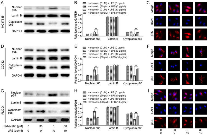
Herbacetin remitted LPS induced inhibition of osteoblast differentiation

                                                                               Phosphatase Color Develop-
                                                                               ment Kit (Beyotime, Haimen,
                                                                               China) according to the manu-
                                                                               facturer’s protocol.

                                                                               ALP activity assay

                                                                               ALP enzymatic activity was
                                                                               examined by the ALP activity
                                                                               assay kit (Beyotime, Haimen,
                                                                               China). The cells were seeded
                                                                               in 96-well plates at a density
                                                                               of 5 × 10³ cells/well. ALP
                                                                               activity was analyzed on cul-
                                                                               ture days 3, 7, 14 and 21,
                                                                               according to the manufactur-
                                                                               er’s instructions. The results
                                                                               were measured at 405 nm by
                                                                               spectrophotometry (BioTek).

                                                                               Immunofluorescence assay

                                                                                  MC3T3-E1/C2C12 cells were
                                                                                  fixed with 4% paraformalde-
                                                                                  hyde for 15 min. After washed
                                                                                  with PBS, cells were permea-
                                                                                  bilized with 0.5% NP-40
                                                                                  (Sigma-Aldrich, St Louis, MO,
                                                                                  USA) for 10 min. Then cells
                                                                                  were blocked with 10% nor-
                                                                                  mal goat serum for 30 min,
                                                                                  incubated with anti-NF-κB
Figure 1. Cytotoxicity of Herbacetin treatment on MC3T3-E1/C2C12/PMCO
cells. A. MC3T3-E1 cells were treated with various concentrations of Her-
                                                                                  p65 at 4°C overnight. The
bacetin for 0 day, 2 days, 4 days, 6 days and 8 days. Cell viability was de-      second day, sections were
tected by CCK-8 assay. B. Effect of Herbacetin treatment on C2C12 cells. C.       incubated with Alexa Fluor®
Effect of Herbacetin treatment on the viability of PMCO cells. Representative     555 goat anti-rabbit IgG (H+L)
data from at least 3 independent experiments are shown. Data are shown as         (Invitrogen, Carlsbad, CA) at
mean ± SD. NS, not significant; *P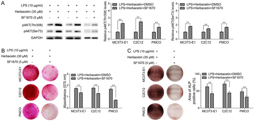
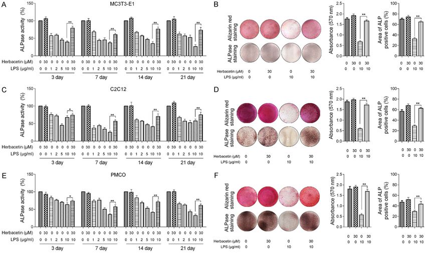
Herbacetin remitted LPS induced inhibition of osteoblast differentiation

Figure 2. Herbacetin treatment remitted LPS induced inhibition of osteoblast differentiation. MC3T3-E1/C2C12/PMCO cells were treated with various concentra-
tions of LPS in the presence or absence of Herbacetin in a time-course experiment. Quantification assay of ALPase activity was presented in (A) (MC3T3-E1), (C)
(C2C12) and (E) (PMCO). ALP staining and Alizarin Red S staining was presented and quantified in (B) (MC3T3-E1), (D) (C2C12) and (F) (PMCO). Representative data
from at least 3 independent experiments are shown. Data are shown as mean ± SD. *P
Herbacetin remitted LPS induced inhibition of osteoblast differentiation

Figure 3. Effect of Herbacetin on the expression of osteoblastic genes in MC3T3E1/C2C12/PMCO cells. A-C.
MC3T3E1 cells were treated with LPS or/and Herbacetin for 7 days. Total RNA was extracted and the expression of
Runx2, osterix, and osteocalcin was quantified by qPCR. D-F. C2C12 cells were treated with LPS or/and Herbacetin
for 7 days. The mRNA levels of Runx2, osterix, and osteocalcin was quantified by qPCR. G-I. Herbacetin restored
the expression of Runx2, osterix, and osteocalcin in PMCO cells. Representative data from at least 3 independent
experiments are shown. Data are shown as mean ± SD. *P
Herbacetin remitted LPS induced inhibition of osteoblast differentiation

Figure 4. Effect of Herbacetin on NF-κB signaling pathway. (A, B) MC3T3E1 cells were treated with LPS or/and
Herbacetin for 7 days. Protein was extracted and the nuclear/cytoplasmic NF-κB p65 was detected by western
blot assay. (C) Cytoplasmic-nuclear translocation of NF-κB p65 was detected by immunofluorescence assay. (D, E)
Nuclear/cytoplasmic protein was extracted after C2C12 cells were treated with LPS or/and Herbacetin for 7 days.
NF-κB p65 in subcellular fractions was detected by western blot assay. (F) Subcellular localization of NF-κB p65 in
C2C12 cells. (G, H) Nuclear/cytoplasmic NF-κB p65 in PMCO cells was detected by western blot assay and the result
was confirmed by immunofluorescence assay (I). Representative data from at least 3 independent experiments are
shown. Data are shown as mean ± SD. *P
Herbacetin remitted LPS induced inhibition of osteoblast differentiation

                                                                             noma cell line SK-MEL-5,
                                                                             which provided a reasonable
                                                                             explanation for Herbacetin
                                                                             mediated AKT inhibition [6]. In
                                                                             this study, we confirmed that
                                                                             Herbacetin could also bind to
                                                                             AKT in MC3T3-E1/C2C12/
                                                                             PMCO cells by pull-down as-
                                                                             say (Figure 5A). To further cla-
                                                                             rify the effect of Herbacetin
                                                                             on AKT signaling pathway
                                                                             in MC3T3-E1/C2C12/PMCO
                                                                             cells, we then detected the
                                                                             phosphorylation levels of AKT
                                                                             under Herbacetin treatment.
                                                                             The result of western blot
                                                                             assay revealed that Herba-
                                                                             cetin treatment could signifi-
                                                                             cantly suppress the phos-
                                                                             phorylation of pAKT Thr308 and
                                                                             pAKTSer473 (Figure 5B and
                                                                             5C). Taken together, Herba-
                                                                             cetin induced AKT inactiva-
Figure 5. Effect of Herbacetin on AKT signaling pathway. A. MC3T3E1/         tion might be involved in the
C2C12/PMCO cell lysates were incubated with herbacetin-conjugated Sep-       repression of NF-κB signal-
harose 4B beads or Sepharose 4B beads alone. Proteins were pulled down
and detected by anti-AKT. B. MC3T3E1/C2C12/PMCO cells were treated
                                                                             ing pathway and osteoblast
with Herbacetin for 7 days. Protein was extracted and the phosphorylation    differentiation.
level of AKT was detected by western blot assay. C. The result of western
blot assay was quantified. Representative data from at least 3 independent   Reactivation of AKT abrogat-
experiments are shown. Data are shown as mean ± SD. *P
Herbacetin remitted LPS induced inhibition of osteoblast differentiation Figure 6. Reactivation of AKT signaling pathway abrogated the effect of Herbacetin in osteoblast differentia- tion. MC3T3-E1/C2C12/PMCO cells were treated with LPS (10 μg/ml), Herbacetin (30 μM) together with/without SF1670 (5 μM). A. The protein levels of pAKT was detected by western blot and the result showed SF1670 treatment promoted the levels of pAKT. B. Alizarin Red S staining showed lower absorbance at 570 nm in cells treated with SF1670. C. ALP staining showed significantly decreased proportion of ALP positive cells in SF1670 treated group. Representative data from at least 3 independent experiments are shown. Data are shown as mean ± SD. *P
Herbacetin remitted LPS induced inhibition of osteoblast differentiation

differentiation, we first screened Herbace-          Herbacetin in MC3T3-E1/C2C12/PMCO cells.
tin-related articles. We noticed that the anti-      Herbacetin exhibited great biocompatibility
inflammatory role of Herbacetin was tightly cor-     with osteoblasts, and have the potential to be
related with the activation of NF-κB signaling       developed as a drug for bone and tissue engi-
pathway. In RAW264.7 and mouse bone mar-             neering in the future.
row-derived macrophages, LPS induced activa-
tion of the JNK and NF-κB pathway could be           Disclosure of conflict of interest
blocked by Herbacetin treatment [9]. Me-
anwhile, previous literatures also revealed that     None.
Herbacetin suppressed AKT pathway in breast          Address correspondence to: Tao Liu, Department of
cancer, melanoma, hepatocellular carcinoma           Orthopedics, Henan Provincial People’s Hospital,
and lung cancer cells [6-8]. Thus, we first          People’s Hospital of Zhengzhou University, 7 Weiwu
extracted cytoplasm and nuclear proteins             Road, Zhengzhou 450000, Henan Province, China.
respectively to confirm LPS stimulation could        Tel: 86-0371-65580172; E-mail: liutaozzu2000@
induce p65 nuclear translocation in MC3T3-E1/        outlook.com
C2C12/PMCO cells. The results of western blot
assay suggested that LPS stimulation upregu-         References
lated nuclear p65 level, which could be minified
by Herbacetin treatment. These results were          [1]   Gomez-Barrena E, Rosset P, Lozano D, Stanovi-
further confirmed by immunofluorescence as-                ci J, Ermthaller C and Gerbhard F. Bone frac-
say. Interestingly, we also observed that Her-             ture healing: cell therapy in delayed unions
bacetin treatment suppressed the phosphory-                and nonunions. Bone 2015; 70: 93-101.
                                                     [2]   Einhorn TA and Gerstenfeld LC. Fracture heal-
lation and activation of AKT, while re-activation
                                                           ing: mechanisms and interventions. Nat Rev
of AKT abrogated the effect of Herbacetin in
                                                           Rheumatol 2015; 11: 45-54.
MC3T3-E1/C2C12/PMCO cells. As is well                [3]   Deng Y, Wu A, Li P, Li G, Qin L, Song H and Mak
reported that AKT was an upstream regulator of             KK. Yap1 regulates multiple steps of chondro-
NF-κB [22, 23], we concluded that Herbacetin               cyte differentiation during skeletal develop-
exhibited the anti-inflammatory effect in                  ment and bone repair. Cell Rep 2016; 14:
MC3T3-E1/C2C12 cells through inhibiting AKT/               2224-2237.
NF-κB pathway.                                       [4]   Daigang L, Jining Q, Jinlai L, Pengfei W, Chuan
                                                           S, Liangku H, Ding T, Zhe S, Wei W, Zhong L
It should be noted that there are some limita-             and Kun Z. LPS-stimulated inflammation inhib-
tions of our work. Firstly, we didn’t explore the          its BMP-9-induced osteoblastic differentiation
role of Herbacetin on osteoblast differentiation           through crosstalk between BMP/MAPK and
in vivo due to lack of suitable animal models.             Smad signaling. Exp Cell Res 2016; 341: 54-
                                                           60.
Our future study will be focused on seeking for
                                                     [5]   Huang RL, Yuan Y, Zou GM, Liu G, Tu J and Li Q.
a suitable animal model to explore the possibil-           LPS-stimulated inflammatory environment in-
ity of developing Herbacetin as a therapeutic              hibits BMP-2-induced osteoblastic differentia-
drug. Additionally, the biological process of              tion through crosstalk between TLR4/MyD88/
osteoblast differentiation involved in the cross-          NF-kappaB and BMP/Smad signaling. Stem
talk of complicated signaling pathways. Lu et al.          Cells Dev 2014; 23: 277-289.
concluded that LPS-mediated inflammatory             [6]   Kim DJ, Lee MH, Liu K, Lim DY, Roh E, Chen H,
environment activated the intrinsic association            Kim SH, Shim JH, Kim MO, Li W, Ma F, Fredimo-
of BMP/MAPK and Smad signaling [4]. In anoth-              ses M, Bode AM and Dong Z. Herbacetin sup-
er study, Huang et al. reported that crosstalk             presses cutaneous squamous cell carcinoma
                                                           and melanoma cell growth by targeting AKT
between TLR4/MyD88/NF-κB and BMP/smad
                                                           and ODC. Carcinogenesis 2017; 38: 1136-
Signaling activated by LPS stimulation inhibited           1146.
osteoblastic differentiation [5]. Thus, the cross-   [7]   Kim DJ, Roh E, Lee MH, Oi N, Lim DY, Kim MO,
talk between AKT/NF-κB signaling pathway and               Cho YY, Pugliese A, Shim JH, Chen H, Cho EJ,
these reported pathways during osteoblast dif-             Kim JE, Kang SC, Paul S, Kang HE, Jung JW,
ferentiation still need further investigation.             Lee SY, Kim SH, Reddy K, Yeom YI, Bode AM
                                                           and Dong Z. Herbacetin is a novel allosteric in-
In conclusion, our current work uncovered                  hibitor of ornithine decarboxylase with antitu-
the anti-inflammatory role and mechanism of                mor activity. Cancer Res 2016; 76: 1146-1157.

873                                                             Am J Transl Res 2019;11(2):865-874
Herbacetin remitted LPS induced inhibition of osteoblast differentiation
[8] Qiao Y, Xiang Q, Yuan L, Xu L, Liu Z and Liu X. [15] Aoyama K, Yamane A, Suga T, Suzuki E, Fukui T
Herbacetin induces apoptosis in HepG2 cells: and Nakamura Y. Bone morphogenetic pro-
involvements of ROS and PI3K/Akt pathway. tein-2 functions as a negative regulator in the
Food Chem Toxicol 2013; 51: 426-433. differentiation of myoblasts, but not as an in-
[9] Li L, Sapkota M, Kim SW and Soh Y. Herbacetin ducer for the formations of cartilage and bone
inhibits inducible nitric oxide synthase via JNK in mouse embryonic tongue. BMC Dev Biol
and nuclear factor-kappaB in LPS-stimulated 2011; 11: 44.
RAW264.7 cells. Eur J Pharmacol 2015; 765: [16] Tak PP and Firestein GS. NF-kappaB: a key role
115-123. in inflammatory diseases. J Clin Invest 2001;
[10] Livak KJ and Schmittgen TD. Analysis of rela- 107: 7-11.
tive gene expression data using real-time [17] Baker RG, Hayden MS and Ghosh S. NF-kap-
quantitative PCR and the 2(-Delta Delta C(T)) paB, inflammation, and metabolic disease.
Method. Methods 2001; 25: 402-408. Cell Metab 2011; 13: 11-22.
[11] Quarles LD, Yohay DA, Lever LW, Caton R and [18] Ghosh S and Hayden MS. New regulators of
Wenstrup RJ. Distinct proliferative and differ- NF-kappaB in inflammation. Nat Rev Immunol
entiated stages of murine MC3T3-E1 cells in 2008; 8: 837-848.
culture: an in vitro model of osteoblast devel- [19] Hayden MS, West AP and Ghosh S. NF-kappaB
opment. J Bone Miner Res 1992; 7: 683-692. and the immune response. Oncogene 2006;
[12] Katagiri T, Yamaguchi A, Komaki M, Abe E, 25: 6758-6780.
Takahashi N, Ikeda T, Rosen V, Wozney JM, Fu- [20] Mao CY, Wang YG, Zhang X, Zheng XY, Tang TT
jisawa-Sehara A and Suda T. Bone morphoge- and Lu EY. Double-edged-sword effect of IL-
netic protein-2 converts the differentiation 1beta on the osteogenesis of periodontal liga-
pathway of C2C12 myoblasts into the osteo- ment stem cells via crosstalk between the NF-
blast lineage. J Cell Biol 1994; 127: 1755- kappaB, MAPK and BMP/Smad signaling
1766. pathways. Cell Death Dis 2016; 7: e2296.
[13] Lee KS, Kim HJ, Li QL, Chi XZ, Ueta C, Komori T, [21] Chen S, Guttridge DC, Tang E, Shi S, Guan K
Wozney JM, Kim EG, Choi JY, Ryoo HM and Bae and Wang CY. Suppression of tumor necrosis
SC. Runx2 is a common target of transforming factor-mediated apoptosis by nuclear factor
growth factor beta1 and bone morphogenetic kappaB-independent bone morphogenetic
protein 2, and cooperation between Runx2 protein/Smad signaling. J Biol Chem 2001;
and Smad5 induces osteoblast-specific gene 276: 39259-39263.
expression in the pluripotent mesenchymal [22] Zhou BP, Hu MC, Miller SA, Yu Z, Xia W, Lin SY
precursor cell line C2C12. Mol Cell Biol 2000; and Hung MC. HER-2/neu blocks tumor necro-
20: 8783-8792. sis factor-induced apoptosis via the Akt/NF-
[14] Sasa K, Yoshimura K, Yamada A, Suzuki D, Mi- kappaB pathway. J Biol Chem 2000; 275:
yamoto Y, Imai H, Nagayama K, Maki K, Yama- 8027-8031.
moto M and Kamijo R. Monocarboxylate trans- [23] Romashkova JA and Makarov SS. NF-kappaB
porter-1 promotes osteoblast differentiation is a target of AKT in anti-apoptotic PDGF signal-
via suppression of p53, a negative regulator of ling. Nature 1999; 401: 86-90.
osteoblast differentiation. Sci Rep 2018; 8:
10579.
874 Am J Transl Res 2019;11(2):865-874You can also read