Knockdown of circ_0003204 alleviates oxidative low-density lipoprotein-induced human umbilical vein endothelial cells injury: Circulating RNAs ...
←
→
Page content transcription
If your browser does not render page correctly, please read the page content below
Open Medicine 2021; 16: 558–569
Research Article
Qiuxia Su#, Xianhua Dong#, Chonghui Tang, Xiaojie Wei, Youguo Hao, Jun Wu*
Knockdown of circ_0003204 alleviates oxidative low-density
lipoprotein-induced human umbilical vein endothelial cells
injury: Circulating RNAs could explain atherosclerosis disease
progression
https://doi.org/10.1515/med-2021-0209 HUVECs. circ_0003204 was highly expressed in ox-LDL-
received February 16, 2020; accepted December 17, 2020 induced HUVECs, and its silencing could inhibit ox-LDL-
Abstract: Atherosclerosis (AS) is a serious cardiovascular induced HUVECs injury. miR-330-5p could be sponged by
disease. Circular RNAs (circRNAs) play an important role circ_0003204, and its inhibitor could reverse the inhibition
in the progression of many diseases, including AS. However, effect of silenced circ_0003204 on ox-LDL-induced HUVECs
the role of circ_0003204 in AS is not clear. Oxidized low- injury. Further, TLR4 could be targeted by miR-330-5p, and
density lipoprotein (ox-LDL)-induced human umbilical vein its overexpression could invert the suppression effect of
endothelial cells (HUVECs) were used to construct an AS cell miR-330-5p on ox-LDL-induced HUVECs injury. The activity
model in vitro. Cell viability was assessed using cell counting of the NF-κB signaling pathway was regulated by the
kit 8 (CCK8) assay. Flow cytometry and caspase-3 activity circ_0003204/miR-330-5p/TLR4 axis. Our results indicated
were used to measure cell apoptosis. The contents of inflam- that circ_0003204 silencing could alleviate ox-LDL-induced
matory cytokines were measured using enzyme-linked HUVECs injury, suggesting that circ_0003204 might be a
immunosorbent assay (ELISA). Oxidative stress marker novel target for AS treatment.
expression and cell injury marker activity were detected by Keywords: AS, ox-LDL, circ_0003204, miR-330-5p, TLR4,
their corresponding Assay Kits. Besides, the expression levels NF-κB
of circ_0003204, miR-330-5p, and toll-like receptor 4 (TLR4)
were tested by real-time polymerase chain reaction (qPCR).
The interaction between miR-330-5p and circ_0003204 or
TLR4 was examined by dual-luciferase reporter assay and 1 Introduction
RNA pull-down assay. Western blot (WB) analysis was used
to determine the levels of TLR4 protein and nuclear factor- Atherosclerosis (AS) is a harmful condition caused by the
kappa B (NF-κB) signaling pathway-related protein. Our data accumulation of fat, blood clots, connective tissue, and
suggested that ox-LDL could suppress viability and promote calcium carbonate in blood vessels [1,2]. As the early
apoptosis, inflammatory response, and oxidative stress in stage of AS is not easy to detect, it is usually diagnosed
based on clinical examination when the disease has pro-
gressed to a certain level [3]. The etiology of AS is still
unknown, but apoptosis and injury of endothelial cells
# These authors contributed equally to this work. caused by various factors are considered to be the main
cause of AS [4,5]. The occurrence of chronic inflamma-
tion and oxidative stress is an important basis for judging
* Corresponding author: Jun Wu, Department of Neurology, Central endothelial cell injury [6,7]. Oxidized low-density lipo-
Hospital of Xianyang, No. 78, East People Road, Xianyang 712000,
protein (ox-LDL) has been proved to be a vital risk factor
Shanxi, China, e-mail: nn6l2v@163.com, tel: +86-029-3328-5737
Qiuxia Su: University Healthcare Branch II, The First Affliated for AS, and ox-LDL-induced human umbilical vein endothe-
Hospital of Xiamen University, Xiamen, China lial cells (HUVECs) apoptosis and injury have been used as
Xianhua Dong: Department of Neurosurgery, The First People’s a good way to construct an in vitro cell model of AS [8,9].
Hospital of Jiangxia District, Xiehe, Wuhan, Hubei, China Therefore, elucidating the causes affecting ox-LDL-induced
Chonghui Tang, Xiaojie Wei: Department of Neurosurgery, Cixi
HUVECs apoptosis and injury is of great clinical significance
Hospital, Wenzhou Medical University, Cixi, Zhejiang, China
Youguo Hao: Department of Rehabilitation, Shanghai Putuo
to reveal the pathogenesis of AS.
People’s Hospital, Putuo People’s Hospital Affiliated to Tongji Circular RNA (circRNA) is non-coding RNA that has
University, Shanghai, China been researched in recent years. It has attracted much
Open Access. © 2021 Qiuxia Su et al., published by De Gruyter. This work is licensed under the Creative Commons Attribution 4.0
International License.
circ_0003204 regulates the progression of atherosclerosis 559
attention because of its relatively stable closed-loop struc- LDL (Solarbio, Beijing, China), cells were transferred into
ture [10,11]. However, there are few studies on circRNAs in the 96-well plate. When the cells grew to 50% confluences,
the development of AS at present. Through microarray HUVECs were treated with the different concentrations of
analysis, Li et al. found that there were 943 differentially ox-LDL (50, 100, and 200 μg/mL) for 24 h to screen the
expressed circRNAs in ox-LDL-treated HUVECs and normal optimal treatment concentration, and cells were treated
HUVECs, among which circ_0003204 was significantly with 100 μg/mL ox-LDL for 12, 24, and 48 h to screen the
upregulated, and qPCR detection results showed good con- optimal treatment time. Cell transfection could be done
sistency with microarray analysis results [12]. However, the when the cells reached 50% confluences, followed by
role of circ_0003204 in the development of AS has not treatment with 100 μg/mL ox-LDL for 24 h. All plasmids
been studied. and oligonucleotides were purchased from GenePharma
Increasing evidence suggests that circRNA can act (Shanghai, China) and transfected into HUVECs by
as a competitive endogenous RNA (ceRNA) for microRNA Lipofectamine 3000 (Invitrogen, Carlsbad, CA, USA).
(miRNA) to indirectly mediate messenger RNA (mRNA) They were listed as follows: circ_0003204 small interfering
expression [13,14]. miR-330-5p has lower expression in RNA and overexpression plasmid (si-circ_0003204 and
many diseases and cancers, and its expression is closely circ_0003204) or their negative controls (si-NC and pcDNA),
related to the progression of the disease [15–17]. Liu et al. miR-330-5p mimic and inhibitor (miR-330-5p and in-miR-
showed that miR-330-5p overexpression could inhibit oxi- 330-5p) or their negative controls (miR-NC and in-miR-
dative stress and inflammatory response of ox-LDL-induced NC), TLR4 overexpression plasmid (TLR4) and its negative
macrophages, suggesting that miR-330-5p might be related control (pcDNA).
to AS progression [18]. Toll-like receptor 4 (TLR4) has been
shown to be highly expressed in the progression of AS [19].
Nuclear factor-kappa B (NF-κB) is an important downstream
signaling pathway mediated by TLR4, which is mainly 2.2 Cell viability assay
related to cellular inflammation and apoptosis [20,21].
Therefore, TLR4 is an important regulator in AS progression. Cell counting kit-8 (CCK-8) Assay Kit was bought from
Here, our research aims to explore the role of GlpBio (Montclair, CA, USA). After treating with ox-LDL
circ_0003204 in AS progression and clarify its potential or transfecting with plasmids and oligonucleotides for
molecular mechanism through bioinformatics prediction 24 h, HUVECs were digested with trypsin (Solarbio) and
and experimental verification. The research on the func- inoculated into 96-well plates. After 24 h, CCK8 solution
tion of circ_0003204 can enrich the deficiencies of circRNA was added into cells and further cultured for 4 h.
research in the progression of AS. In addition, elucidating Finally, the absorbance at the wavelength of 450 nm
circ_0003204 molecular mechanism may provide a new was detected by a microplate reader, and the cell viabi-
theoretical target for the prevention and treatment of AS. lity was calculated.
2.3 Cell apoptosis assay
2 Materials and methods
Annexin V-fluorescein isothiocyanate (FITC) Apoptosis
2.1 Cell culture, treatment, and transfection Detection Kit and Caspase-3 Activity Assay Kit (Beyotime,
Shanghai, China) were used to determine the apoptosis rate
HUVECs were obtained from China Center For Type Culture and caspase-3 activity of cells, respectively. Briefly, HUVECs
Collection (CCTCC, Wuhan, China) and cultured in were digested with trypsin and collected the suspension
Dulbecco’s modified Eagle’s medium (DMEM; Gibco, after treatment or transfection. For apoptosis rate detection,
Grand Island, NY, USA) containing 10% fetal bovine serum HUVECs lysates were stained with Annexin V-FITC and
(FBS; Gibco) and 1% penicillin/streptomycin (Gibco) at 37℃ propidium iodide, and then the apoptosis rate of HUVECs
with 5% CO2 incubator. When the cells reached 90% con- was assessed using a flow cytometer. For caspase-3 activity
fluences, they could be transferred into suitable Petri dishes detection, HUVECs lysates were incubated with acetyl-Asp-
for subsequent experiments. For the screening of the appro- Glu-Val-Asp p-nitroanilide (Ac-DEVD-pNA) for 2 h, and the
priate treatment concentration and treatment time of ox- absorbance at 405 nm was detected by a microplate reader.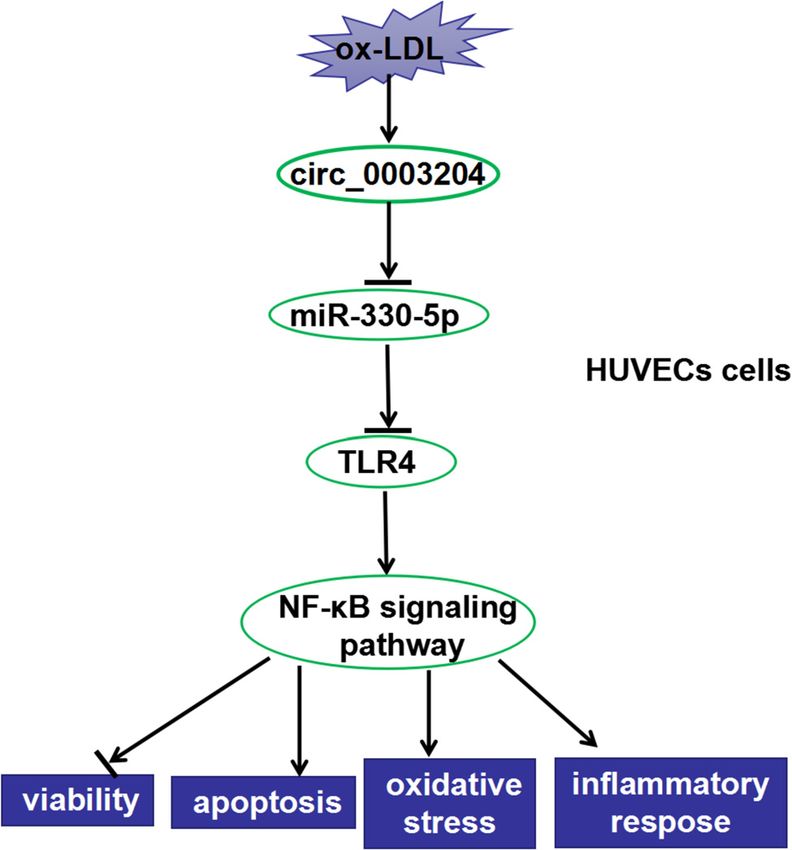
560 Qiuxia Su et al.
2.4 Enzyme-linked immunosorbent assay (MUT) binding sites for miR-330-5p were cloned into
(ELISA) the pGL3 reporter vectors (Promega, Madison, WI, USA),
yielding the circ_0003204 WT/MUT or TLR4 3′UTR WT/
HUVECs were inoculated into 6-well plates, treated with MUT reporter vector. miR-330-5p mimic or inhibitor was
ox-LDL or transfected with plasmids or oligonucleotides co-transfected with the reporter vectors into HUVECs. The
for 24 h, and cell supernatant was collected. The contents luciferase activity was measured using Dual-Lucy Assay
of interleukin-6 (IL-6), interleukin-1β (IL-1β), and tumor Kit (Solarbio).
necrosis factor (TNF-α) were detected using IL-6, IL-1β,
and TNF-α ELISA Kits (MSK, Wuhan, China), respectively.
2.8 RNA pull-down assay
2.5 Measurement of the expression of Pierce RNA 3′ End Desthiobiotinylation Kit (Thermo Fisher
reactive oxygen species (ROS) and Scientific, Waltham, MA, USA) was used for this assay.
malondialdehyde (MDA) and the activity Biotin-labeled miR-NC (Bio-miR-NC) probe, Bio-miR-330-
5p probe, or Bio-miR-330-5p mutant (Bio-miR-330-5p
of lactic dehydrogenase (LDH)
MUT; binding sites were mutated to the complementary
sequences) probe were transfected into HUVECs for
After treating with ox-LDL or transfecting with plasmids
48 h. Then, HUVECs were lysed and the cell lysates
or oligonucleotides, the expression of ROS and MDA and
were incubated with the magnetic beads. The expression
the activity of LDH of HUVECs were determined using
of circ_0003204 or TLR4 was determined using qPCR.
ROS, MDA, and LDH Assay Kits (Wanleibio, Wuhan,
China), respectively.
2.9 WB analysis
2.6 Real-time polymerase chain
reaction (qPCR) The protein was lysed using RIPA lysis buffer (Beyotime),
separated on a 10% sodium dodecyl sulfate-polyacryla-
Total RNAs were extracted using TRIzol reagent (Invitrogen) mide gel electrophoresis gel, and transferred to polyviny-
and reverse-transcribed into cDNA using miScript Reverse lidene difluoride membrane (Roche, Basel, Switzerland).
Transcription Kit (Qiagen, Dusseldorf, Germany). Next, The membrane was blocked with 5% nonfat milk and
SYBR Green (Takara, Tokyo, Japan) was used to perform incubated with primary antibodies against TLR4 (1:1,000,
qPCR analysis. β-Actin and U6 were used as internal con- Beyotime), p65 (1:500, Beyotime), phosphorylated-p65 (p-
trols. All primers were exhibited as below: circ_0003204: p65; 1:1,000, Beyotime), IκBα (1:1,000, Beyotime), p-IκBα
F, 5′-CCCCAAGATGCTGTTGTCCC-3′, R, 5′-TCCGTGGTTCTG (1:750, Beyotime) or β-actin (1:1,000, Beyotime) at 4℃ over-
ACGTCCC-3′; TLR4: F, 5′-AACCACCTCCACGCAGGGCT-3′, night. After incubating with secondary antibody (1:2,000,
R, 5′-TGATGTCTGCCTCGCGCCTG-3′; β-actin: F, 5′-GAGCGC Beyotime), the protein bands were visualized using
GGCTACAGCTT-3′, R, 5′-TCCTTAATGTCACGCACGATTT-3′; BeyoECL Plus (Beyotime).
miR-330-5p: F, 5′-GCCTCTCTGGGCCTGTGTC-3′, R, 5′-CAGT
GCAGGGTCCGAGGTAT-3′; U6: F, 5′-CTCGCTTCGGCAGCAC
ATATACT-3′, R, 5′-CGCTTCACGAATTTGCGTGT-3′. Relative
expression was calculated using the 2−ΔΔCt method. 2.10 Statistical analysis
All data were presented as mean ± standard deviation.
The collected data were processed using SPSS19.0 soft-
2.7 Dual-luciferase reporter assay ware (SPSS, Inc., Chicago, IL, USA). Statistical analysis
was performed using Student’s t-test or one-way analysis
The fragments of circ_0003204 or TLR4 3′UTR containing of variance analysis. P < 0.05 was defined as statistically
the putative wild-type (WT) binding sites and mutated significant.circ_0003204 regulates the progression of atherosclerosis 561 Figure 1: Effects of ox-LDL on HUVECs injury and circ_0003204 expression. (a) HUVECs were treated with 50, 100, and 200 μg/mL ox-LDL for 24 h, and the viability of HUVECs was measured by CCK8 assay. (b) HUVECs were treated with 100 μg/mL ox-LDL for 12, 24, and 48 h, respectively. CCK8 assay was used to detect the viability of HUVECs. (c–k) HUVECs were treated with 100 μg/mL ox-LDL for 24 h. (c) The apoptosis of HUVECs was assessed by flow cytometry. (d) The activity of caspase-3 was determined using Caspase-3 Activity Assay Kit. (e–g) The contents of IL-6, IL-1β, and TNF-α were tested via the IL-6, IL-1β, and TNF-α ELISA Assay Kits, respectively. (h–j) The expression of ROS and MDA and the activity of LDH were measured using the ROS, MDA, and LDH Assay Kits, respectively. (k) The expression of circ_0003204 was determined by qPCR. *P < 0.05. Figure 2: Effects of circ_0003204 silencing on ox-LDL-induced HUVECs injury. HUVECs were transfected with si-circ_0003204 or si-NC, followed by treatment with ox-LDL. (a) The qPCR was used to detect circ_0003204 expression. (b) CCK8 assay was used to determine the viability of HUVECs. (c) Flow cytometry was performed to test the apoptosis of HUVECs. (d) The activity of caspase-3 was measured using Caspase-3 Activity Assay Kit. (e–g) IL-6, IL-1β, and TNF-α ELISA Assay Kits were used to assess the contents of IL-6, IL-1β, and TNF-α in ox- LDL-induced HUVECs. (h–j) The expression of ROS and MDA and the activity of LDH were evaluated using the ROS, MDA, and LDH Assay Kits, respectively. *P < 0.05.
562 Qiuxia Su et al.
3 Results confirmed that ox-LDL could induce the release of inflam-
matory cytokines (Figure 1e–g). Moreover, we also found
that ox-LDL was able to increase the ROS and MDA
3.1 ox-LDL induced HUVECs injury and
expression and the activity of LDH, suggesting that ox-LDL
promoted circ_0003204 expression could enhance the oxidative stress and cell injury of
HUVECs (Figure 1h–j). Interestingly, we found that
To determine whether ox-LDL could induce cell injury, circ_0003204 expression was significantly upregulated
we examined the biological function of HUVECs after when ox-LDL-induced HUVECs injury (Figure 1k).
ox-LDL treatment. By measuring cell viability, we found
that ox-LDL could markedly inhibit the viability of HUVECs
in a dose-dependent manner and a time-dependent manner
(Figure 1a and b). Therefore, 100 μg/mL ox-LDL was used
to treat HUVECs for 24 h in further experiments. The 3.2 Interference of circ_0003204 alleviated
results of flow cytometry showed that ox-LDL could ox-LDL-induced HUVECs injury
remarkably promote the apoptosis of HUVECs (Figure 1c),
and the significant increase of caspase-3 activity also For exploring the function of circ_0003204 in ox-LDL-
confirmed this (Figure 1d). Furthermore, the detection induced HUVECs, we silenced circ_0003204 expression
of the IL-6, IL-1β, and TNF-α contents by ELISA assay using si-circ_0003204. The results confirmed that
Figure 3: circ_0003204 served as a sponge of miR-330-5p. (a) The fragments of circ_0003204 containing the binding sites or mutant
binding sites of miR-330-5p were exhibited. (b and c) Dual-luciferase reporter assay was performed to determine the luciferase activity of
circ_0003204 WT or MUT vector in ox-LDL-induced HUVECs. (d) RNA pull-down assay was used to verify the interaction between
circ_0003204 and miR-330-5p. The relative expression of circ_0003204 in Bio-miR-330-5p probe or Bio-miR-330-5p MUT probe was
detected using qPCR. (e) The miR-330-5p expression in ox-LDL-induced HUVECs or normal HUVECs was assessed by qPCR. (f) HUVECs were
transfected with circ_0003204 overexpression plasmid or si-circ_0003204 or their negative controls (pcDNA or si-NC), followed by treat-
ment with ox-LDL. The qPCR was used to test the expression of miR-330-5p. *P < 0.05.circ_0003204 regulates the progression of atherosclerosis 563 si-circ_0003204 had a good inhibitory effect on circ_0003204 3.3 circ_0003204 served as a sponge of expression (Figure 2a). CCK8 assay results demonstrated miR-330-5p that the viability of ox-LDL-induced HUVECs was markedly enhanced by circ_0003204 knockdown (Figure 2b). Besides, Subsequently, we performed bioinformatics analysis using silenced circ_0003204 suppressed the apoptosis of ox-LDL- the Circinteractome tool to predict the targeted miRNAs induced HUVECs, as determined by detection of the apop- and found that miR-330-5p contained the binding sites tosis rate and caspase-3 activity of cells (Figure 2c and d). for circ_0003204, as demonstrated in Figure 3a. Then, Further, we also uncovered that the silencing of dual-luciferase reporter assay results showed that miR- circ_0003204 could inhibit the contents of the inflamma- 330-5p mimic could significantly inhibit the luciferase tory cytokines IL-6, IL-1β, and TNF-α in ox-LDL-induced activity driven by circ_0003204 WT vector, while miR- HUVECs (Figure 2e–g). By measuring the expression of 330-5p inhibitor could markedly promote its luciferase ROS and MDA and the activity of LDH, we suggested activity, but neither of them affected the luciferase activity that circ_0003204 knockdown could repress the oxidative driven by circ_0003204 MUT vector (Figure 3b and c). stress and cell injury of ox-LDL-induced HUVECs (Figure 2h–j). Further, RNA pull-down assay results suggested that Therefore, our data indicated that circ_0003204 knock- circ_0003204 was pulled down by the Bio-miR-330-5p down might be an effective way to inhibit ox-LDL-induced probe but not the Bio-miR-330-5p MUT probe, indicating HUVECs injury. that there was an interaction between circ_0003204 and Figure 4: Effects of miR-330-5p inhibitor on ox-LDL-induced HUVECs injury. HUVECs were transfected with si-NC, si-circ_0003204, si-circ_0003204 + in-miR-NC, or si-circ_0003204 + in-miR-330-5p, followed by treatment with ox-LDL. (a) The expression of miR-330-5p was measured using qPCR. (b) CCK8 assay was performed to test the viability of HUVECs. (c) The apoptosis of HUVECs was determined via flow cytometry. (d) Caspase-3 Activity Assay Kit was used to assess the activity of caspase-3. (e–g) The contents of IL-6, IL-1β, and TNF-α were detected using the IL-6, IL-1β, and TNF-α ELISA Assay Kits, respectively. (h–j) The ROS, MDA, and LDH Assay Kits were used to measure the expression of ROS and MDA and the activity of LDH. *P < 0.05.
564 Qiuxia Su et al.
miR-330-5p (Figure 3d). Next, we observed that miR-330- injury, we carried out the rescue experiments using miR-
5p was remarkably lower expressed in ox-LDL-induced 330-5p inhibitor. As shown in Figure 4a, in-miR-330-5p
HUVECs (Figure 3e). In addition, miR-330-5p expression could reverse the promoting effect of si-circ_0003204 on
was hindered by circ_0003204 overexpression and enhanced miR-330-5p expression, indicating that its transfection
by circ_0003204 silencing (Figure 3f). These results implied efficiency was better. CCK8 assay suggested that the
that miR-330-5p could serve as a target of circ_0003204. increasing effect of circ_0003204 silencing on the viability
of ox-LDL-induced HUVECs could be inverted by miR-330-
5p inhibitor (Figure 4b). Moreover, miR-330-5p inhibitor
also could recover the decreasing effect of circ_0003204
3.4 circ_0003204 regulated ox-LDL-induced knockdown on the apoptosis rate and caspase-3 activity of
HUVECs injury by targeting miR-330-5p ox-LDL-induced HUVECs (Figure 4c and d). Besides, the
contents of IL-6, IL-1β, and TNF-α suppressed by circ_0003204
For exploring whether miR-330-5p was involved in the silencing could be reversed by miR-330-5p inhibitor, sug-
regulation of circ_0003204 on ox-LDL-induced HUVECs gesting that miR-330-5p inhibitor increased the inflammatory
Figure 5: TLR4 was a target of miR-330-5p. (a) The sequences of TLR4 3′UTR contained the binding sites or mutant binding sites of miR-330-
5p were presented. (b and c) The luciferase activity of TLR4 3′UTR WT or MUT vector in ox-LDL-induced HUVECs was measured using dual-
luciferase reporter assay. (d) The interaction between TLR4 and miR-330-5p was confirmed via RNA pull-down assay, and the relative expression
of TLR4 in Bio-miR-330-5p probe or Bio-miR-330-5p MUT probe was determined using qPCR. (e) The protein level of TLR4 in ox-LDL-induced
HUVECs or normal HUVECs was detected by WB analysis. (f) The protein level of TLR4 was tested using WB analysis to evaluate the effect of miR-
330-5p mimic on TLR4 expression. (g) HUVECs were transfected with si-NC, si-circ_0003204, si-circ_0003204 + in-miR-NC, or si-circ_0003204 +
in-miR-330-5p, followed by treatment with ox-LDL. WB analysis was performed to assess the expression of TLR4. *P < 0.05.circ_0003204 regulates the progression of atherosclerosis 565
response of ox-LDL-induced HUVECs (Figure 4e–g). At the (Figure 5b and c). Further, TLR4 also could be pulled
same time, the inhibitory effect of silenced circ_0003204 on down by the Bio-miR-330-5p probe rather than the Bio-
the expression levels of ROS and MDA, as well as the LDH miR-330-5p MUT probe (Figure 5d). Through measuring
activity in ox-LDL-induced HUVECs could also be inverted by the expression of TLR4, we discovered that ox-LDL could
miR-330-5p inhibitor (Figure 4h–j). Our data revealed that remarkably promote TLR4 expression in HUVECs (Figure 5e).
circ_0003204 modulated the viability, apoptosis, inflamma- Furthermore, miR-330-5p overexpression could repress the
tory response, and oxidative stress of ox-LDL-induced protein level of TLR4 in ox-LDL-induced HUVECs (Figure 5f).
HUVECs via sponging miR-330-5p. In addition, we found that circ_0003204 silencing could
inhibit TLR4 expression, while this effect could be reversed
by miR-330-5p inhibitor (Figure 5g). Together, we suggested
that miR-330-5p could target TLR4.
3.5 TLR4 was a target of miR-330-5p
Using the DIANA tool, conserved binding sites between
miR-330-5p and TLR4 are shown in Figure 5a. The results 3.6 TLR4 overexpression reversed the
of dual-luciferase reporter assay indicated that the luci- regulation of miR-330-5p mimic on
ferase activity driven by TLR4 3′UTR WT vector could be ox-LDL-induced HUVECs injury
restrained by miR-330-5p mimic and enhanced by miR-
330-5p inhibitor, while the luciferase activity driven by To verify that miR-330-5p regulated ox-LDL-induced
TLR4 3′UTR MUT vector was not affected by any factor HUVECs injury was achieved by regulating TLR4, we
Figure 6: Effects of miR-330-5p mimic and TLR4 overexpression on ox-LDL-induced HUVECs injury. HUVECs were transfected with miR-NC,
miR-330-5p, miR-330-5p + pcDNA, or miR-330-5p + TLR4, followed by treatment with ox-LDL. (a) The protein level of TLR4 was determined
using WB analysis. (b) The viability of HUVECs was detected via CCK8 assay. (c) The apoptosis of HUVECs was measured using flow
cytometry. (d) Caspase-3 Activity Assay Kit was used to determine the activity of caspase-3. (e–g) The IL-6, IL-1β, and TNF-α ELISA Assay Kits
were used to detect the contents of IL-6, IL-1β, and TNF-α, respectively. (h–j) The expression of ROS and MDA and the activity of LDH were
assessed using the ROS, MDA, and LDH Assay Kits, respectively. *P < 0.05.566 Qiuxia Su et al.
co-transfected miR-330-5p mimic and TLR4 overexpres- this effect (Figure 6e–g). In addition, the oxidative stress
sion plasmid into HUVECs, followed by treatment with markers ROS and MDA levels suppressed by miR-330-5p
ox-LDL. The detection of the TLR4 protein level results overexpression could be recovered by elevated TLR4
showed that TLR4 overexpression plasmid could recover expression in ox-LDL-induced HUVECs (Figure 6h–i),
the inhibition effect of miR-330-5p overexpression on and the inhibition effect of miR-330-5p on the activity
TLR4 expression, which revealed that the transfection of LDH also could be recovered by TLR4 overexpression
of miR-330-5p mimic and TLR4 overexpression plasmid (Figure 6j). These data suggested that miR-330-5p could
was successful (Figure 6a). The results of the CCK8 assay regulate ox-LDL-induced HUVECs injury via targeting TLR4.
suggested that miR-330-5p overexpression increased the
viability of ox-LDL-induced HUVECs, while this effect
could be reversed by overexpression of TLR4 (Figure 6b).
In addition, overexpressed TLR4 could invert the sup-
3.7 The circ_0003204/miR-330-5p/TLR4
pression effect of miR-330-5p overexpression on the axis regulated the activity of the NF-κB
apoptosis rate and the caspase-3 activity of ox-LDL- signaling pathway
induced HUVECs (Figure 6c and d). Moreover, we found
that overexpression of miR-330-5p restrained the con- The phosphorylation of p65 and IκBα is the hallmark
tents of inflammatory cytokines, including IL-6, IL-1β, event of activation of the NF-κB signaling pathway [22].
and TNF-α, and TLR4 overexpression also could reverse To investigate whether the circ_0003204/miR-330-5p/
Figure 7: The circ_0003204/miR-330-5p/TLR4 axis regulated the activity of the NF-κB signaling pathway. (a) HUVECs were transfected with
si-NC, si-circ_0003204, si-circ_0003204 + in-miR-NC, or si-circ_0003204 + in-miR-330-5p, followed by treatment with ox-LDL. The relative
expression of p-p65/p65 and p-IκBα/IκBα was measured using WB analysis. (b) HUVECs were transfected with miR-NC, miR-330-5p, miR-
330-5p + pcDNA, or miR-330-5p + TLR4, followed by treatment with ox-LDL. WB analysis was used to determine the relative expression of
p-p65/p65 and p-IκBα/IκBα. *P < 0.05.circ_0003204 regulates the progression of atherosclerosis 567
TLR4 axis could modulate the activity of the NF-κB sig- a binding site with circ_0003204. Previous studies have
naling pathway, we detected the relative expression of p- shown that increased miR-330-5p expression could lead
p65/p65 and p-IκBα/IκBα in ox-LDL-induced HUVECs. As to the instability of carotid plaques, indicating that miR-
shown in Figure 7a, silenced circ_0003204 restrained the 330-5p might be a biomarker for AS treatment [30].
relative expression of p-p65/p65 and p-IκBα/IκBα, while Herein, miR-330-5p inhibitor reversed the suppression
this effect could be reversed by miR-330-5p inhibitor. effect of circ_0003204 knockdown on ox-LDL-induced
Similarly, the inhibition effect of miR-330-5p mimic on HUVECs injury, which further verified that miR-330-5p
the relative expression of p-p65/p65 and p-IκBα/IκBα also could be targeted by circ_0003204. Similar to the results
could be inverted by TLR4 overexpression (Figure 7b). of Liu et al. [18], we also found that miR-330-5p over-
These indicated that circ_0003204/miR-330-5p/TLR4 axis expression could restrain the HUVECs injury induced by
regulated the viability, apoptosis, oxidative stress, and ox-LDL. Our further studies determined that TLR4 was a
inflammatory response of ox-LDL-induced HUVECs by target of miR-330-5p, which was also confirmed by the
mediating the NF-κB signaling pathway (Figure 8). reversal effect of TLR4 overexpression on miR-330-5p
inhibiting ox-LDL-induced HUVECs injury.
The NF-κB signaling pathway is a classical signaling
pathway involved in regulating immune response, inflam-
4 Discussion matory response, cell differentiation, and apoptosis [22],
and its activity is often associated with the expression of
At present, preventing the further development of AS TLR4, so it is also called the TLR4/NF-κB signaling pathway
from extending the life of patients is the basic policy of [31]. In our study, we demonstrated that circ_0003204
AS treatment. Studies have confirmed that anti-inflam- regulated ox-LDL-induced HUVECs injury by regulating
matory agents and sonodynamic therapy may be poten- the NF-κB signaling pathway activation through the miR-
tial treatments for AS [23,24]. With the deepening of 330-5p/TLR4 axis, which was agreed with our previously
research, researchers are also exploring new targets for anticipated results. Of course, we also considered some
the treatment of AS. circRNA has received a lot of atten-
tion as a potential therapeutic target for many diseases,
mainly because its expression is closely related to disease
progression [25,26]. For example, circ-YOD1 was consid-
ered as a biomarker for coronary artery disease [27], and
circDLPAG4 was identified as a potential therapeutic
target for myocardial ischemia [28]. In AS, Yang et al.
confirmed that circCHFR was abnormally expressed in
ox-LDL-modulated vascular smooth muscle cells and
was involved in cell proliferation and migration, sug-
gesting that it might be associated with the progression
of AS [29]. Li et al. reported that circ_0003575 partici-
pated in the proliferation and angiogenesis ability of
ox-LDL-induced HUVECs [12].
Our study confirmed the high expression of circ_0003204
in ox-LDL-induced HUVECs, which was agreed with the
results of previous studies [12]. Subsequently, circ_0003204
knockdown promoted the viability and inhibited the apop-
tosis, inflammatory response, and oxidative stress of
ox-induced HUVECs, which confirmed that circ_0003204
expression had a positive effect on the progression of AS.
More importantly, these results also suggested that the
silencing of circ_0003204 might be an effective treatment
Figure 8: The schematic diagram of the main findings of this
for AS.
research. In ox-LDL-induced HUVECs, circ_0003204 inhibited the
As previously described, circRNA could function as a viability and promoted the apoptosis, oxidative stress, and inflam-
ceRNA to sponge miRNA [13,14]. Through performing matory response of cells via mediating the activity of NF-κB sig-
bioinformatics analysis, we found that miR-330-5p had naling pathway by regulating the miR-330-5p/TLR4 axis.568 Qiuxia Su et al.
limitations of this study. In our research, we noticed that the global burden of atherothrombotic disease. Circ Res.
the reversal of miR-330-5p inhibitor on the function of 2016;118:535–46.
circ_0003204 silencing is partial. This indicates that [6] Kattoor AJ, Pothineni NVK, Palagiri D, Mehta JL. Oxidative stress
in atherosclerosis. Curr Atheroscler Rep. 2017;19:42.
there may be other miRNAs involved in the regulation of
[7] Geovanini GR, Libby P. Atherosclerosis and inflammation:
circ_0003204 on ox-LDL-induced HUVECs injury, which overview and updates. Clin Sci (Lond). 2018;132:1243–52.
requires further investigation. In addition, our research has [8] Mitra S, Goyal T, Mehta JL. Oxidized LDL, LOX-1 and athero-
only been verified at the cellular level, and the development sclerosis. Cardiovasc Drugs Ther. 2011;25:419–29.
of in vivo experiments will be our further research direction. [9] Trpkovic A, Resanovic I, Stanimirovic J, Radak D, Mousa SA,
Cenic-Milosevic D, et al. Oxidized low-density lipoprotein as a
To conclude, our study suggested that circ_0003204
biomarker of cardiovascular diseases. Crit Rev Clin Lab Sci.
increased TLR4 expression to regulate the viability, apop- 2015;52:70–85.
tosis, inflammation response, and oxidative stress of ox- [10] Liu J, Li D, Luo H, Zhu X. Circular RNAs: the star molecules in
LDL-induced HUVECs by targeting miR-330-5p. cancer. Mol Aspects Med. 2019;70:141–52.
[11] Yin Y, Long J, He Q, Li Y, Liao Y, He P, et al. Emerging roles of
circRNA in formation and progression of cancer. J Cancer.
2019;10:5015–21.
Abbreviations [12] Li CY, Ma L, Yu B. Circular RNA hsa_circ_0003575 regulates
oxLDL induced vascular endothelial cells proliferation and
AS atherosclerosis angiogenesis. Biomed Pharmacother. 2017;95:1514–9.
[13] Song W, Fu T. Circular RNA-associated competing endogenous
ox-LDL oxidized low-density lipoprotein
RNA network and prognostic nomogram for patients with
HUVECs human umbilical vein endothelial cells colorectal cancer. Front Oncol. 2019;9:1181.
CCK8 cell counting kit 8 [14] Yuan W, Peng S, Wang J, Wei C, Ye Z, Wang Y, et al.
Identification and characterization of circRNAs as competing
endogenous RNAs for miRNA-mRNA in colorectal cancer.
PeerJ. 2019;7:e7602.
[15] Lei B, He A, Chen Y, Cao X, Zhang P, Liu J, et al. Long non-coding
Acknowledgment: None.
RNA RPPH1 promotes the proliferation, invasion and migration
of human acute myeloid leukemia cells through down-regu-
Funding information: This work was supported by Ningbo lating miR-330-5p expression. EXCLI J. 2019;18:824–37.
Natural Science Foundation (grant No. 2015A610192) and [16] Chen S, Chen JZ, Zhang JQ, Chen HX, Qiu FN, Yan ML, et al.
Zhejiang Provincial Department of Health Project (grant Silencing of long noncoding RNA LINC00958 prevents tumor
initiation of pancreatic cancer by acting as a sponge of microRNA-
No. 2018KY739).
330-5p to down-regulate PAX8. Cancer Lett. 2019;446:49–61.
[17] Su BB, Zhou SW, Gan CB, Zhang XN. miR-330-5p regulates
Conflict of interest: The authors declare that they have no tyrosinase and PDIA3 expression and suppresses cell prolif-
financial conflicts of interest. eration and invasion in cutaneous malignant melanoma.
J Surg Res. 2016;203:434–40.
Data availability statement: The datasets used and/or [18] Liu J, Huang GQ, Ke ZP. Silence of long intergenic noncoding
RNA HOTAIR ameliorates oxidative stress and inflammation
analyzed during the current study are available from
response in ox-LDL-treated human macrophages by upregu-
the corresponding author on reasonable request. lating miR-330-5p. J Cell Physiol. 2019;234:5134–42.
[19] Pasterkamp G, Van Keulen JK, De Kleijn DP. Role of Toll-like
receptor 4 in the initiation and progression of atherosclerotic
disease. Eur J Clin Invest. 2004;34:328–34.
[20] Roy A, Srivastava M, Saqib U, Liu D, Faisal SM, Sugathan S,
References et al. Potential therapeutic targets for inflammation in toll-like
receptor 4 (TLR4)-mediated signaling pathways. Int
[1] Holmstedt CA, Turan TN, Chimowitz MI. Atherosclerotic intra- Immunopharmacol. 2016;40:79–89.
cranial arterial stenosis: risk factors, diagnosis, and treat- [21] Baker RG, Hayden MS. Ghosh S. NF-kappaB, inflammation,
ment. Lancet Neurol. 2013;12:1106–14. and metabolic disease. Cell Metab. 2011;13:11–22.
[2] Lusis AJ. Atherosclerosis. Nature. 2000;407:233–41. [22] Taniguchi K, Karin M. NF-kappaB, inflammation, immunity and
[3] Rafieian-Kopaei M, Setorki M, Doudi M, Baradaran A, Nasri H. cancer: coming of age. Nat Rev Immunol. 2018;18:309–24.
Atherosclerosis: process, indicators, risk factors and new [23] Geng C, Zhang Y, Hidru TH, Zhi L, Tao M, Zou L, et al.
hopes. Int J Prev Med. 2014;5:927–46. Sonodynamic therapy: a potential treatment for athero-
[4] Tabas I, Garcia-Cardena G, Owens GK. Recent insights into sclerosis. Life Sci. 2018;207:304–13.
the cellular biology of atherosclerosis. J Cell Biol. 2015;209:13–22. [24] Chistiakov DA, Melnichenko AA, Grechko AV, Myasoedova VA,
[5] Herrington W, Lacey B, Sherliker P, Armitage J, Lewington S. Orekhov AN. Potential of anti-inflammatory agents for treat-
Epidemiology of atherosclerosis and the potential to reduce ment of atherosclerosis. Exp Mol Pathol. 2018;104:114–24.circ_0003204 regulates the progression of atherosclerosis 569
[25] Li JJ, Wang W, Wang XQ, He Y, Wang SS, Yan YX. A novel [29] Yang L, Yang F, Zhao H, Wang M, Zhang Y. Circular RNA
strategy of identifying circRNA biomarkers in cardiovascular circCHFR facilitates the proliferation and migration of vascular
disease by meta-analysis. J Cell Physiol. 2019;234:21601–12. smooth muscle via miR-370/FOXO1/cyclin D1 pathway.
[26] Han B, Chao J, Yao H. Circular RNA and its mechanisms in Mol Ther Nucleic Acids. 2019;16:434–41.
disease: from the bench to the clinic. Pharmacol Ther. [30] Wei X, Sun Y, Han T, Zhu J, Xie Y, Wang S, et al. Upregulation of
2018;187:31–44. miR-330-5p is associated with carotid plaque’s stability by
[27] Miao L, Yin RX, Zhang QH, Liao PJ, Wang Y, Nie RJ, et al. A novel targeting Talin-1 in symptomatic carotid stenosis patients.
circRNA-miRNA-mRNA network identifies circ-YOD1 as a bio- BMC Cardiovasc Disord. 2019;19:149.
marker for coronary artery disease. Sci Rep. 2019;9:18314. [31] Guo J, Zheng L, Chen L, Luo N, Yang W, Qu X, et al.
[28] Chen L, Luo W, Zhang W, Chu H, Wang J, Dai X, et al. Lipopolysaccharide activated TLR4/NF-kappaB signaling
circDLPAG4/HECTD1 mediates ischaemia/reperfusion injury in pathway of fibroblasts from uterine fibroids. Int J Clin Exp
endothelial cells via ER stress. RNA Biol. 2020;17:240–53. Pathol. 2015;8:10014–25.You can also read