Effect of focal adhesion kinase inhibition on osteoblastic cells grown on titanium with different topographies - SciELO
←
→
Page content transcription
If your browser does not render page correctly, please read the page content below
Original Article
                                                                                                            http://dx.doi.org/10.1590/1678-7757-2019-0156

                                                Effect of focal adhesion kinase
                                                inhibition on osteoblastic cells
                                                grown on titanium with different
                                                topographies

                                                Abstract

             Helena Bacha LOPES1                    Objective: The present study aimed to investigate the participation of focal

Alann Thaffarell Portilho SOUZA1                adhesion kinases (FAK) in interactions between osteoblastic cells and titanium
                                                (Ti) surfaces with three different topographies, namely, untreated (US),
        Gileade Pereira FREITAS1
                                                microstructured (MS), and nanostructured (NS). Methodology: Osteoblasts
              Carlos Nelson ELIAS²
                                                harvested from the calvarial bones of 3-day-old rats were cultured on US,
              Adalberto Luiz ROSA1              MS and NS discs in the presence of PF-573228 (FAK inhibitor) to evaluate
           Marcio Mateus BELOTI1                osteoblastic differentiation. After 24 h, we evaluated osteoblast morphology
                                                and vinculin expression, and on day 10, the following parameters: gene
                                                expression of osteoblastic markers and integrin signaling components, FAK
                                                protein expression and alkaline phosphatase (ALP) activity. A smooth surface,
                                                porosities at the microscale level, and nanocavities were observed in US,
                                                MS, and NS, respectively. Results: FAK inhibition decreased the number of
                                                filopodia in cells grown on US and MS compared with that in NS. FAK inhibition
                                                decreased the gene expression of Alp, bone sialoprotein, osteocalcin, and
                                                ALP activity in cells grown on all evaluated surfaces. FAK inhibition did not
                                                affect the gene expression of Fak, integrin alpha 1 (Itga1) and integrin beta
                                                1 (Itgb1) in cells grown on MS, increased the gene expression of Fak in
                                                cells grown on NS, and increased the gene expression of Itga1 and Itgb1 in
                                                cells grown on US and NS. Moreover, FAK protein expression decreased in
                                                cells cultured on US but increased in cells cultured on MS and NS after FAK
                                                inhibition; no difference in the expression of vinculin was observed among
                                                cells grown on all surfaces. Conclusions: Our data demonstrate the relevance
                                                of FAK in the interactions between osteoblastic cells and Ti surfaces regardless
                                                of surface topography. Nanotopography positively regulated FAK expression
                                                and integrin signaling pathway components during osteoblast differentiation.
                                                In this context, the development of Ti surfaces with the ability to upregulate
                                                FAK activity could positively impact the process of implant osseointegration.

               Submitted: March 12, 2019            Keywords: Bone. Focal adhesion kinase. Osteoblast. Titanium.
             Modification: August 16, 2019
            Accepted: September 23, 2019

                    Corresponding address:      1
                                                  Universidade de São Paulo, Faculdade de Odontologia de Ribeirão Preto, Bone Research
                       Marcio Mateus Beloti     Laboratory, Ribeirão Preto, São Paulo, Brasil.
Faculdade de Odontologia de Ribeirão Preto -
                Universidade de São Paulo
                                                2
                                                  Instituto Militar de Engenharia, Laboratório de Biomateriais, Rio de Janeiro, Rio de Janeiro, Brasil.
               Av do Café s/n, 14040-904 -
                Ribeirão Preto - SP - Brasil.
                 Phone: +55 16 3315 4785
                  e-mail: mmbeloti@usp.br

                                                         J Appl Oral Sci. 1/10 2020;28:e20190156
Effect of focal adhesion kinase inhibition on osteoblastic cells grown on titanium with different topographies

Introduction                                                                              interactions between osteoblastic cells and Ti surfaces
                                                                                          with different topographies by using a specific FAK

   Excellent mechanical and biological properties                                         inhibitor.

render titanium (Ti) to be the most frequently used
biomaterial for manufacturing dental implants.1 The
bone−Ti contact is influenced by several parameters,                                      Methodology
including topography and surface chemistry of the
dental implants. Osseointegration of implants is                                          Cell culture
strongly associated with the responses of osteoblasts                                           Osteoblastic cells were obtained according to
to the surface of the biomaterial, and signaling                                          a method previously described. 17 All procedures
pathways involved in osteoblastic differentiation, such                                   involving animals were approved by the animal care
as integrin signaling, are known to play an important                                     guidelines of the School of Dentistry of Ribeirão Preto
role in this process.                                                                     (CEUA/FORP Protocol #2015.1.581.58.1). Briefly,
   Integrins are heterodimeric transmembrane                                              3-day-old Wistar rats were decapitated with a scalpel
proteins composed of α and β subunits forming a family                                    blade, and their calvaria were removed with the use
of membrane receptors whose primary function is                                           of scissors. Osteoblastic cells were isolated through
adhesion of cells to extracellular matrix proteins, such                                  sequential enzymatic digestion with 0.25% trypsin
as collagen and fibronectin; some of these receptors                                      solution (Gibco-Invitrogen, Grand Island, NY, USA) and
involved in osteoblastic differentiation.1-3 The integrin                                 collagenase type II 0.20% (Gibco-Invitrogen). Only
signaling pathway acts in both directions of the cell                                     cells isolated in the 2nd and 3rd digestion were used.
membrane, i.e., binding of integrins to components
of the extracellular matrix triggers an intracellular                                     Selection of FAK inhibitor concentration
signaling cascade and the activation of integrins is                                            A potent and specific FAK inhibitor, PF-573228
modulated by intracellular signals and, consequently,                                     (3,4-dihydro-6-[[4-[[[3-(methylsulfonyl) phenyl]
their affinity to the ligand present in the extracellular                                 methyl] amino]-5-(trifluoromethyl)-2-pyrimidinyl]
matrix.4                                                                                  a m i n o ] - 2 ( 1 H ) - q u i n o l i n o n e ) ( S i g m a -A l d r i c h ,
   Focal adhesion kinases (FAK) or Src family                                             Darmstadt, Germany), which interacts with ATP
kinases are the main integrin-activated protein                                           binding sites and blocks their catalytic activity, was
tyrosine kinases that play a key role in this signaling                                   used in this study.18-20 PF-573228 was reconstituted in
pathway.   1,4-6
                   The associations of extracellular matrix                               dimethylsulfoxide (DMSO) (Sigma-Aldrich) to produce
ligands, integrins, and cytoskeletal components form                                      a 20 mg/mL stock solution, which was then diluted to
a focal adhesion complex, where FAK is recruited and                                      final concentrations of 0.1, 1 and 10 µM according to
interacts directly or indirectly with these complexes                                     a previous study.20 Next, a non-cytotoxic concentration
causing their activation by autophosphorylation                                           of PF-573228 was determined. Here, osteoblastic
and consequent binding to Src kinase. Src kinases                                         cells were cultured on Thermanox® coverslips (Nunc,
phosphorylate several components of focal adhesion                                        Rochester, NY, USA) at a density of 1×10⁴ cells/well
sites participating in FAK signaling to generate the                                      in 24-well polystyrene plates (Corning Incorporated,
signal transduction mechanism.               7,8
                                                                                          Corning, NY, USA) containing osteogenic medium
   The development of biomaterials modulating the                                         (OM) prepared with alpha-minimum essential medium
interaction between integrins and the extracellular                                       (α-MEM, Gibco-Invitrogen), 10% fetal calf serum
matrix represents an important strategy for therapies                                     (Gibco-Invitrogen), 50 µg/mL gentamicin (Gibco-
related to bone tissue in dentistry and medicine.                                         Invitrogen), 0.3 mg/mL fungisone (Gibco-Invitrogen),
Several studies have demonstrated the participation                                       5 μg/mL ascorbic acid (Gibco-Invitrogen), and 7 mM
of the integrin signaling pathway, through FAK, in                                        β-glycerophosphate (Sigma-Aldrich) for up to 3 days
response of osteoblastic cells to different surface                                       and exposed to three different concentrations of PF-
topographies.        9-16
                            Thus, the role of specific FAK                                573228 or to vehicle (DMSO, control). On day 3, cells
inhibitors in the osteogenic potential of cells on Ti                                     were counted by staining of cell nuclei with 300 nM
surfaces is an interesting research topic. The present                                    4′,6-diamidino-2-phenylindole, dihydrochloride (DAPI;
study aimed to investigate the participation of FAK in                                    Molecular Probes, Thermo Fischer Scientific, Waltham,

                                                           J Appl Oral Sci. 2/10 2020;28:e20190156
LOPES HB, SOUZA AT, FREITAS GP, ELIAS CN, ROSA AL, BELOTI MM
MA, USA) as described elsewhere.21 An AxioCam MRM cells were immersed in a solution of 1% tannic acid
digital camera (Carl Zeiss Inc., Oberkochen, Germany) (Sigma-Aldrich) and 0.1 M sodium cacodylate solution
coupled with an AxioImager M2 Zeiss light microscope for 60 min and rinsed with 0.2 M sodium cacodylate.
(Carl Zeiss Inc.) was used to analyze the samples. The The samples were dehydrated by crescent alcohol
cell nuclei were counted from three different fields on concentrations followed by hexamethyldisiloxane
three different coverslips for each treatment (n=9). prior to sputter-coating (20 nm gold/palladium) and
evaluation by using a Zeiss Sigma FE-SEM microscope
Preparation of Ti surfaces (Carl Zeiss Inc.).
Discs of pure grade 2 Ti (Realum, São Paulo,
SP, Brazil; diameter, 12 mm; thickness, 1.5 mm) Gene expression of osteoblastic markers and
were polished with up to 600 grit silicon carbide integrin signaling pathway components
and treated with either HNO3-H2SO4-HCl to produce On day 10 after culture, quantitative real-time
the microstructured surface (MS) or H2SO4-H2O2 to PCR was performed, using TaqMan (Life Technologies-
produce the nanostructured surface (NS). 22,23
Ti with Invitrogen, Carlsbad, CA, USA) probes (Figure 1)
an untreated surface (US) was used as the control. to evaluate the gene expression of the osteoblastic
All samples were rinsed several times with deionized markers alkaline phosphatase (Alp), bone sialoprotein
H2O, air-dried, and then autoclaved. The surfaces (Bsp), and osteocalcin (Oc), as well as that of the
were evaluated by field emission scanning electron integrin signaling pathway components integrin β1
microscopy (SEM) operated at 5 kV (Inspect S50, FEI, (Itgb1), integrin α1 (Itga1), and Fak. The total RNA
Hillsboro, OR, USA). of cells cultured on Ti surfaces with or without PF-
573228 was extracted to synthesize complementary
Effect of FAK inhibition in osteoblastic cells DNA (cDNA) as previously described.9 The expression
grown on Ti surfaces of the housekeeping gene β-actin was selected to
The cells were plated on the Ti surfaces at a density normalize the expression of genes of interest and it
of 2×10⁴ cells/disc in 24-well polystyrene plates was relative to the cells cultured on US, MS, or NS
(Corning Incorporated) for up to 10 days, using OM surfaces in the absence of PF-573228 (control) using
containing PF-573228 0.1 µM, which was selected in a the comparative threshold 2-ΔΔCt method.24
previous experiment. The parameters described below
were then evaluated. ALP activity
On day 10, cells cultured on Ti surfaces with or
Scanning electron microscopy without PF-573228 were lysed, and a commercial
Exactly 24 h after culture on the Ti surface, kit (Labtest Diagnostica, Lagoa Santa, MG, Brazil)
SEM was carried out to observe cells grown with was used to detect their ALP activity as previously
or without PF-573228. The samples were fixed in described.25 The absorbance (n=4) was evaluated
4% glutaraldehyde (Electron Microscopy Sciences, at 590 nm by using the plate reader μQuant (Bio-
Washington, PA, USA), rinsed with 0.2 M sodium Tek Instruments Inc., Winooski, VT, USA), and ALP
cacodylate buffer, pH 7.4 (Sigma-Aldrich), post-fixed activity was calculated and expressed as μmol of
with 1% osmium tetroxide (Sigma-Aldrich), and then thymolphthalein/h/mg protein as previously described.
rinsed in sodium cacodylate buffer. Thereafter, the
Gene Gene name Identification
Runx2 Runt-related transcription factor 2 Rn01512298_m1
Alpl Alkaline phosphatase (Alp) Rn01516028_m1
Ibsp Integrin binding sialoprotein (Bone sialoprotein/Bsp) Rn00561414_m1
Bglap Bone gamma-carboxyglutamate protein (Osteocalcin/Oc) Rn00566386_g1
Gapdh Glyceraldehyde-3-phosphate dehydrogenase Rn01775763_g1
Itga1 Integrin subunit alpha 1 Rn0057864_m1
Itgb1 Integrin subunit beta 1 Rn00566727_m1
Ptk2 Protein tyrosine kinase 2 (Focal adhesion kinase/Fak) Rn01505115_m1
Figure 1- TaqMan (Life Technologies-Invitrogen) probes for real-time PCR
J Appl Oral Sci. 3/10 2020;28:e20190156Effect of focal adhesion kinase inhibition on osteoblastic cells grown on titanium with different topographies
FAK protein detection Cambridge, UK).
On day 10, cells cultured on Ti surfaces with or
without PF-573228 were lysed, and the protein of Statistical analysis
each cell was extracted and transferred to PVDF The data of cell counting were analyzed by a one-
membrane as previously described 9 to evaluate way analysis of variance, followed by the Student−
FAK protein expression by Western Blotting assay. Newman−Keuls post hoc test. Student’s t-test was
Blocking of non-specific sites was performed with used to analyze the data of gene expression and ALP
5% Non-Fat Dry Milk Blotting Grade Blocker (Bio-Rad activity. The significance level used was set to 5%
Laboratories, Hercules, CA, USA) for 2 h. Cells were (p≤0.05).
incubated overnight at 4°C with a rabbit polyclonal
antibody to FAK (1:1000, Cell Signaling Technology,
Denver, MA, USA) and a mouse anti-glyceraldehyde- Results
3-phosphate dehydrogenase (GAPDH) monoclonal
antibody (1:2000, Santa Cruz Biotechnology), Selection of FAK inhibitor concentration
which was used as a control. Secondary antibodies Fluorescence labeling of cell nuclei stained with
conjugated to HRP (1:2000, Santa Cruz Biotechnology) DAPI in the control treatment (Figure 2A) was similar
were used for immunodetection with Western to those of cultures grown in the presence of 0.1 μM
Lightning Chemiluminescence Reagent (PerkinElmer (Figure 2B) and 1 μM (Figure 2C) PF-573228 but lower
Life Sciences, Waltham, MA, USA), and images were in cultures grown in 10 μM PF-573228 (Figure 2D).
captured by a G:Box gel imaging system (Syngene, Quantification of stained nuclei (Figure 2E) showed a
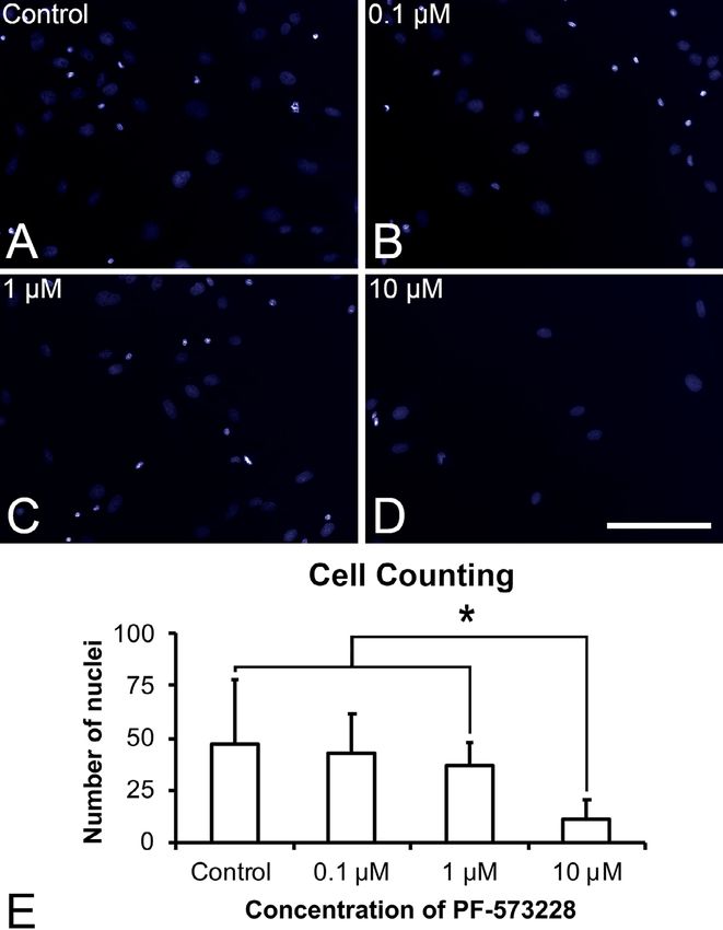
Figure 2- Cell counting of DAPI-positive cells in the control (A), 0.1 µM (B), 1µM (C), and 10 µM (D) PF-573228. The data (E) are
presented as mean ± standard deviation (n=9), and asterisks (*) indicate statistically significant differences (p≤0.05). Scale bar: 200 µm
J Appl Oral Sci. 4/10 2020;28:e20190156LOPES HB, SOUZA AT, FREITAS GP, ELIAS CN, ROSA AL, BELOTI MM
statistically significantly lower number of nuclei in cells
treated with 10 μM PF-573228 compared with those
in cells from the control, 1 μM PF-573228, and 0.1
μM PF-573228 treatments (p=0.001, p=0.003, and
p=0.046, respectively), among which no statistically
significant difference was found (p>0.05). Based on
this finding and taking into account that 0.1 μM is the
lowest concentration of the FAK inhibitor that did not
induce cytotoxic effects, further experiments were
carried out using 0.1 μM PF-573228.
Ti surface characteristics
The methods used in this experiment effectively
produced surfaces with different topographies (Figure
3). US exhibited a smooth surface (Figure 3A), MS
presented porosities at the microscale level (Figure
3B), and NS presented nanocavities (Figure 3C).
Effect of FAK inhibition on morphology of
osteoblastic cells grown on Ti surfaces
SEM imaging showed that the cells remained viable
and well spread in all Ti surfaces (Figure 4A-C), with
higher number of filopodia on them (Figure 4D-F). FAK
inhibition decreased the number of filopodia in cells
grown on US and MS (Figures 4G and 4H, respectively,
and Figures 4J and 4K, respectively) but demonstrated
no change in cells grown on NS (Figures 4 and 4L).
Figure 3- High-resolution scanning electron micrographs of US
(A), MS (B), and NS (C). Scale bar: 500 nm
Figure 4- High-resolution scanning electron micrographs of cells cultured on US (A, D, G, J), MS (B, E, H, K), and NS (C, F, I, L) in
presence or absence of 0.1 µM PF-573228 at 24 h. Scale bars: A, B, C, G, H, and I=10 µm; D, E, F, J, K, and L=1 µm
J Appl Oral Sci. 5/10 2020;28:e20190156Effect of focal adhesion kinase inhibition on osteoblastic cells grown on titanium with different topographies
Effect of FAK inhibition on the gene expression expression of Itga1 (Figure 7D, p=0.015) and Itgb1
of osteogenic markers of osteoblastic cells (Figure 7G, p=0.005) in cells cultured on US. FAK
grown on Ti surfaces inhibition did not affect the gene expression of Fak
FAK inhibition reduced the gene expression of all (Figure 7B, p=0.708), Itga1 (Figure 7E, p=0.176), and
evaluated osteoblastic markers in cells cultured on Itgb1 (Figure 7H, p=0.835) in cells cultured on MS.
US, MS, and NS (Figure 5). Specifically, FAK inhibition Finally, FAK inhibition increased the gene expression of
decreased the gene expression of Alp (Figures 5A−5C, Fak, Itga1, and Itgb1 in cells cultured on NS (Figures
p=0.001 for all surfaces), Bsp (Figures 5D−5F, 7C, 7F, and 7I, respectively, p=0.001 for all genes).
p=0.001 for all surfaces), and Oc (Figures 5G−5I,
p=0.001 for US and MS; p=0.002 for NS) in cells Effect of FAK inhibition on FAK protein
grown on all evaluated surfaces. expression in osteoblastic cells grown on Ti
surfaces
Effect of FAK inhibition on ALP activity of FAK inhibition slightly reduced FAK expression in
osteoblastic cells grown on Ti surfaces cells grown on US (1.07-fold) (Figure 8A) but increased
Similar to the gene expression findings, FAK this expression in cells grown on MS and NS; of these
inhibition decreased the ALP activity in cells grown on two surfaces, a more pronounced effect was noted in
US (Figure 6A, p=0.001), MS (Figure 6B, p=0.013), the latter (1.3-fold and 2.5-fold, respectively) (Figures
and NS (Figure 6C, p=0.001). 8B and 8C, respectively).
Effect of FAK inhibition on the gene expression
of integrin signaling pathway components in
osteoblastic cells grown on Ti surfaces
FAK inhibition did not affect the gene expression
of Fak (Figure 7A, p=0.249) but increased the gene
Figure 5- Gene expression of the osteoblastic markers alkaline phosphatase (Alp, A−C), bone sialoprotein (Bsp, D−F), and osteocalcin
(Oc, G−I) in cells cultured on US, MS, and NS in presence or absence of 0.1 µM PF-573228 on day 10. The data are presented as mean
± standard deviation (n=3), and asterisks (*) indicate statistically significant differences (p≤0.05)
J Appl Oral Sci. 6/10 2020;28:e20190156LOPES HB, SOUZA AT, FREITAS GP, ELIAS CN, ROSA AL, BELOTI MM
Osseointegration of Ti depends mostly on the
interactions between the material surface and cells,
in which cell signaling pathways play an important
role. FAK is involved in several signaling pathways
due to its ability to bind to several proteins involved
in these pathways; its function is related to many
cellular processes such as migration, growth factor
signaling, cell cycle progression, and cell survival.26,27
The present study focused on the role of an FAK
inhibitor in the responses of osteoblastic cells to Ti.
We selected an FAK inhibitor concentration of 0.1 μM
because this value is the lowest concentration that did
not significantly interfere with cell counting among
the tested concentrations. This finding is supported
by the observation that the same concentration of
PF-573228 does not prevent cell proliferation but
efficiently inhibits FAK activity.20
Cells grown on MS and NS showed a higher number
of slender cytoplasmic projections attached to the
surfaces compared with those grown on a smooth
surface. In support of this finding, a previous study
observed higher numbers of pseudopodia and more
cell spreading on nanotextured Ti compared with those
on a smooth surface.28 The FAK inhibitor reduced
cell spreading on both US and MS surfaces without
significantly affecting the morphology of cells grown on
NS. This finding may be due to the higher wettability of
NS compared with those of US and MS; such wettability
could inactivate or compensate the inhibitory effect
produced by the FAK inhibitor.22,29
Figure 6- Alkaline phosphatase (ALP) activity of cells cultured
on US (A), MS (B), and NS (C) in the presence or absence of In this study, FAK inhibition downregulated the gene
0.1 µM PF-573228 on day 10. The data are presented as mean expression of key bone markers and ALP activity in cells
± standard deviation (n=3), and asterisks (*) indicate statistically
significant differences (p≤0.05) grown on all evaluated surfaces. FAK is a component
of the focal adhesion complex and is essential for the
Discussion development of integrin signaling. The participation
of integrins in osteoblastic cell behavior has been
This study aimed to evaluate the participation of
extensively discussed in the literature. Indeed, we
FAK in interactions between osteoblastic cells and Ti
previously demonstrated the role of integrins α1,
surface with three different topographies, namely,
β1, and β3 in the osteogenic potential of NS.10,11
US, MS, and NS. The results indicated that FAK is
The use of an FAK inhibitor impaired the genotypic
relevant to osteoblastic differentiation of cells grown
and phenotypic expression of osteoblasts cultured
on Ti surfaces regardless of topographic characteristics
on all evaluated Ti surfaces, thereby corroborating a
since the inhibition of FAK reduced the differentiation
previous study showing that FAK inhibition adversely
of cells grown on all three surfaces. However, among
affects the development of osteoblastic phenotype in
the surfaces studied, NS was the surface in which the
the same culture model used in the present work.30
integrin signaling pathway was most affected by FAK
Moreover, FAK inhibition increased the gene expression
inhibition.
of integrins α1 and β1 in cells cultured on US and NS
Ti surface modification can improve implant
but not on MS. These data suggest the presence of a
wettability and increase the available surface for
compensatory mechanism upregulating the expression
bone growth and fixation and blood clotting. 22
J Appl Oral Sci. 7/10 2020;28:e20190156Effect of focal adhesion kinase inhibition on osteoblastic cells grown on titanium with different topographies
Figure 7- Gene expression of the integrin signaling pathway components focal adhesion kinase (Fak, A), integrin a1 (Itga1, B), and
integrin b1 (Itgb1, C) in cells cultured on US, MS, and NS in presence or absence of 0.1 µM PF-573228 on day 10. The data are presented
as mean ± standard deviation (n=3), and asterisks (*) indicate statistically significant differences (p≤0.05)
of these integrins in response of FAK inhibition that vinculin expression by immunofluorescence but we did
is dependent on surface topography. In fact, this not find a correspondence between the topography
compensatory phenomenon has been previously and vinculin expression of cells grown with or without
described for other molecular mechanisms.31,32 the FAK inhibitor (data not shown). This result may be
The presence of PF-573228 did not affect the Fak due to the effects of the trial periods chosen for the
gene expression of cells grown on US and MS but evaluation or the methodology used.39
increased Fak gene and protein expression on NS. This
finding may be explained by the positive modulation
of the NS topography of FAK expression even in the
Conclusion
presence of the FAK inhibitor and/or the ability of PF-
573228 to physically bind FAK and inhibit its catalytic
Our results demonstrated the relevance of FAK
activity rather than its synthesis process. Previous
to the interactions between osteoblastic cells and
studies using osteoblastic and fibroblastic cells
Ti surfaces regardless of surface topography. We
revealed remarkable FAK expression and activation
also observed that nanotopography upregulates
on nanostructured surfaces featuring 14 and 29 nm
FAK expression and integrin signaling pathway
nanopits, similar to the pore size (22 nm on average)
components during osteoblastic differentiation.
of the NS used in the present research14,15,33,34.
Thus, the development of Ti surfaces with the ability
Focal adhesion complexes are key structures
to regulate FAK activity could positively impact the
participating in the interactions between cells
process of implant osseointegration.
and surfaces of biomaterials and may affect cell
morphology, proliferation, differentiation, and
Acknowledgements
apoptosis.35 Vinculin detection has been conducted
This study was supported by the State of São Paulo
to identify these complexes, but distinct data have
Research Foundation (FAPESP, Brazil, # 2013/05181-
been described36-38. In the present work, we evaluated
J Appl Oral Sci. 8/10 2020;28:e20190156LOPES HB, SOUZA AT, FREITAS GP, ELIAS CN, ROSA AL, BELOTI MM
5- Yan YX, Gong YW, Guo Y, Lv Q, Guo C, Zhuang Y, et al. Mechanical
strain regulates osteoblast proliferation through integrin-mediated ERK
activation. PLoS One 2012;7:e35709.
6- Shekaran A, García AJ. Extracellular matrix-mimetic adhesive
biomaterials for bone repair. J Biomed Mater Res. 2011;96(1):261-72.
7- Salmela M, Jokinen J1, Tiitta S, Rappu P, Cheng RH, Heino J. Integrin
α2β1 in nonactivated conformation can induce focal adhesion kinase
signaling. Sci Rep. 2017;7(1):3414.
8- Docheva D, Popov C, Alberton P, Aszodi A. Integrin signaling in
skeletal development and function. Birth Defects Res C Embryo Today.
2014;102(1):13-36.
9- Lopes HB, Freitas GP, Elias CN, Tye C, Stein JL, Stein GS, et al.
Participation of integrin β3 in osteoblast differentiation induced
by titanium with nano or microtopography. J Biomed Mater Res A.
2019;107(6):1303-13.
10- Rosa AL, Kato RB, Castro Raucci LM, Teixeira LN, Oliveira FS,
Bellesini LS, et al. Nanotopography drives stem cell fate toward
osteoblast differentiation through α1β1 integrin signaling pathway. J
Cell Biochem. 2014;115(3):540-8.
11- Wang W, Zhao L, Wu K, Ma Q, Mei S, Chu PK, et al. The role of
integrin-linked kinase/β- catenin pathway in the enhanced MG63
differentiation by micro/nano-textured topography. Biomaterials.
2013;34(3):631-40.
12- Lu Z, Zreiqat H. The osteoconductivity of biomaterials is regulated
by bone morphogenetic protein 2 autocrine loop involving α2β1 integrin
and mitogen-activated protein kinase/extracellular related kinase
signaling pathways. Tissue Eng Part A. 2010;16(10):3075-84.
13- Olivares-Navarrete R, Raz P, Zhao G, Chen J, Wieland M, Cochran
DL, et al. Integrin alpha2beta1 plays a critical role in osteoblast
response to micron-scale surface structure and surface energy of
titanium substrates. Proc Natl Acad Sci U S A. 2008;105(41):15767-72.
14- Zambuzzi WF, Bonfante EA, Jimbo R, Hayashi M, Andersson M, Alves
G, et al. Nanometer scale titanium surface texturing are detected by
signaling pathways involving transient FAK and Src activations. PLoS
One. 2014;9(7):e95662.
15- Costa Fernandes CJ, Bezerra FJ, Campos Souza B, Campos
MA, Zambuzzi WF. Titanium-enriched medium drives low profile of
ECM remodeling as a pre-requisite to pre-osteoblast viability and
proliferative phenotype. J Trace Elem Med Biol. 2018;50:339-46.
16- Salasznyk RM, Klees RF, Boskey A, Plopper GE. Activation of FAK
Figure 8- Protein expression of focal adhesion kinase (FAK) in
cells cultured on US (A), MS (B), and NS (C) in the presence or is necessary for the osteogenic differentiation of human mesenchymal
absence of 0.1 µM PF-573228 on day 10 stem cells on laminin-5. J Cell Biochem. 2007;100(2):499-514.
17- Moura J, Teixeira LN, Ravagnani C, Peitl O, Zanotto ED, Beloti
3, 2014/08443-1 and 2016/21116-5). Fabiola S. de MM, et al. In vitro osteogenesis on a highly bioactive glass-ceramic
Oliveira, Adriana L. G. de Almeida, Roger R. Fernandes (Biosilicate). J Biomed Mater Res A. 2007;82(3):545-57.
18- Webber PJ, Park C, Qui M, Ramalingam SS, Khuri FR, Fu H, et
and Milla S. Tavares are acknowledged for technical
al. Combination of heat shock protein 90 and focal adhesion kinase
assistance during the experiments and ENAGO (www. inhibitors synergistically inhibits the growth of non-small cell lung
enago.com) for the English language review. cancer cells. Oncoscience. 2015;2(9):765-76.
19- Kim JB, Leucht P, Luppen CA, Park YJ, Beggs HE, Damsky CH, et
al. Reconciling the roles of FAK in osteoblast differentiation, osteoclast
remodeling, and bone regeneration. Bone. 2007;41(1):39-51.
20- Slack-Davis JK, Martin KH, Tilghman RW, Iwanicki M, Ung EJ, Autry
REFERENCES
C, et al. Cellular characterization of a novel focal adhesion kinase
inhibitor. Biol Chem. 2007;282(20):14845-52.
1- Schneider GB, Zaharias R, Seabold D, Stanford C. Integrin-
21- Kepner RL Jr, Pratt JR. Use of fluorochromes for direct enumeration
associated tyrosine kinase FAK affects Cbfa1 expression. J Orthop
of total bacteria in environmental samples: past and present. Microbiol
Res. 2011;29(9):1443-7.
Rev. 1994;58(4):603-15.
2- Jikko A, Harris SE, Chen D, Mendrick DL, Damsky CH. Collagen
22- Elias CN, Gravina PA, Silva Filho C, Nascente PA. Preparation of
integrin receptors regulate early osteoblast differentiation induced by
bioactive titanium surfaces via fluoride and fibronectin retention. Int
BMP-2. J Bone Miner Res. 1999;14(7):1075-83.
J Biomat. 2012;2012:290179.
3- Hynes RO. Integrins: a family of cell surface receptors. Cell.
23- Oliveira PT, Zalzal SF, Beloti MM, Rosa AL, Nanci A. Enhancement
1987;48(4):549-54.
of in vitro osteogenesis on titanium by chemically produced
4- Giancotti FG, Ruoslahti E. Integrin signaling. Science
nanotopography. J Biomed Mater Res A. 2007;80(3):554-64.
1999;285(5430):1028-32.
J Appl Oral Sci. 9/10 2020;28:e20190156Effect of focal adhesion kinase inhibition on osteoblastic cells grown on titanium with different topographies
24- Livak KJ, Schmittgen TD. Analysis of relative gene expression data 33- Lim JY, Dreiss AD, Zhou Z, Hansen JC, Siedlecki CA, Hengstebeck
using realtime quantitative PCR and the 2(-Delta Delta C(T)). Methods. RW, et al. The regulation of integrin-mediated osteoblast focal adhesion
2001;25(4):402-8. and focal adhesion kinase expression by nanoscale topography.
25- Franco RL, Chiesa R, Beloti MM, de Oliveira PT, Rosa AL. Human Biomaterials. 2007;28(10):1787-97.
osteoblastic cell response to a Ca- and P-enriched titanium surface 34- Takebe J, Miyata K, Miura S, Ito S. Effects of the nanotopographic
obtained by anodization. J Biomed Mater Res A. 2009;15;88(4):841-8. surface structure of commercially pure titanium following anodization-
26- Schlaepfer DD, Mitra SK. Multiple connections link FAK to cell hydrothermal treatment on gene expression and adhesion in gingival
motility and invasion. Curr Opin Genet Dev. 2004;14(1):92-101. epithelial cells. Mater Sci Eng C Mater Biol Appl. 2014;42:273-9.
27- Parsons JT. Focal adhesion kinase: the first ten years. J Cell Sci. 35- Dalby MJ, Gadegaard N, Oreffo ROC. Harnessing nanotopography
2003;116(Pt 8):1409-16. and integrin-matrix interactions to influence stem cell fate. Nat Mater.
28- Elkhidir Y, Lai R, Feng Z. The impact of photofunctionalized gold 2014;13(6):558-69.
nanoparticles on osseointegration. Heliyon. 2018;4(7):e00662. 36- Peng X, Nelson ES, Maiers JL, DeMali KA. New insights into vinculin
29- Gittens RA, Olivares-Navarrete R, Cheng A, Anderson DM, function and regulation. Int Rev Cell Mol Biol. 2011;287:191-231.
McLachlan T, Stephan I, et al. The roles of titanium surface micro/ 37- Schulte C, Podestà A, Lenardi C, Tedeschi G, Milani P. Quantitative
nanotopography and wettability on the differential response of human control of protein and cell interaction with nanostructured surfaces by
osteoblast lineage cells. Acta Biomater. 2013;9(4):6268-77. cluster assembling. Acc Chem Res. 2017;50(2):231-9.
30- Liu Y, Ma Y, Zhang J, Xie Q, Wang Z, Yu S, et al. MBG-modified β-TCP 38- Sisti KE, de Andrés MC, Johnston D, Almeida-Filho E, Guastaldi
scaffold promotes mesenchymal stem cells adhesion and osteogenic AC, Oreffo RO. Skeletal stem cell and bone implant interactions are
differentiation via a FAK/MAPK signaling pathway. ACS Appl Mater enhanced by LASER titanium modification. Biochem Biophys Res
Interfaces. 2017;9(36):30283-96. Commun. 2016;473(3):719-25.
31- Ahmad DS, Srivastava PP, Varghese T, Irfan Rasool S, Anand 39- Dhawan U, Pan HA, Shie MJ, Chu YH, Huang GS, Chen PC, et al. The
G, Gupta S, et al. Regulation of compensatory growth by molecular spatiotemporal control of osteoblast cell growth, behavior, and function
mechanism in Labeo rohita juveniles under different feeding regimes. dictated by nanostructured stainless-steel artificial microenvironments.
Gen Comp Endocrinol. 2018;261:89-96. Nanoscale Res Lett. 2017;12(1):86.
32- Zhang D, Li X, Wang Z, Zhang Y, Guo K, Wang S, et al. Hailey-
Hailey disease: investigation of a possible compensatory SERCA2
up-regulation and analysis of SPCA1, p63, and IRF6 expression. Arch
Dermatol Res. 2015;307(2):143-9.
J Appl Oral Sci. 10/10 2020;28:e20190156You can also read