Close Association of Intraepithelial Accumulation of M2-Skewed Macrophages with Neoplastic Epithelia of the Esophagus
←
→
Page content transcription
If your browser does not render page correctly, please read the page content below
Kobe J. Med. Sci.,   Vol. 67, No. 1, pp. E18-E33,    2021

      Close Association of Intraepithelial Accumulation of M2-Skewed
          Macrophages with Neoplastic Epithelia of the Esophagus
                           YUMI ICHIHARA and HIROSHI YOKOZAKI*
 Division of Pathology, Department of Pathology, Kobe University Graduate School of Medicine, Kobe, Japan
                                          *Corresponding author

                                 Received 6 April 2021/ Accepted 11 May 2021

Keywords: Macrophage, Squamous intraepithelial neoplasia, Esophageal cancer

    Tumor-associated macrophages (TAMs) are the most abundant cancer stromal cells and are directed
by the tumor microenvironment to acquire trophic functions facilitating angiogenesis, matrix breakdown
and cancer cell motility. TAMs have anti-inflammatory or alternatively activated (M2) phenotypes
expressing CD204 and/or CD163. We previously reported that infiltration of a large number of CD204-
positive TAMs are associated with angiogenesis, progression and poor disease-free survival of human
esophageal squamous cell carcinomas (ESCCs). In this study, we investigated the initraepithelial
distribution of TAMs in the early human esophageal carcinogenesis. We found that the numbers of CD68-,
CD163- or CD204-positive macrophages within the unit length of 38 lesions of carcinoma in situ (CIS)
excised by endoscopic mucosal dissection were significantly higher than those in the corresponding non-
neoplastic squamous epithelia. Mapping of the infiltrating number of CD204-positive macrophages per 5
mm unit length within the whole epithelial area of 5 resected cancer laden esophagi demonstrated that the
areas with high CD204-positive macrophage infiltration were significantly associated with CIS or squamous
intraepithelial neoplasia. These results may suggest that macrophages with the M2-skewed phenotype have
some biological roles in the early squamous carcinogenesis of the esophagus.

    Esophageal cancer is the eighth most common cancer and the sixth leading cause of cancer-related deaths
worldwide, with an estimated 604,100 new cases and 544,076 deaths in 2020 (9). Despite recent advances in
multidisciplinary treatment strategies, the outcomes of esophageal cancer treatment remain unsatisfactory (34).
The survival of esophageal cancer patients after treatment depends largely on the clinical stage (cStage) of the
tumor. The five-year survival rates of the esophageal cancer patients after surgery with clinical stage 0 and I were
reported to be 91.7% and 77.5%, respectively, while that in esophageal cancer patients with advanced cStages
declined to less than 60% (30). Therefore, early detection with appropriate care is mandatory to improve the
outcome of the esophageal cancer treatment. To this end, endoscopy with narrow-band imaging (38) and
endoscopic submucosal dissection (ESD) (22) have provided a new modality for the management of early-stage
esophageal cancers.
    Squamous cell carcinoma (SCC) and adenocarcinoma (AC) are the most common histological types of
esophageal epithelial malignancy (21). Shibata et al. reported that the mean percentages of SCC and AC in Japan
were 73.3% and less than 3%, respectively (24). Esophageal squamous cell carcinogenesis is known to progress
in a stepwise manner from normal squamous epithelia to low-grade to high-grade intraepithelial neoplasia and
finally to invasive SCC. Although the major genetic and epigenetic abnormalities or risk factors of the esophageal
SCCs have been elucidated, the pathological features and the molecular characteristics of their precursor lesions
remain areas for further research (21).
    Macrophages differentiate into two different phenotypes depending on their microenvironment: inflammatory
or tumor suppressive (M1) and tumor promotive (M2). Macrophages present in the tumor microenvironment,
known as tumor-associated macrophages (TAMs), have been reported to show the M2-phenotype and frequently
accelerate the tumor malignancy (28, 31). TAMs express specific molecules, such as the cluster of differentiation
(CD)163 and CD204, which are used as markers for the M2-macrophages (15, 16). It was reported that TAMs
expressing the M2-phenotype were associated with tumor aggressiveness of esophageal SCC (27). However, the
roles of macrophages in the early squamous cell carcinogenesis of the esophagus remain obscure. The present
study aimed to elucidate the morphological association of M2-skewed macrophages and intraepithelial neoplastic
lesions of the esophagus using step-cut ESD and surgical specimens.

Phone: +81-78-382-5460        Fax: +81-78-382-5479       E-mail: hyoko@med.kobe-u.ac.jp

E18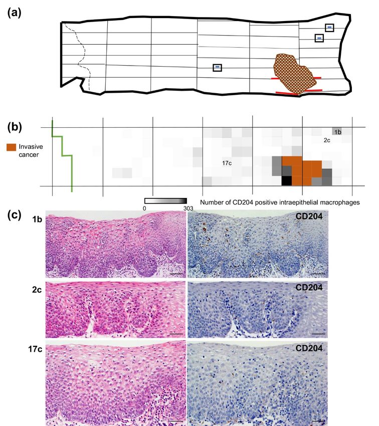
MACROPHAGES IN THE EARLY ESOPHAGEAL CARCINOGENESIS
MATERIALS AND METHODS
Tissue samples
A total of 38 carcinoma in situ (CIS) removed from 36 patients by ESD and 5 cases of invasive SCC of the
esophagus resected by surgery at Kobe University Hospital (Kobe, Japan) were employed. The ESD cases
consisted of 28 men and 8 women with an age range of 47 to 83 years and mean age of 66.1 years. The patients
receiving esophagectomy consisted of 3 men and 2 women with an age range of 49 to 73 years and mean age of
63.0 years. None of them received adjuvant chemotherapy or radiotherapy before surgery. Informed consent was
obtained from all patients, and the study was approved by the Kobe University Institutional Review Board.
All resected specimens were fixed in 10% buffered formalin. The whole specimens of 38 ESD samples and 5
cases of surgically resected esophageal squamous cell carcinoma were step-cut at 3-mm and 5-mm intervals,
respectively. Four-μm thick sections were cut from the paraffin-embedded tissues and subjected to hematoxylin
and eosin (H&E) staining and immunohistochemistry. Histological and clinicopathological evaluations were
performed according to the Japanese Classification of Esophageal Cancer proposed by the Japan Esophageal
Society (10, 11) along with the TNM classification of the Union for International Cancer Control (UICC) (29).
Immunohistochemistry
A modified version of the immunoglobulin enzyme bridge technique using a Linked Streptavidin-Biotin kit
(DakoCytomation, Glostrup, Denmark) was applied to visualize the immunoreactivities of macrophage antigens
in the tissue sections as described elsewhere (27). Specific mouse monoclonal antibodies to Ki-67 (MIB-1, 1:100;
DakoCytomation), CD68 (Kp-1, 1:100; DakoCytomation), CD163 (10D6, 1:100; Novocastra, Newcastle upon
Tyne, UK) and CD204 (SRA-E5, 1:50; Trans Genic Inc., Kobe, Japan) were used for the primary reaction. After
gentle rinsing with 0.05 mol/L of Tris-HCl, the sections were incubated with biotinylated goat anti-mouse IgG and
streptavidin conjugated to horseradish peroxidase. Chromogenic fixation was carried out by immersing the
sections in a solution of 3,3'-diaminobenzidine. Sections were counterstained with Mayer’s hematoxylin.
Macrophage count
CD68-, CD163- or CD204-positive round cells above the extrapolated line of the basement membrane were
counted in each specimen. The macrophage count was performed 3 times separately. In the case of ESD samples,
the number of macrophages per μm length of the CIS and adjacent non-neoplastic squamous epithelia in the
representative section of each sample were calculated. The number of CD204- or CD163-positive cells per 5 mm
unit length of the epithelial area was counted and plotted on each step-cut whole area of the resected esophagus to
make a density map of intraepithelial macrophages.
Statistical analysis
The significance of differences in the mean macrophage count between the neoplastic and non-neoplastic
squamous epithelia in ESD specimens was tested using the paired Student's t-test. Differences of the mean
macrophage count among the unit area of squamous intraepithelial neoplasia, carcinoma in situ and non-neoplastic
squamous epithelia within the resected esophagus were determined by the Mann-Whitney U-test. A P-value less
than 0.05 was considered significant. The statistical analyses were conducted using EZR software (12).
RESULTS
Macrophages were frequently found in neoplastic squamous epithelia of the esophagus excised by ESD
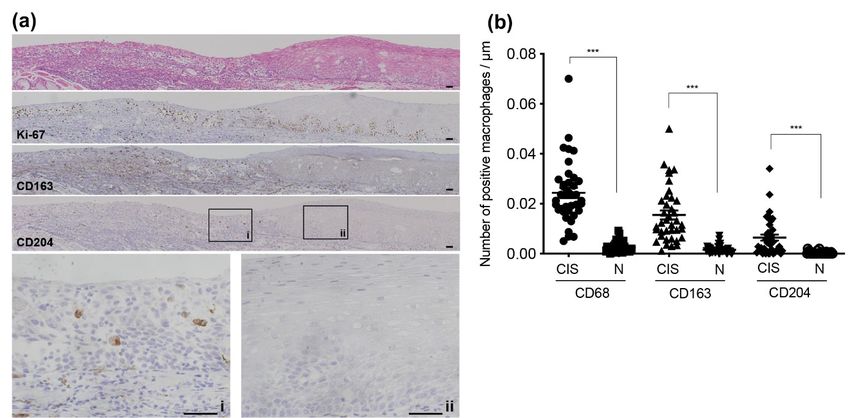
Figure 1a demonstrates the representative results of immunohistochemistry of CIS lesion excised by ESD.
H&E staining of a CIS specimen demonstrates that proliferative basal and parabasal-like cells replace the whole
epithelial layer in the left which is demarcated with the non-neoplastic squamous epithelia in the right by so-called
oblique-line. Ki-67 immunoreactive nuclei are irregularly distributed throughout the neoplastic epithelia, while
they are limited to the para-basal part of the non-neoplastic epithelia. CD163- or CD204-positive macrophages are
more frequently observed in the neoplastic than in the comparison with non-neoplastic squamous epithelia of the
esophagus.
The number of CD68-positive macrophages per μm of CIS lesion ranged from 0.07 to 0.005 (mean, 0.024; SD,
0.0123) and that of non-neoplastic squamous epithelia ranged from 0.09 to 0.0004 (mean, 0.0025; SD, 0.00209).
The number of CD163-positive macrophages per μm of CIS lesion ranged from 0.05 to 0.001 (mean, 0.016; SD,
0.0111) and that of non-neoplastic squamous epithelia ranged from 0.007 to 0 (mean, 0.0015; SD, 0.00157). The
number of CD204-positive macrophages per μm of CIS lesion ranged from 0.03 to 0 (mean, 0.006; SD, 0.0074)
and that of non-neoplastic squamous epithelia ranged from 0.002 to 0 (mean, 0.0003; SD, 0.00054). The mean
numbers of CD68-, CD163- and CD204-positive macrophages per μm of CIS lesion were significantly higher than
those of non-neoplastic squamous epithelia (each P < 0.001, Figure 1b).
E19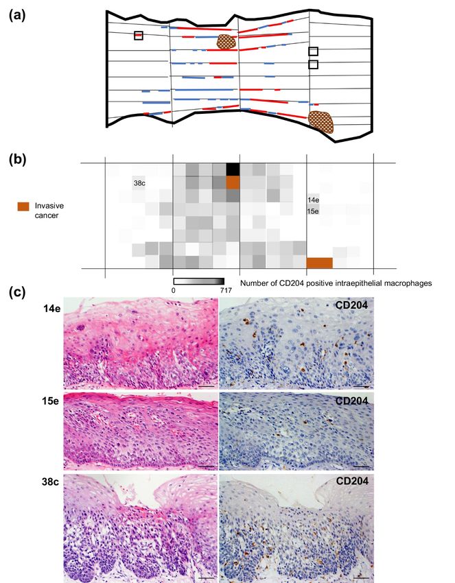
Y. ICHIHARA and H. YOKOZAKI

Figure 1. Frequent macrophage infiltration within the neoplastic squamous epithelia of the esophagus. (a) Hematoxylin and
eosin specimen of a carcinoma in situ (CIS) excised by endoscopic submucosal dissection (top panel). Immunoreactivities of
Ki-67, CD163 and CD204 are demonstrated in the following panels. i: Higher-power view of CD204 immunoreaction in the
CIS (square, i). ii, Higher power view of CD204 immunoreaction in the non-neoplastic mucosa (square, ii). Bars, 100μm.
(reproduced from Yokozaki, H., Koma, Y.-I., Shigeoka, M., Nishio, M. Cancer as a tissue: The significance of cancer-stromal
interactions in the development, morphogenesis and progression of human upper digestive tract cancer. Pathol. Int., 2018; 68:
334–352. DOI: 10.1111/pin.12674) (37). (b) Numbers of intraepithelial CD68-, CD163- or CD204-positive macrophages per
μm within 38 lesions of CIS excised by endoscopic submucosal dissection are compared with those of the corresponding non-
neoplastic squamous epithelia (N). Student’s t-test; ***, significant difference (P < 0.001).

Esophageal epithelial areas with high CD204- or CD163-positive macrophage density were morphologically
neoplastic
    To clarify the distribution of intraepithelial macrophages with CD204 or CD163 immunoreactivity within the
whole squamous cell mucosal area, we investigated in 5 cases of surgically resected SCCs. Figures 2 to 6 show
the mappings of the neoplastic lesions and the density of intraepithelial CD204-positive macrophages with
representative histopathological findings of the neoplastic lesions outside the main SCC(s) of each case.
    A total of 238 units of the mucosal area of the esophagus was observed for the CD204-positive intraepithelial
macrophage density in case 1 (Figure 2). The maximum number of immunoreactive cells per unit area was 260.
There were 12 intraepithelial neoplasia and 6 CIS independent of the invasive squamous cell carcinomas. The
mean number of CD204-positive macrophages within the unit area of these intraepithelial neoplastic lesions was
10.1 ± 10.1 (SD), while that of the 20 units of the mucosal area of the non-neoplastic esophageal mucosa was 1.4
± 1.6 (SD). There was a significant difference in the number of infiltrated CD204-positive macrophages between
the neoplastic and non-neoplastic epithelia (P = 0.011). Figure 2c shows the representative images of the CIS
indicated at the initial histopathological diagnosis. The mean numbers of intraepithelial CD204-positive
macrophages in the units 29c, 32c and 46c were 20.0, 2.0 and 28.0, respectively (Supplemental Figure S1a). The
distribution of CD163-positive macrophages within the same esophagus was almost parallel with that of CD204
(Supplemental Figures S2a and S3a).
    A total of 172 units of the mucosal area of the esophagus was observed for the CD204-positive intraepithelial
macrophage density in case 2 (Figure 3). The maximum number of immunoreactive cells per unit area was 434.
There were 22 intraepithelial neoplasia and 19 CIS independent of the invasive squamous cell carcinomas. The
mean number of CD204-positive macrophages within the unit area of these intraepithelial neoplastic lesions was
20.3 ± 25.8 (SD), while that of the 20 units of the mucosal area of the non-neoplastic esophageal mucosa was
101.4 ± 123.4 (SD). There was a significant difference in the number of infiltrated CD204-positive macrophages
between the neoplastic and non-neoplastic epithelia (P < 0.001). Figure 3c shows the representative histological
findings of the 3 intraepithelial neoplasia diagnosed at the initial histopathological examination. The mean
numbers of intraepithelial CD204-positive macrophages in the units 14e, 27d and 33e were 8.0, 15.3 and 33.7,
respectively (Supplemental Figure S1b). The distribution of CD163-positive macrophages within the same
esophagus was almost parallel with that of CD204 (Supplemental Figures S2b and S3b).

E20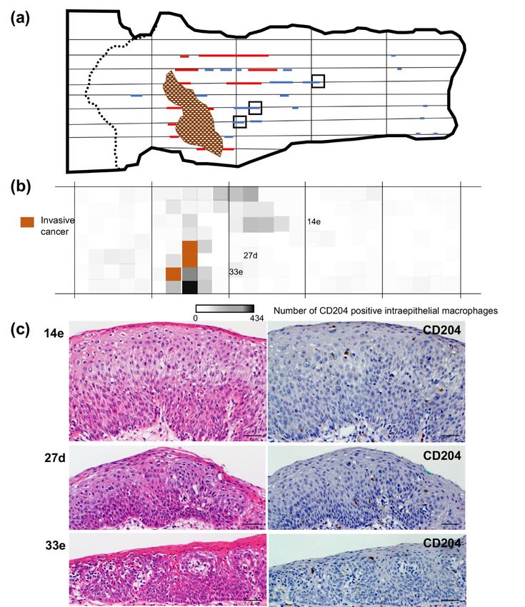
MACROPHAGES IN THE EARLY ESOPHAGEAL CARCINOGENESIS

Figure 2. Intraepithelial macrophage density mapping of whole resected esophagus (case 1). (a) Mapping of the
histopathological findings of the esophagus. There were two invasive cancers; a 30 x 30 mm plateau type elevated tumor with
well-differentiated squamous cell carcinoma invading to the submucosa in the proximal side (I), and a 19 x 18 mm
predominantly subepithelial type protruding lesion with moderately differentiated squamous cell carcinoma invading to the
submucosa in the distal side (II). Scattered intraepithelial neoplasia (blue lines) and carcinoma in situ (red lines) were indicated
by the step-cut histological examination. (b) Density mapping of CD204-positive intraepithelial macrophages. Number of
CD204-positive cells per 5 mm unit length of epithelial area (0 to 260) is illustrated as white to black gradation. (c)
Representative histopathological and immunohistochemical findings of the unit area with relatively high intraepithelial
macrophage densities. Number of each panel law is corresponded to that in (b). Bars, 200 μm.

                                                                                                                               E21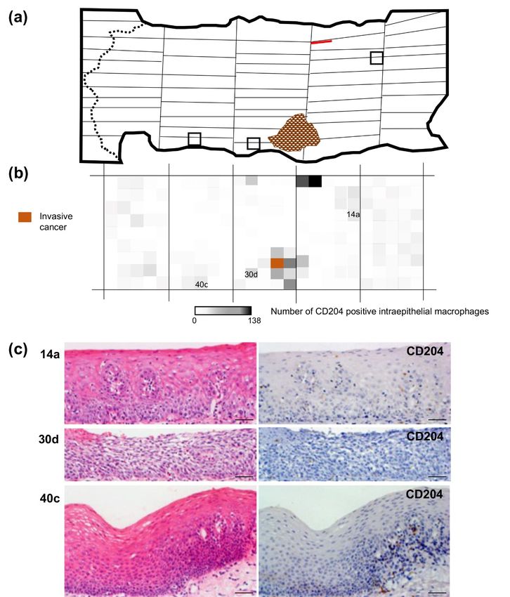
Y. ICHIHARA and H. YOKOZAKI

Figure 3. Intraepithelial macrophage density mapping of whole resected esophagus (case 2). (a) Mapping of the
histopathological findings of the esophagus. There was an invasive cancer; a 45 x 30 mm slightly elevated type lesion with
moderately differentiated squamous cell carcinoma invading to the submucosa. Scattered intraepithelial neoplasia (blue lines)
and carcinoma in situ (red lines) were indicated by the step-cut histological examination. (b) Density mapping of CD204-
positive intraepithelial macrophages. Number of CD204 positive cells per 5 mm unit length of epithelial area (0 to 434) is
illustrated as white to black gradation. (c) Representative histopathological and immunohistochemical findings of the unit area
with relatively high intraepithelial macrophage densities. Number of each panel law is corresponded to that in (b). Bars, 200
μm.

E22
MACROPHAGES IN THE EARLY ESOPHAGEAL CARCINOGENESIS

Figure 4. Intraepithelial macrophage density mapping of whole resected esophagus (case 3). (a) Mapping of the
histopathological findings of the esophagus. There was an invasive cancer; a 25 x 19 mm superficial and flat plus slightly
depressed type lesion with moderately differentiated squamous cell carcinoma invading to the muscularis mucosae. A
carcinoma in situ (red lines) were indicated by the step-cut histological examination. (b) Density mapping of CD204-positive
intraepithelial macrophages. Number of CD204-positive cells per 5 mm unit length of epithelial area (0 to 138) is illustrated as
white to black gradation. (c) Representative histopathological and immunohistochemical findings of the unit area with relatively
high intraepithelial macrophage densities. Number of each panel law is corresponded to that in (b). Bars, 200 μm.

                                                                                                                           E23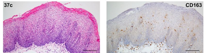
Y. ICHIHARA and H. YOKOZAKI

Figure 5. Intraepithelial macrophage density mapping of whole resected esophagus (case 4). (a) Mapping of the
histopathological findings of the esophagus. There was an invasive cancer; a 70 x 40 mm flat plus slightly elevated type lesion
with well-differentiated squamous cell carcinoma invading to the lamina propria mucosae. Scattered intraepithelial neoplasia
(blue lines) and carcinoma in situ (red lines) were indicated by the step-cut histological examination. (b) Density mapping of
CD204-positive intraepithelial macrophages. Number of CD204-positive cells per 5 mm unit length of epithelial area (0 to 717)
is illustrated as white to black gradation. (c) Representative histopathological and immunohistochemical findings of the unit
area with relatively high intraepithelial macrophage densities. Number of each panel law is corresponded to that in (b). Bars,
200 μm.

E24
MACROPHAGES IN THE EARLY ESOPHAGEAL CARCINOGENESIS

Figure 6. Intraepithelial macrophage density mapping of whole resected esophagus (case 5). (a) Mapping of the
histopathological findings of the esophagus. There was an invasive cancer; a 33 x 20 mm slightly elevated plus flat type lesion
with moderately differentiated squamous cell carcinoma invading to the submucosa. Three intraepithelial neoplasia (blue lines)
were indicated by the step-cut histological examination. (b) Density mapping of CD204-positive intraepithelial macrophages.
Number of CD204-positive cells per 5 mm unit length of epithelial area (0 to 303) is illustrated as white to black gradation. (c)
Representative histopathological and immunohistochemical findings of the unit area with relatively high intraepithelial
macrophage densities. Number of each panel law is corresponded to that in (b). Bars, 200 μm.

                                                                                                                            E25
Y. ICHIHARA and H. YOKOZAKI
A total of 236 units of the mucosal area of the esophagus was observed for the CD204-positive intraepithelial
macrophage density in case 3 (Figure 4). The maximum number of immunoreactive cells per unit area was 138.
There were 2 CIS independent of the invasive squamous cell carcinomas. Although the mean number of CD204-
positive macrophages within the unit area of these intraepithelial neoplastic lesions (115.7 ± 31.1 [SD]) was larger
than that of the 20 units of the mucosal area of the non-neoplastic esophageal mucosa (0.9 ± 0.9 [SD]), the
difference was not statistically significant (P = 0.5) because of the low number of CIS lesions. The representative
histological findings of the units with higher numbers of macrophages than surrounding units are demonstrated in
Figure 4c. Although they were not diagnosed as neoplastic lesions at the initial pathological diagnosis, each lesion
shows definite neoplastic epithelial growth. The mean number of intraepithelial CD204-positive macrophages in
the units 14a, 30a and 40c were 12.7, 22.7 and 12.3, respectively (Supplemental Figure S1c). The distribution of
CD163-positive macrophages within the same esophagus was almost paralleled with that of CD204 (Supplemental
Figures S2c and S3c).
A total of 165 units of the mucosal area of the esophagus was observed for the CD204-positive intraepithelial
macrophage density in case 4 (Figure 5). The maximum number of immunoreactive cells per unit area was 717.
There were 39 intraepithelial neoplasia and 37 CIS independent of the invasive squamous cell carcinomas. The
mean number of CD204-positive macrophages within the unit area of these intraepithelial neoplastic lesions was
120.2 ± 100.7 (SD), while that of the 20 units of the mucosal area of the non-neoplastic esophageal mucosa was
1.0 ± 1.1 (SD). There was a significant difference in the number of infiltrated CD204-positive macrophages
between the neoplastic and non-neoplastic epithelia (P < 0.001). Figure 5c depicts the representative histological
findings of 2 units with higher numbers of macrophages than surrounding units (14e and 15e) and a unit with CIS
(38e). Both units 14e and 15e demonstrate neoplastic growth within the basal part of the epithelia. The mean
numbers of intraepithelial CD204-positive macrophages in the units 14e, 15e and 38e were 64.3, 119.3 and 241.7,
respectively (Supplemental Figure S1d). The distribution of CD163-positive macrophages within the same
esophagus was almost parallel with that of CD204 (Supplemental Figures S2a and S3a).
A total of 167 units of the mucosal area of the esophagus was observed for the CD204-positive intraepithelial
macrophage density in case 5 (Figure 6, Supplemental Figure S1e). The maximum number of immunoreactive
cells per unit area was 303. There were 3 intraepithelial neoplasia and 6 CIS independent of the invasive squamous
cell carcinomas. The mean number of CD204-positive macrophages within the unit area of these intraepithelial
neoplastic lesions was 136.6 ± 93.3 (SD), while that of the 20 units of mucosal area of the non-neoplastic
esophageal mucosa was 0.9 ± 1.1 (SD). There was a significant difference in the number of infiltrated CD204-
positive macrophages between the neoplastic and non-neoplastic epithelia (P < 0.001). The representative
histological findings of the 3 intraepithelial neoplasia pointed out at the initial histopathological diagnosis are
demonstrated in Figure 6c. The mean numbers of intraepithelial CD204-positive macrophages in the units 1b, 2c
and 17c were 92.5, 2.0 and 14.0, respectively (Supplemental Figure S1b). The distribution of CD163-positive
macrophages within the same esophagus was almost parallel with that of CD204 (Supplemental Figures S2e and
S3e) in all units except the unit 37c (Supplemental Figure S4).
DISCUSSION
There is accumulating evidence of the clinicopathological significance of TAMs for the progression of SCCs
arising not only in the esophagus but also in the other human organs has been accumulated. A high number of
CD204-positive TAMs in the stroma was a significant prognostic factor of lung SCCs that was closely associated
with an advanced pathological stage, T factor, N factor, vascular and pleural invasion (7). An increase in CD68-
positive macrophages in the tumor stroma has been significantly associated with lymphatic metastasis of SCCs of
the uterine cervix (2). A high number of CD163-positive TAMs in head and neck SCCs has been associated with
advanced T stage, increased rates of nodal positivity, presence of vessel invasion, and poor patient outcome (3,
18). In addition, several investigators have investigated macrophage infiltration into the initial intraepithelial
neoplastic growth of the squamous epithelia.
Hammes et al. counted CD68-positive macrophages in biopsy specimens of the normal uterine cervix, low-
grade intraepithelial lesions, high-grade intraepithelial lesions, and invasive SCCs and found that the number of
macrophages infiltrating the lesions was closely associated with the progression of cervical intraepithelial
neoplasia independent of the background inflammation (6). Tongue leukoplakia tissues with high numbers of
intraepithelial CD163-positive macrophages significantly associated with higher degrees of epithelial dysplasia,
abnormal Ki-67 expression, and cytokeratin 13 loss in comparison with those with low CD163-positive
macrophage infiltration (26). Pre-operative biopsy specimens of tongue leukoplakia progressed to invasive SCC
have been shown to contain a significant number of CD163-positive intraepithelial macrophages (25). In another
report, the number of CD163-positive macrophages infiltrating into early-stage SCCs of the esophagus was
E26MACROPHAGES IN THE EARLY ESOPHAGEAL CARCINOGENESIS
significantly associated with the neo-angiogenic potential estimated by the increased microvessel density within
the hot spots of the lesions (17).
Infiltration of a significant number of macrophages, especially macrophages expressing M2 markers, in the
esophageal CIS lesions in comparison with the corresponding non-neoplastic squamous epithelia was also
confirmed in the present study using histopathologically defined ESD specimens. Statistically significant
associations of intraepithelial accumulation of M2-skewed macrophages with CIS and squamous intraepithelial
neoplasia in the step-cut esophagectomy specimens were also demonstrated in 4 out of 5 esophageal cancer cases.
A retrospective histopathological review of the mucosal units with a high number of intraepithelial CD204-positive
macrophages in cases 3 and 4 or CD163-positive macrophages in case 5 also revealed intraepithelial neoplastic
growth that was overlooked at the initial diagnosis of the esophagectomy specimens. These findings of the present
study may suggest that macrophages have some biological roles in the early squamous carcinogenesis of the
esophagus.
Wound-associated basal keratinocytes of mice were reported to secrete Ccl2 through activated Nrf2 and
induced the production of EGF in macrophages to promote keratinocyte proliferation (33). It has been established
that TAMs, especially those skewed to the M2-phenotype, promote growth and motility of cancer cells by secreting
growth factors (20, 32) and chemokines (4, 8, 13). Macrophages activated by defined cytokines were reported to
promote the growth and migration of an immortalized human esophageal squamous epithelial cell line with the
induction of IL6 and p38MAPK signals by co-culture experiments (14). These reports and our present observations
suggest that the macrophages found within the initial proliferative lesions of the squamous epithelia may play
biological roles in their progression.
In terms of the triggers of macrophage recruitment into the pre-neoplastic squamous epithelia of the esophagus,
several studies suggest that these triggers may consist of responses to the DNA damage signals elicited by the
exposure to the carcinogen(s). Acetaldehyde associated with alcoholic beverages is carcinogenic to humans (Group
1) and confirmed the Group 1 classification of alcohol consumption and of ethanol in alcoholic beverages. (23) It
has been well-established that the aldehyde dehydrogenase 2 (ALDH2) variant allele confers higher relative risk
of esophageal SCC (36). Amanuma et al. demonstrated that significantly elevated acetaldehyde-derived DNA
damage signals represented by N2-ethylidene-2'-deoxyguanosine were higher in the esophagus of Aldh2-knockout
mice than in wild-type mice upon ethanol consumption (1). Another study found that infiltration of macrophages
into the squamous epithelia of ethanol-treated Aldh2-knockuout mice was significantly elevated compared to that
in wild-type animals (37). In addition, p38MAPK is known to be involved in the DNA damage stress responses
during cancer development (5). Macrophages may receive the DNA damage signals from and recruited into the
injured squamous epithelia of the esophagus establishing the cell-to-cell interactions to promote the pre-neoplastic
growth and the accumulation of the replication stress that leads to the mutation of the driver gene(s) (37). In fact,
intensive sequencing of micro-scale human esophageal samples demonstrated the progressive age-related
expansion of clones carrying mutations of driver genes even in physiologically normal squamous epithelia, which
was substantially accelerated by alcohol consumption and smoking (19, 35).
The limitation of the present study is that the results just demonstrate the close association of intraepithelial
accumulation of M2-skewed macrophages with neoplastic epithelia of the human esophagus. Further molecular
pathological analysis to elucidate the detailed biological roles of intraepithelial M2-macrophages in the
development of esophageal SCC will be required. While detection of the intraepithelial M2-macrophages may
assist in the histopathological assessment of esophageal mucosal lesions at risk.
ACKNOWLEDGEMENTS
The experimental work presented here was supported in part by JSPS KAKENHI Grants (nos. 23590397,
26460418, JP17K08693, and JP20K07373) to HY. We thank Atsuko Kawashima, Yumi Hashimoto, Nobuo Kubo,
and Miki Yamazaki for their excellent technical assistance. The mapping study of macrophages in the resected
esophageal tissue sections was initiated by Dr. Yasue Morita-Sato in 2011, when she was a first-year medical
student at Kobe University.
AUTHOR CONTRIBUTIONS
Y.I. and H.Y. conceived and performed the experiments; Y.I. and H.Y. wrote the manuscript.
CONFLICTS OF INTEREST
The authors declare that they have no conflict of interest.
E27Y. ICHIHARA and H. YOKOZAKI
REFERENCES
1. Amanuma, Y., Ohashi, S., Itatani, Y., Tsurumaki, M., Matsuda, S., Kikuchi, O., Nakai, Y., Miyamoto,
S., Oyama, T., Kawamoto, T., Whelan, K. A., Nakagawa, H., Chiba, T., Matsuda, T., and Muto, M. 2015.
Protective role of ALDH2 against acetaldehyde-derived DNA damage in oesophageal squamous epithelium.
Sci Rep 5: 14142.
2. Ding, H., Cai, J., Mao, M., Fang, Y., Huang, Z., Jia, J., Li, T., Xu, L., Wang, J., Zhou, J., Yang, Q., and
Wang, Z. 2014. Tumor-associated macrophages induce lymphangiogenesis in cervical cancer via interaction
with tumor cells. APMIS 122: 1059–1069.
3. Fujii, N., Shomori, K., Shiomi, T., Nakabayashi, M., Takeda, C., Ryoke, K., and Ito, H. 2012. Cancer-
associated fibroblasts and CD163-positive macrophages in oral squamous cell carcinoma: their
clinicopathological and prognostic significance. J Oral Pathol Med 41: 444–451.
4. Fujikawa, M., Koma, Y. I., Hosono, M., Urakawa, N., Tanigawa, K., Shimizu, M., Kodama, T.,
Sakamoto, H., Nishio, M., Shigeoka, M., Kakeji, Y., and Yokozaki, H. 2021. CCL1 derived from tumor-
associated macrophages contributes to esophageal squamous cell carcinoma progression via CCR8-mediated
Akt/PRAS40/mTOR pathway. Am J Pathol 191: 686–703.
5. Gorgoulis, V. G., Pefani, D. E., Pateras, I. S., and Trougakos, I. P. 2018. Integrating the DNA damage and
protein stress responses during cancer development and treatment. J Pathol 246: 12–40.
6. Hammes, L. S., Tekmal, R. R., Naud, P., Edelweiss, M. I., Kirma, N., Valente, P. T., Syrjanen, K. J., and
Cunha-Filho, J. S. 2007. Macrophages, inflammation and risk of cervical intraepithelial neoplasia (CIN)
progression--clinicopathological correlation. Gynecol Oncol 105: 157–165.
7. Hirayama, S., Ishii, G., Nagai, K., Ono, S., Kojima, M., Yamauchi, C., Aokage, K., Hishida, T., Yoshida,
J., Suzuki, K., and Ochiai, A. 2012. Prognostic impact of CD204-positive macrophages in lung squamous
cell carcinoma: possible contribution of Cd204-positive macrophages to the tumor-promoting
microenvironment. J Thorac Oncol 7: 1790–1797.
8. Hosono, M., Koma, Y. I., Takase, N., Urakawa, N., Higashino, N., Suemune, K., Kodaira, H., Nishio,
M., Shigeoka, M., Kakeji, Y., and Yokozaki, H. 2017. CXCL8 derived from tumor-associated macrophages
and esophageal squamous cell carcinomas contributes to tumor progression by promoting migration and
invasion of cancer cells. Oncotarget 8: 106071–106088.
9. International Agency for Research on Cancer, Cancer Today (Globocan 2020). 2020. Population Fact
Sheets, World. Retrieved from https://gco.iarc.fr/today/data/factsheets/populations/900-world-fact-sheets.
pdf
10. Japan Esophageal Society. 2017. Japanese Classification of Esophageal Cancer, 11th Edition: part I.
Esophagus 14: 1–36.
11. Japan Esophageal Society. 2017. Japanese Classification of Esophageal Cancer, 11th Edition: part II and
III. Esophagus 14: 37–65.
12. Kanda, Y. 2013. Investigation of the freely available easy-to-use software 'EZR' for medical statistics. Bone
Marrow Transplant 48: 452–458.
13. Kodama, T., Koma, Y. I., Arai, N., Kido, A., Urakawa, N., Nishio, M., Shigeoka, M., and Yokozaki, H.
2020. CCL3-CCR5 axis contributes to progression of esophageal squamous cell carcinoma by promoting cell
migration and invasion via Akt and ERK pathways. Lab Invest 100: 1140–1157.
14. Koma, Y. I., Nishio, M., Shigeoka, M., and Yokozaki, H. 2017. Role of macrophage in ESCC
carcinogenesis. Bessatsu Bio Clinica 6: 123–127. (in Japanese)
15. Komohara, Y., Jinushi, M., and Takeya, M. 2014. Clinical significance of macrophage heterogeneity in
human malignant tumors. Cancer Sci 105: 1–8.
16. Komohara, Y., Ohnishi, K., Kuratsu, J., and Takeya, M. 2008. Possible involvement of the M2 anti-
inflammatory macrophage phenotype in growth of human gliomas. J Pathol 216: 15–24.
17. Kumagai, Y., Sobajima, J., Higashi, M., Ishiguro, T., Fukuchi, M., Ishibashi, K.-i., Mochiki, E., Yakabi,
K., Kawano, T., Tamaru, J.-i., and Ishida, H. 2016. Tumor-associated macrophages and angiogenesis in
early stage esophageal squamous cell carcinoma. Esophagus 13: 245–253.
18. Kumar, A. T., Knops, A., Swendseid, B., Martinez-Outschoom, U., Harshyne, L., Philp, N., Rodeck, U.,
Luginbuhl, A., Cognetti, D., Johnson, J., and Curry, J. 2019. Prognostic significance of tumor-associated
macrophage content in head and neck squamous cell carcinoma: A meta-analysis. Front Oncol 9: 656.
19. Martincorena, I., Fowler, J. C., Wabik, A., Lawson, A. R. J., Abascal, F., Hall, M. W. J., Cagan, A.,
Murai, K., Mahbubani, K., Stratton, M. R., Fitzgerald, R. C., Handford, P. A., Campbell, P. J., Saeb-
Parsy, K., and Jones, P. H. 2018. Somatic mutant clones colonize the human esophagus with age. Science
362: 911–917.
20. O'Sullivan, C., Lewis, C. E., Harris, A. L., and McGee, J. O. 1993. Secretion of epidermal growth factor
by macrophages associated with breast carcinoma. Lancet 342: 148–149.
E28MACROPHAGES IN THE EARLY ESOPHAGEAL CARCINOGENESIS
21. Odze, R. D., Lam, A. K., Ochiai, A., and Washington, M. K. 2019. Tumours of the oesophagus, p.22–53.
In WHO Classification of Tumor Editorial Board (Ed.), Digestive System Tumours (WHO Classification of
Tumours, 5th ed. vol 1). International Agency for Research on Cancer, Lyon.
22. Ono, H., Kondo, H., Gotoda, T., Shirao, K., Yamaguchi, H., Saito, D., Hosokawa, K., Shimoda, T., and
Yoshida, S. 2001. Endoscopic mucosal resection for treatment of early gastric cancer. Gut 48: 225–229.
23. Secretan, B., Straif, K., Baan, R., Grosse, Y., El Ghissassi, F., Bouvard, V., Benbrahim-Tallaa, L., Guha,
N., Freeman, C., Galichet, L., Cogliano, V., and WHO International Agency for Research on Cancer
Monograph Working Group. 2009. A review of human carcinogens--Part E: tobacco, areca nut, alcohol,
coal smoke, and salted fish. Lancet Oncol 10: 1033–1034.
24. Shibata, A., Matsuda, T., Ajiki, W., and Sobue, T. 2008. Trend in incidence of adenocarcinoma of the
esophagus in Japan, 1993–2001. Jpn J Clin Oncol 38: 464–468.
25. Shigeoka, M., Koma, Y. I., Kodama, T., Nishio, M., Akashi, M., and Yokozaki, H. 2019. Intraepithelial
CD163+ macrophages in tongue leukoplakia biopsy: A promising tool for cancer screening. Oral Dis 26: 527–
536.
26. Shigeoka, M., Koma, Y. I., Nishio, M., Komori, T., and Yokozaki, H. 2019. CD163+ macrophages
infiltration correlates with the immunosuppressive cytokine interleukin 10 expression in tongue leukoplakia.
Clin Exp Dent Res 5: 627–637.
27. Shigeoka, M., Urakawa, N., Nakamura, T., Nishio, M., Watajima, T., Kuroda, D., Komori, T., Kakeji,
Y., Semba, S., and Yokozaki, H. 2013. Tumor associated macrophage expressing CD204 is associated with
tumor aggressiveness of esophageal squamous cell carcinoma. Cancer Sci 104: 1112–1119.
28. Sica, A., Schioppa, T., Mantovani, A., and Allavena, P. 2006. Tumour-associated macrophages are a
distinct M2 polarised population promoting tumour progression: potential targets of anti-cancer therapy. Eur
J Cancer 42: 717–727.
29. Sobin, L.H., Gospodarowicz, M.K., and Wittekind, C. (eds). 2011. TNM Classification of Malignant
Tumours (7th ed.). Hoboken, NJ: Wieley-Blackwell.
30. Tachimori, Y., Ozawa, S., Numasaki, H., Fujishiro, M., Matsubara, H., Oyama, T., Shinoda, M., Toh,
Y., Udagawa, H., Uno, T., and Registration Committee for Esophageal Cancer of the Japan Esophageal
Society. 2016. Comprehensive registry of esophageal cancer in Japan, 2009. Esophagus 13: 110–137.
31. Takeya, M., and Komohara, Y. 2016. Role of tumor-associated macrophages in human malignancies: friend
or foe? Pathol Int 66: 491–505.
32. Urakawa, N., Utsunomiya, S., Nishio, M., Shigeoka, M., Takase, N., Arai, N., Kakeji, Y., Koma, Y. I.,
and Yokozaki, H. 2015. GDF15 derived from both tumor-associated macrophages and esophageal squamous
cell carcinomas contributes to tumor progression via Akt and Erk pathways. Lab Invest 95: 491-503.
33. Villarreal-Ponce, A., Tiruneh, M. W., Lee, J., Guerrero-Juarez, C. F., Kuhn, J., David, J. A., Dammeyer,
K., Mc Kell, R., Kwong, J., Rabbani, P. S., Nie, Q., and Ceradini, D. J. 2020. Keratinocyte-macrophage
crosstalk by the Nrf2/Ccl2/EGF signaling axis orchestrates tissue repair. Cell Rep 33: 108417.
34. Watanabe, M., Otake, R., Kozuki, R., Toihata, T., Takahashi, K., Okamura, A., and Imamura, Y. 2020.
Recent progress in multidisciplinary treatment for patients with esophageal cancer. Surg Today 50: 12–20.
35. Yokoyama, A., Kakiuchi, N., Yoshizato, T., Nannya, Y., Suzuki, H., Takeuchi, Y., Shiozawa, Y., Sato, Y.,
Aoki, K., Kim, S. K., Fujii, Y., Yoshida, K., Kataoka, K., Nakagawa, M. M., Inoue, Y., Hirano, T.,
Shiraishi, Y., Chiba, K., Tanaka, H., Sanada, M., Nishikawa, Y., Amanuma, Y., Ohashi, S., Aoyama, I.,
Horimatsu, T., Miyamoto, S., Tsunoda, S., Sakai, Y., Narahara, M., Brown, J. B., Sato, Y., Sawada, G.,
Mimori, K., Minamiguchi, S., Haga, H., Seno, H., Miyano, S., Makishima, H., Muto, M., and Ogawa,
S. 2019. Age-related remodelling of oesophageal epithelia by mutated cancer drivers. Nature 565: 312–317.
36. Yokoyama, A., and Omori, T. 2003. Genetic polymorphisms of alcohol and aldehyde dehydrogenases and
risk for esophageal and head and neck cancers. Jpn J Clin Oncol 33: 111–121.
37. Yokozaki, H., Koma, Y. I., Shigeoka, M., and Nishio, M. 2018. Cancer as a tissue: The significance of
cancer-stromal interactions in the development, morphogenesis and progression of human upper digestive
tract cancer. Pathol Int 68: 334–352.
38. Yoshida, T., Inoue, H., Usui, S., Satodate, H., Fukami, N., and Kudo, S. E. 2004. Narrow-band imaging
system with magnifying endoscopy for superficial esophageal lesions. Gastrointest Endosc 59: 288–295.
E29Y. ICHIHARA and H. YOKOZAKI
Supplemental Figure S1 Number of CD204-positive cells per 5 mm unit length of epithelial area of the resected esophagus of case 1 (a), case 2 (b), case 3 (c), case 4 (d) and
case 5 (e). Mean number with SD of the triplicate counts per unit are described.
E30MACROPHAGES IN THE EARLY ESOPHAGEAL CARCINOGENESIS
Supplemental Figure S2 Density mappings of CD163-positive intraepithelial macrophages of the resected esophagus of case
1 (a), case 2 (b), case 3 (c), case 4 (d) and case 5 (e). Number of CD163-positive cells per 5 mm unit length of epithelial area
is plotted as white to black gradation.
E31Y. ICHIHARA and H. YOKOZAKI
Supplemental Figure S3 Number of CD163-positive cells per 5 mm unit length of epithelial area of the resected esophagus of case 1 (a), case 2 (b), case 3 (c), case 4 (d) and
case 5 (e). Mean number with SD of the triplicate counts per unit are described.
E32MACROPHAGES IN THE EARLY ESOPHAGEAL CARCINOGENESIS
Supplemental Figure S4 Histopathological findings of unit 37c of the case 5. Thickened squamous epithelia shows surface
parakeratosis and neoplastic growth of the one third of the basal area with elongated vascular papillae (left). Scattered CD163-
positive cells are observed within the epithelial area (right). The mean number of triplicate counts was 267.7 (Supplemental
Figure 3e). Bars, 200 μm.
E33You can also read