SMAD3 promotes ELK3 expression following transforming growth factor β mediated stimulation of MDA MB231 cells - Spandidos Publications
←
→
Page content transcription
If your browser does not render page correctly, please read the page content below
ONCOLOGY LETTERS 19: 2749-2754, 2020

      SMAD3 promotes ELK3 expression following transforming
      growth factor β‑mediated stimulation of MDA‑MB231 cells
                                          JI‑HOON PARK and KYUNG‑SOON PARK

                                Department of Biomedical Science, College of Life Science,
                            CHA University, Seongnam‑si, Gyeonggi‑do 463‑400, Republic of Korea

                                     Received October 1, 2019; Accepted December 19, 2019

                                                   DOI: 10.3892/ol.2020.11375

Abstract. Transforming growth factor‑β (TGFβ) is a secreted         Introduction
cytokine whose aberrant spatiotemporal expression is related
to cancer progression and metastasis. While TGFβ acts as a          Cancer metastasis is the process of cancer cells disseminating
tumor suppressor in normal and premalignant stages, TGFβ            from the primary tumor to a distal site through lymphatic
functions as a tumor promoter during the malignant phases           tissue and blood vessels. Cancer metastasis is responsible
of tumor progression by prompting cancer cells to undergo           for approximately 90% of cancer deaths, indicating that it
epithelial‑mesenchymal transition (EMT), which enhances             is the primary cause of morbidity and mortality (1). Even
tumor cell invasion and ultimately promotes metastasis to           though most solid tumors are now manageable or curable
other organs. Extensive studies have been performed to              by advances in early cancer detection and treatment,
uncover the molecular and cellular mechanisms underlying            cancers spreading beyond the initial primary site are usually
TGFβ inducing EMT in cancer cells. Here, we suggested               highly incurable (2). Lack of understanding of the mecha-
that ELK3, which encodes a protein that orchestrates inva-          nism underlying the metastatic process has meant that the
sion and metastasis of triple negative breast cancer cells, is a    predominant cancer treatments focus on inhibition of cancer
downstream target of TGFβ‑SMAD3 in MDA‑MB231 cells.                 growth with little emphasis on metastasis, meaning that the
ELK3 expression was increased in a time‑dependent manner            overall survival of metastatic cancer patients has not been
upon TGFβ treatment. Chemical and molecular inhibition              improved significantly.
of the TGFβ receptor blocked the ability of TGFβ to induce              Transforming growth factor‑β (TGFβ) is one of the master
ELK3 expression. Small interfering RNA‑mediated suppres-            factors of metastasis in that it induces the epithelial‑mesen-
sion analysis revealed that SMAD3 induces TGFβ signaling to         chymal transition (EMT), which is associated with cancer.
express ELK3. Moreover, the results of the luciferase reporter      EMT is the reversible orchestrated transcriptional program in
assay and chromatin immunoprecipitation analysis showed             which well‑organized, tightly connected epithelial cells trans-
that SMAD3 directly binds to the SMAD‑binding element on            differentiate into disorganized and motile mesenchymal cells.
the promoter of ELK3 to activate gene expression following          TGF‑β signaling mediated by SMAD or non‑SMAD pathways
TGFβ stimulation. We concluded that ELK3 is a novel down-           plays a fundamental role in activating the transcriptional
stream target of TGFβ‑SMAD3 signaling in aggressive breast          network to induce the expression of mesenchymal components
cancer cells.                                                       and to suppress the expression of epithelial genes (3,4). As a
                                                                    result, epithelial cancer cells undergo dramatic remodeling
                                                                    of the cytoskeleton along with dissolution of tight junctions
                                                                    to acquire mesenchymal features that exhibit a significantly
Correspondence to: Professor Kyung‑Soon Park, Department            enhanced metastatic dissemination potential into distal
of Biomedical Science, College of Life Science, CHA University,     organs. This event is induced by the activity of master regula-
Pangyo‑Ro 335, Bundang‑gu, Seongnam‑si, Gyeonggi‑do 463‑400,        tors of EMT, which include SNAIL, SLUG, ZEB1/delta EF1
Republic of Korea                                                   and ZEB2/SIP1 (5‑8).
E‑mail: kspark@cha.ac.kr                                                ELK3 is an ETS domain‑containing protein capable of
                                                                    forming a ternary complex with DNA and serum response
Abbreviations: TGFβ, transforming growth factor‑β; SBE, SMAD        factor (9). ELK3 is reported to be involved in the migration
binding element; TNBCs, triple negative breast cancer cells; EMT,
                                                                    and invasion of various cancer cells including aggressive
epithelial‑mesenchymal transition; siTGFR1, small interfering RNA
targeting to TGFβ receptor R1; CA‑ALK5, constitutively active       basal‑like breast cancer cells and liver cancer stem cells (10,11).
form of TGFβ receptor‑I                                             Previously, we reported that ELK3 suppression impairs the
                                                                    ability of TGFβ signaling to activate the expression of mesen-
Key words: TGFβ, SMAD3, ELK3, MDA‑MB‑231, MCF7,                     chymal markers such as Vimentin, Slug and SNAIL in the
luciferase assay, chromatin immunoprecipitation                     triple negative breast cancer cells, which suggests that ELK3
                                                                    is implicated in the TGFβ signaling pathway to regulate the
                                                                    metastatic process of aggressive cancer cells (12,13).
2750 PARK and PARK: REGULATION OF ELK3 EXPRESSION BY TGFβ‑Smad3
In the present study, to extend our understanding of the using the Dual‑Luciferase Reporter Assay System (Promega)
molecular implication of ELK3 to the TGFβ signaling pathway according to the manufacturer's protocols. The values of firefly
in cancer cells, we analyzed the regulatory mechanism of luciferase were normalized to the respective values of Renilla
TGFβ signaling on ELK3 expression. We found that TGFβ luciferase.
stimulates the transcriptional expression of ELK3 in the repre-
sentative triple negative breast cancer cell line, MDA‑MB231. Chromatin immunoprecipitation. In brief, 37% formaldehyde
Furthermore, based on the biochemical and molecular biology was added to the cell culture medium to a final concentration
study, we demonstrated that TGFβ ‑mediated phosphoryla- of 1% and incubated for 15 min at RT. Glycine was added
tion of SMAD3 functions as a transcriptional activator of to a final concentration of 125 mM for 5 min at RT, and
ELK3. Taken together, our data reveal that ELK3 is a direct the cells were washed three times with cold PBS. The cells
downstream target of TGFβ ‑SMAD3 signaling pathway in were lysed in 400 µl of 1X cell lysis buffer (Cell Signaling)
MDA‑MB231 cells. containing protease/phosphatase inhibitor cocktail (Pierce
Biotechnology). After eight rounds of sonication, the lysates
Materials and methods were cleared by centrifugation at 13,000 rpm for 15 min at
4˚C. The supernatants were mixed with 40 µl of Dynabead
Plasmids, siRNA and primers. Information on the plas- protein G and 2 µg of primary antibodies for 2 h at RT or
mids and siRNAs is summarized in the supplementary overnight at 4˚C. The complexes were washed sequentially
Tables SI and SII. with 1X RIPA buffer, 1X RIPA buffer (500 mM NaCl), LiCl
buffer and TE buffer twice for 10 min each. Then, 3 µl of 10%
Cell culture and transfection. The triple negative breast SDS and 5 µl of 20 mg/ml proteinase K were added to sepa-
cancer cell line MDA‑MB231 and the human breast adeno- rate the DNA‑protein complex. The DNA was purified by the
carcinoma cell line MCF7 and the human embryonic kidney phenol/chloroform extraction method, and then it was used in
293T cells were purchased from American Type Culture PCR with primers targeting the ELK3 promoter.
Collection (Manassas, VA, USA). These cells were main-
tained in DMEM (Gibco BRL Life Technologies, Rockville, Statistical analysis. Samples were analyzed with Student's
MD, USA) containing 10% (v/v) heat‑inactivated fetal bovine t‑test or ANOVA with Duncan's multiple range procedure for
serum (Gibco BRL). 293T cells were used for the luciferase multiple comparisons. All statistical analyses were performed
assay with the pGL3‑ELK3 plasmid. Transient transfection of using GraphPad Prism 5 (GraphPad Prism, USA) or the
plasmid DNA or siRNA was performed with Lipofectamine SigmaPlot 11.2 program (Systat Software, USA). All statistical
2000 (Invitrogen, Carlsbad, CA, USA) according to the manu- analyses were performed using GraphPad Prism 5 (GraphPad
facturer's protocols. Prism, USA). The error bars represent the standard errors from
three independent experiments, which were each performed
RNA extraction and reverse transcription‑quantitative (RT‑q) using triplicate samples. P‑values less than 0.05 were consid-
PCR. Total RNA was extracted by manual methods using ered statistically significant.
TRIZol (Invitrogen), and 1 µg of cDNA was synthesized
using the LeGene Express 1st Strand cDNA Synthesis System Results
(LeGene Biosciences Inc., San Diego, CA, USA) according
to the manufacturer's instructions. RT‑qPCR was performed TGFβ induces accumulation of ELK3 in the nucleus of
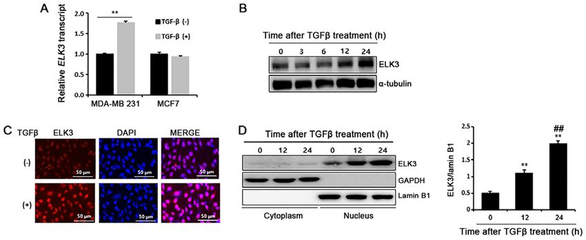
using synthetic cDNAs and TOPrealTM qPCR 2X PreMIX MDA‑MB231 cells, but not in MCF7 cells. Cancer cells
(Enzynomics, Daejeon, Korea). The expression of the target treated with TGFβ undergo the EMT process by developing
genes was normalized to that of glyceraldehyde 3‑phosphate a fibroblast‑like morphological appearance and changing
dehydrogenase (GAPDH). The PCR primers are listed in epithelial and mesenchymal phenotype marker expression.
Table SIII. Unlike MDA‑MB231 cells, TGFβ ‑treated MCF7 cells that
display morphological changes of EMT do not show suppres-
Immunoblot analysis. Cells were lysed with RIPA buffer sion of E‑cadherin, a typical epithelial phenotype marker (14).
(Cell Signaling Technology, Beverly, MA, USA) and total cell Recently, we reported that ELK3 is highly expressed in
lysates were separated by sodium dodecyl sulfate‑polyacryl- TNBC‑like MDA‑MB231 cells, where it functions as a tran-
amide gel electrophoresis (SDS‑PAGE) and then transferred to scriptional repressor of E‑cadherin by collaborating with
polyvinylidene difluoride membranes (Bio‑Rad, Hercules, CA, ZEB1 (15). Therefore, we hypothesized that ELK3 is the
USA). The membranes were blotted with the indicated primary missing link that explains the different molecular responses
antibodies at 4˚C overnight. After washing with TBST, the of MDA‑MB231 and MCF7 cells when they are treated with
membranes were incubated for 1 h at room temperature with TGFβ. We first compared the expression of ELK3 between
secondary antibodies. Immunoreactivity was detected with an MDA‑MB231 and MCF7 cells following TGFβ treatment. As
ECL kit (Thermo Scientific, Rochester, NY, USA). The anti- expected, TGFβ stimulated ELK3 expression in MDA‑MB231
bodies used in this study are summarized in Table SIV. cells but not in MCF7 cells (Fig. 1A). Consistently, ELK3
protein was also accumulate in the TGFβ‑treated MDA‑MB231
Luciferase assay. The 293T cells were transfected with the cells (Fig. 1B). Immunocytochemical analysis and subcellular
indicated plasmids using Lipofectamine 2000 (Invitrogen) fractionation assays of the cytosol and nucleus confirmed
according to the manufacturer's protocols. Cells were harvested that ELK3 accumulates in the TGFβ ‑treated MDA‑MB231
48 h after transfection, and luciferase activity was measured cells (Fig. 1C and D). Overall, these data indicate that TGFβONCOLOGY LETTERS 19: 2749-2754, 2020 2751 Figure 1. TGFβ induces accumulation of ELK3 in the nuclei of MDA‑MB231 cells. (A) Effect of TGFβ on the expression of ELK3 in MDA‑MB231 and MCF7 cells was compared by RT‑qPCR of cancer cells treated with TGFβ (5 ng/ml) for 24 h. **P
2752                                 PARK and PARK: REGULATION OF ELK3 EXPRESSION BY TGFβ‑Smad3

Figure 3. SMAD3 binds to the ELK3 promoter to activate transcription upon TGFβ treatment. (A) Schematics of luciferase assay promoter construct of ELK3
(‑92 bp ‑ +345 bp) (left panel). The right panel shows the DNA binding motif of SMAD3 in the ELK3 promoter region of human (+225 bp ‑ +257 bp) and mouse
(+112 bp ‑ +144 bp) genomes. (B) Effect of cotransfection of SMAD3 and constitutively active form of the TGFβ receptor‑I (CA‑ALK5) on the activity of the
ELK3 promoter. Reporter plasmid containing the ELK3 promoter (pGL3‑ELK3) was cotransfected into 293T cells with CA‑ALK5 and SMAD3 expressing
plasmids for 24 hrs. The expression of p‑SMAD3, SMAD3 and CA‑ALK5 was analyzed by immunoblot (left panel), and the activity of the ELK3 promoter
was analyzed by luciferase assay (right panel). (C) Chromatin from TGFβ‑treated or nontreated MDA‑MB231 cells were immunoprecipitated (ChIP) with
antibodies against SMAD3 and IgG. The PCR results for the ELK3 promoter region (+225 bp ‑ +257 bp) are presented. SMAD3 binding to the ID1 promoter
was used as a positive control of the ChIP experiment. The error bars represent the standard errors from three independent experiments, which were each per-
formed using triplicate samples. **P
ONCOLOGY LETTERS 19: 2749-2754, 2020                                               2753

(Fig. 3C). Taken together, we concluded that SMAD3 activates        immunohistochemical studies are needed to demonstrate the
transcription of ELK3 by directly binding to the SBE region of      accumulation of ELK3 at the site of excessive TGFβ expres-
the ELK3 promoter following TGFβ treatment.                         sion on invasive tumors.

Discussion                                                          Acknowledgements

During cancer development and progression in malignancy,            The authors would like to thank Dr Seong‑Jin Kim (Seoul
the TGFβ signaling pathway acts as a tumor promotor by              National University) for providing the expression plasmids
driving EMT, which induces tumor cell migration, invasion           containing FLAG‑SMAD3 and constitutively active ALK5
and ultimately metastasis to distant organs. ELK3 is consti-        (CA‑ALK5).
tutively activated in basal triple negative breast cancer cells
(TNBCs) and functions as a master regulator of cancer metas-        Funding
tasis (10,12). Previously, we suggested that the TGFβ signaling
pathway is interconnected with ELK3 activity, based on the          The present study was supported by The Ministry of Education,
fact that ELK3 knockdown in TNBCs induces collapse of               Science, and Technology (NRF‑2019R1A2C1003581) and
TGFβ signaling (12). In this study, we demonstrated that ELK3       by Basic Science Research Program through The National
is transcriptionally activated by TGFβ treatment in TNBCs.          Research Foundation of Korea (NRF) funded by The Ministry
Pharmacological and molecular analysis revealed that ELK3           of Education (grant no. 2019R1A6A1A03032888).
is a direct downstream target of SMAD3. In addition, TGFβ
induced migration was decreased in ELK3 knockdowned                 Availability of data and materials
MDA‑MB231 cells (data not shown).
    There are numerous reports that the TGF β signaling             The datasets used and/or analyzed during the present study are
pathway is strictly regulated by a finely tuned system of nega-     available from the corresponding author on reasonable request.
tive and positive feedback loops. The expression of SMAD7,
a representative inhibitory SMAD, is stimulated by TGFβ             Authors' contributions
treatment and forms a complex with E3 ubiquitin ligase to
degrade the TGF β receptor, which results in the SMAD               JHP designed the experiment and performed all experiments.
pathway inhibiting hyper activation of TGFβ signaling (17).         KSP made substantial contributions to the analysis and inter-
During late stages of colorectal cancer, TGF β activates            pretation of data. KSP has also been involved in drafting the
miR‑1269a expression targeting SMAD7, hence forming                 manuscript and revising it critically for important intellectual
a positive feedback loop to promote metastasis (18). Since          content. JHP agreed to the final version of the manuscript.
TGFβ signaling is impaired by ELK3 suppression and ELK3
expression is increased by TGFβ treatment, we suggest that          Ethics approval and consent to participate
TGFβ and ELK3 might form a positive autofeedback loop to
promote the EMT process.                                            Not applicable.
    Numerous studies have shown that inhibition of EMT is
considered an appropriate approach towards the prevention of        Patient consent for publication
metastasis of cancer. Since TGFβ functions as an inducer of
EMT, blocking the TGFβ pathway is considered a promising            Not applicable.
strategy to inhibit EMT in cancer cells; cytotoxic drugs such
as paclitaxel, which targets TGFβ receptor kinase, have been        Competing interests
used to target the metastatic potential of breast cancer cells
to colonize the lung (19). In line with this concept, ELK3 can      The authors declare that they have no competing interests.
be a prominent therapeutic target to prevent TGFβ‑mediated
metastasis of cancer cells. The potential value of ELK3 as          References
a target of anticancer drug development is supported by the
fact that TNBCs with reduced ELK3 activity completely lost           1. Seyfried TN and Huysentruyt LC: On the origin of cancer
their metastatic characteristics (12). It was shown that small          metastasis. Crit Rev Oncog 18: 43‑73, 2013.
                                                                     2. Wells A, Grahovac J, Wheeler S, Ma B and Lauffenburger D:
molecule based inhibition of Ras/ERK‑mediated ELK3                      Targeting tumor cell motility as a strategy against invasion and
activity results in the inhibition of prostate cancer progression       metastasis. Trends Pharmacol Sci 34: 283‑289, 2013.
and metastasis in mice (20). It would be interesting to inves-       3. Xu J, Lamouille S and Derynck R: TGF‑beta‑induced epithelial
                                                                        to mesenchymal transition. Cell Res 19: 156‑172, 2009.
tigate whether simultaneous inhibition of the TGFβ pathway           4. Thiery JP and Sleeman JP: Complex networks orchestrate
and ELK3 activity produces clinically effective therapeutic             epithelial‑mesenchymal transitions. Nat Rev Mol Cell Biol 7:
outcomes.                                                               131‑142, 2006.
                                                                     5. Cano A, Pérez‑Moreno MA, Rodrigo I, Locascio A, Blanco MJ,
    In summary, we suggest that ELK3 is a novel down-                   del Barrio MG, Portillo F and Nieto MA: The transcription factor
stream target of the TGFβ ‑SMAD3 signaling pathway and                  snail controls epithelial‑mesenchymal transitions by repressing
that it performs a major role in directing the metastasis of            E‑cadherin expression. Nat Cell Biol 2: 76‑83, 2000.
                                                                     6. Savagner P, Yamada KM and Thiery JP: The zinc‑finger protein
cancer. TGF‑β1 is preferentially expressed at the advancing             slug causes desmosome dissociation, an initial and necessary
tumor edges, where it promotes malignant progression and                step for growth factor‑induced epithelial‑mesenchymal tran-
metastasis (21‑23). To strengthen our findings, follow‑up               sition. J Cell Biol 137: 1403‑1419, 1997.
2754 PARK and PARK: REGULATION OF ELK3 EXPRESSION BY TGFβ‑Smad3
7. Eger A, Aigner K, Sonderegger S, Dampier B, Oehler S, 16. Liang YY, Brunicardi FC and Lin X: Smad3 mediates immediate
Schreiber M, Berx G, Cano A, Beug H and Foisner R: DeltaEF1 is early induction of Id1 by TGF‑beta. Cell Res 19: 140‑148, 2009.
a transcriptional repressor of E‑cadherin and regulates epithelial 17. Kavsak P, Rasmussen RK, Causing CG, Bonni S, Zhu H,
plasticity in breast cancer cells. Oncogene 24: 2375‑2385, 2005. Thomsen GH and Wrana JL: Smad7 binds to Smurf2 to form
8. Comijn J, Berx G, Vermassen P, Verschueren K, van Grunsven L, an E3 ubiquitin ligase that targets the TGF beta receptor for
Bruyneel E, Mareel M, Huylebroeck D and van Roy F: The degradation. Mol Cell 6: 1365‑1375, 2000.
two‑handed E box binding zinc finger protein SIP1 downregulates 18. Bu P, Wang L, Chen KY, Rakhilin N, Sun J, Closa A, Tung KL,
E‑cadherin and induces invasion. Mol Cell 7: 1267‑1278, 2001. King S, Kristine Varanko A, Xu Y, et al: miR‑1269 promotes
9. Buchwalter G, Gross C and Wasylyk B: Ets ternary complex metastasis and forms a positive feedback loop with TGF‑β. Nat
transcription factors. Gene 324: 1‑14, 2004. Commun 6: 6879, 2015.
10. Heo SH, Lee JY, Yang KM and Park KS: ELK3 expression 19. Park SY, Kim MJ, Park SA, Kim JS, Min KN, Kim DK, Lim W,
correlates with cell migration, invasion, and membrane type Nam JS and Sheen YY: Combinatorial TGF‑β attenuation with
1‑matrix metalloproteinase expression in MDA‑MB‑231 breast paclitaxel inhibits the epithelial‑to‑mesenchymal transition and
cancer cells. Gene Expr 16: 197‑203, 2015. breast cancer stem‑like cells. Oncotarget 6: 37526‑37543, 2015.
11. Lee JH, Hur W, Hong SW, Kim JH, Kim SM, Lee EB and 20. Semenchenko K, Wasylyk C, Cheung H, Tourrette Y, Maas P,
Yoon SK: ELK3 promotes the migration and invasion of liver Schalken JA, van der Pluijm G and Wasylyk B: XRP44X, an inhibitor
cancer stem cells by targeting HIF‑1α. Oncol Rep 37: 813‑822, of Ras/Erk activation of the transcription factor Elk3, inhibits tumour
2017. growth and metastasis in mice. PLoS One 11: e0159531, 2016.
12. Kong SY, Kim KS, Kim J, Kim MK, Lee KH, Lee JY, Oh N, 21. Pickup M, Novitskiy S and Moses HL: The roles of TGFβ in the
Park JI, Park JH, Heo SH, et al: The ELK3‑GATA3 axis orches- tumour microenvironment. Nat Rev Cancer 13: 788‑799, 2013.
trates invasion and metastasis of breast cancer cells in vitro and 22. Dalal BI, Keown PA and Greenberg AH: Immunocytochemical
in vivo. Oncotarget 7: 65137‑65146, 2016. localization of secreted transforming growth factor‑beta 1 to the
13. Kim KS, Kim J, Oh N, Kim MY and Park KS: ELK3‑GATA3 advancing edges of primary tumors and to lymph node metastases
axis modulates MDA‑MB‑231 metastasis by regulating cell‑cell of human mammary carcinoma. Am J Pathol 143: 381‑389, 1993.
adhesion‑related genes. Biochem Biophys Res Commun 498: 23. Steiner MS, Zhou ZZ, Tonb DC and Barrack ER: Expression
509‑515, 2018. of transforming growth factor‑beta 1 in prostate cancer.
14. Lv ZD, Kong B, Li JG, Qu HL, Wang XG, Cao WH, Liu XY, Endocrinology 135: 2240‑2247, 1994.
Wang Y, Yang ZC, Xu HM and Wang HB: Transforming growth
factor‑beta 1 enhances the invasiveness of breast cancer cells by
inducing a Smad2‑dependent epithelial‑to‑mesenchymal tran- This work is licensed under a Creative Commons
sition. Oncol Rep 29: 219‑225, 2013. Attribution-NonCommercial-NoDerivatives 4.0
15. Cho HJ, Oh N, Park JH, Kim KS, Kim HK, Lee E, Hwang S, International (CC BY-NC-ND 4.0) License.
Kim SJ and Park KS: ZEB1 collaborates with ELK3 to repress
E‑cadherin expression in triple negative breast cancer cells. Mol
Cancer Res 17: 2257‑2266, 2019.You can also read


