Study on the Dynamic Proliferation of JEV in BHK-21 Cells
←
→
Page content transcription
If your browser does not render page correctly, please read the page content below
Research Article

                                                   Intervirology 2021;64:1–7                                                   Received: April 24, 2020
                                                                                                                               Accepted: July 29, 2020
                                                   DOI: 10.1159/000510585                                                      Published online: January 5, 2021

Study on the Dynamic Proliferation of
JEV in BHK-21 Cells
Fuliang Zhang a, b Jun Luo c Man Teng c Guangxu Xing c Junqing Guo c
Yihua Zhang a
aCollege
         of Veterinary Medicine, Northwest A & F University, Yangling, China; bCollege of Biology and Food
Engineering, Anyang Institute of Technology, Anyang, China; cHenan Provincial Key Laboratory of Animal
Immunology, Key Laboratory of Animal Immunology of the Ministry of Agriculture, Henan Academy of Agriculture
Sciences, Zhengzhou, China

Keywords                                                                            Introduction
Japanese encephalitis virus · Proliferation profile ·
Proliferation curve · Quantitative real-time PCR                                   Epidemic Japanese encephalitis (JE) is one of the most
                                                                                important zoonotic diseases caused by the transmission
                                                                                of Japanese encephalitis virus (JEV) through mosquitoes
Abstract                                                                        and causes central nervous system damage. Approxi-
Introduction: Epidemic Japanese encephalitis is one of the                      mately 70,000 people are infected with JE every year
most important zoonotic diseases that cause central ner-                        worldwide, of which 10,000–15,000 die [1, 2]. Survivors
vous system damage. The vaccination has become the most                         of severe cases often leave severe sequelae. JEV is classi-
effective and economical measure for its control. Hence, re-                    fied within the family Flaviviridae, genus Flavivirus. It is
al-time monitoring of Japanese encephalitis virus (JEV) pro-                    an enveloped virus containing a positive-sense single-
liferation is crucial to optimize virus inoculation, culturing                  stranded ribonucleic acid (RNA) genome approximately
conditions, and virus harvest time. Methods: The prolifera-                     11 kilobases in length. The genome encodes 3 structural
tion dynamics of JEV in BHK-21 cells was studied by combin-                     proteins (capsid, pre-membrane or membrane, and enve-
ing the established quantitative PCR method with the con-                       lope (E)), and 7 non-structural proteins [3–5].
ventional TCID50 assay in this study. Results: The prolifera-                      There are no effective drugs for treating JE currently,
tion curve determined by the 2 methods has a definite                           and vaccination has become the most effective and eco-
parallel relationship, but the quantitative real-time PCR                       nomical measure for epidemic control. In the production
method (4 h) is faster and more sensitive than the TCID50                       of JEV, BHK-21 and Vero cells are mostly used for the
method (3–4 days). The determination results of TCID50                          proliferation of the virus [6–8]. As is known to all, the
showed that the highest viral titer was 105.44 TCID50/0.1 mL                    quality and effect of the vaccine are closely associated
and 104.86 TCID50/0.1 mL in cell suspension and culture su-                     with the proliferation of the virus in the cell. Hence, real-
pernate, respectively, while the virus RNA copies reached the                   time monitoring of JEV proliferation is crucial to opti-
peak at 1.0 × 107.5 copies/µL and 1.0 × 105.6 copies/µL in cell                 mize virus inoculation, culturing conditions, and virus
suspension and culture supernate, respectively. Conclusion:                     harvest time.
The comprehensive analysis showed that the best time for                           TCID50 detection is currently used to determine the
JEV proliferation in BHK-21 cell was 60 h post infection.                       quantitative assay of JEV [9–11]. However, due to the
                                                   © 2021 S. Karger AG, Basel

karger@karger.com     © 2021 S. Karger AG, Basel                                Yihua Zhang
www.karger.com/int                                                              College of Veterinary Medicine
                                                                                Northwest A & F University
                                                                                No. 3 Taicheng Road, Yangling 712100 (China)
                                                                                zyh19620207@163.com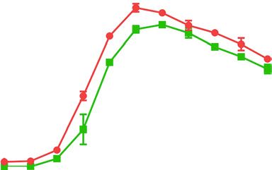
Table 1. Primers and probes used in this
                                                   Name                 Sequences (5′–3′)                                         Amplicon
study
                                                                                                                                  size, bp

                                                   JEV-F*               GCTGGATCC                                                 184
                                                                        CTGAAATGTAGGCTGAAAAT
                                                   JEV-R*               CCAAGCTT
                                                                        TGAGGCTCGCAAGGAAACA
                                                   JEV-R-F**            CAACCTATGGCATGTGTA                                        103
                                                   JEV-R-R**            CCATCACTCCCAGAGTAG
                                                   JEV-probe            FAM-CAATGACAACTGTTCCGTGACCA-BHQ

                                                      JEV, Japanese encephalitis virus. Letters in italics indicate the restriction site in the
                                                   primer. * Primers used for the construction of the standard plasmids and conventional
                                                   PCR. ** Primers used for qRT-PCR.

complexity of the method, the practical application is re-                    Primers and Probes Design
stricted by many conditions. Nowadays, qRT-PCR has                            Multiple sequence alignments of the E gene of JEV available in
                                                                          GenBank were performed with Clustal-W (DNAStar Inc., Madi-
been widely used for the qualitative and quantitative de-                 son, WI, USA), and conserved regions were identified. Primers
tection of the virus due to its specificity, sensitivity, and             and probe were designed to target conserved regions of E gene by
accuracy [12–18], whereas qRT-PCR detects only viral                      Primer Premier 5.0 (Primer Biosoft International, Palo Alto, CA,
nucleic acids, including inactive viruses. Therefore, the                 USA). All primers and probe were synthesized by Sangon Biotech.
TCID50 assay is more valuable for evaluating the prepara-                 Co., Ltd (Shanghai, China). All data related to the primers and
                                                                          probe are shown in Table 1. A BLAST search was also conducted
tion of live virus particles, theoretically. However, for all             to verify the specificities of the primers and probe.
this, if a reliable correlation is found between the above 2
methods, qRT-PCR may be possible to replace or par-                           RNA Extraction and Conventional RT-PCR
tially replace the TCID50, which is more conducive to                         Viral total RNA of JEV vaccine strain SA 14-14-2 were extract-
practical applications. In this study, a TaqMan probe-                    ed using InvitrogenTM TRIzol® Reagent (Life Technologies Corpo-
based qRT-PCR analysis method was developed to mon-                       ration, Carlsbad, CA, USA) under the user guide. Viral RNA was
                                                                          reverse transcribed into cDNA. RT was carried out with Prime-
itor the proliferation of JEV in BHK-21 cells real timely,                ScriptTM1st Strand cDNA Synthesis Kit (TaKaRa, Beijing, China)
and the results of this method were compared with the                     subsequently. The resulting cDNA was used as a template in the
results of TCID50 method. This study provides a new ap-                   following PCR reactions.
proach for studying the in vitro growth laws of JEV and
virus detection during vaccine preparation.                                   qRT-PCR Standard Plasmid Template Preparation
                                                                              PCR assay was carried out in a 20-μL volume containing 2 µL
                                                                          of cDNA template, 2 µL primers (each 0.2 μM), 10 µL Premix
                                                                          TaqTM (TaKaRa, Beijing, China), and 6 µL RNase-free water. Am-
    Materials and Methods                                                 plification began with a 7-min incubation at 95°C, followed by 40
                                                                          cycles of denaturation at 94°C for 1 min, annealing at 56°C for
    Cells and Viruses                                                     30 s, extension at 72°C for 30 s, and 1 cycle of final extension at
    Baby hamster kidney-21 cells (American Type Culture Collec-           72°C for 10 min. After examination by 1% (w/v) agarose gel elec-
tion), JEV vaccine strain SA 14-14-2 (SINOLAND), PPV (porcine             trophoresis, the amplified fragments were digested with BamHI
parvovirus), PCV2 (porcine circovirus type 2), CSFV (classical            and HindIII and inserted into pMDTM19-T-Vector (TaKaRa, Bei-
swine fever virus), PRV (pseudorabies virus), PRRSV (porcine re-          jing, China). The constructed plasmids were confirmed by PCR
productive and respiratory syndrome virus), and TGEV (trans-              and DNA sequencing (Sangon Biotech, China). The extracted
missible gastroenteritis virus) were all kept in the College of Vet-      plasmids were determined by OD260 optical absorption value in
erinary Medicine, Northwest A & F University. All experiments             NanoDrop 2000 spectrophotometer (Thermo Scientific, Waltham,
related to egg drop syndrome virus were done in a P2 biosafety            MA, USA) and diluted at 1.0 × 10¹⁰ copies/µL ultimately.
laboratory and strictly carried out according to the Laboratory
Biosafety Manual in the College of Veterinary Medicine, North-                Fluorescent Quantitative PCR Assay
west A & F University.                                                        The final volume of TaqMan Probe-based fluorescent quantita-
                                                                          tive PCR is 20 μL, which contains 10 μL 2× FastStart Universal

2                      Intervirology 2021;64:1–7                                                  Zhang/Luo/Teng/Xing/Guo/Zhang
                       DOI: 10.1159/000510585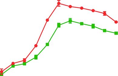
Color version available online
                                                                        Amplification plot
         7.0
         6.5
         6.0
         5.5
         5.0
         4.5                                    1.0 × 10⁸
                                                            1.0 × 10⁷
         4.0                                                            1.0 × 10⁶
         3.5                                                                  1.0 × 10⁵
   ΔRn

                                                                                          1.0 × 10⁴
         3.0
                                                                                                  1.0 × 10³
         2.5
                                                                                                           1.0 × 10²
         2.0
                                                                                                            1.0 × 101
         1.5
         1.0
         0.5
          0

               0   2    4     6      8    10     12    14       16      18    20   22        24       26   28     30    32   34   36   38   40   42
                                                                               Cycle

                         Fig. 1. The sensitivity of qRT-PCR. qRT-PCR amplification plot of 10-fold serial dilutions of standard positive
                         plasmids DNA (from 1.0 × 10⁸ copies/µL to 1.0 × 101 copies/µL). JEV, Japanese encephalitis virus.

Probe Master (Roche), 10 μM upstream and downstream each                         bovine serum (FBS, Hyclone, Logan, UT, USA) and cultivated for
primer 0.5 μL, 10 μM probe 1 µL, templates 1 μL, and RNase-free                  24 h at 37°C in a 5% CO2 incubator routinely. And then, the cells
water 6 μL. PCR cycling parameters were 94°C for 5 min, followed                 were infected with 0.1 mL JEV at a titer of 102 TCID50 in T-25 flasks
by 40 cycles at 95°C for 15 s and 60°C for 1 min.                                for 1 h at 37°C.

    Standard Curve, Sensitivity, Specificity, and Reproducibility of                 Virus Collection
    the Assay                                                                        After incubation, the cells were washed 3 times with PBS (pH
    To evaluate the sensitivity, 10-folds serially diluted standard plas-        7.2) to remove unbound virus particles and then maintained in
mid DNA (from 1.0 × 10⁸ copies/µL to 1.0 × 101 copies/µL) were used              5 mL mediums DMEM containing 2% FBS. At 0, 12, 24, 36, 48, 60,
to perform the PCR. The results were compared with conventional                  72, 96, and 120 h post infection (hpi), 500 μL culture supernate and
RT-PCR. Meanwhile, to generate a standard curve of the assay,                    cell suspension were collected from every 3 samples after frozen
10-folds serially diluted standard plasmid DNA (from 1.0 × 10⁸ cop-              and thawed 3 times.
ies/µL to 1.0 × 10⁴ copies/µL) were used. The standard curve of JEV
detection is established by taking the logarithm of the number of start-            Determination of Virus Titer
ing templates as the X-axis and the Ct value as the Y-axis as the regres-           qRT-PCR assay and TCID50 determination were performed on
sion curve. The specificity was determined using RNA/DNA extract-                samples taken at different time points. According to the average
ed from PPV, PCV2, CSFV, PRV, PRRSV, and TGEV, which were                        determination results, the proliferation curve of the virus was
used as templates in parallel to evaluate the specificity of the assay.          drawn, and comparative analysis was performed to calculate the
    For verifying the reproducibility, 3 different dilutions of the              correlation coefficient.
standard plasmid DNA (1.0 × 10⁷ copies/µL, 1.0 × 10⁶ copies/µL,
and 1.0 × 10⁵ copies/µL) were tested for 10 replicates and statisti-
cally analyzed.
                                                                                     Results
   Comparison of the Dynamic Proliferation of JEV in BHK-21
   Cells
   Cell Culture and Inoculation                                                     Identification of Standard Positive Plasmids
   BHK-21 cells (inoculation concentration of 5.0 × 10⁵ cells/mL)                   PCR products were analyzed by 1% agarose gel elec-
were seeded in DMEM (Gibco, UK) supplemented with 8% fetal                       trophoresis, and a specific fragment of approximately

Dynamic Proliferation of JEV                                                     Intervirology 2021;64:1–7                                            3
                                                                                 DOI: 10.1159/000510585
Color version available online
Amplification plot
8.0
7.5
7.0
6.5
6.0
5.5
5.0
4.5
4.0 1.0 × 10⁷
ΔRn
3.5 1.0 × 10⁶
1.0 × 10⁵
3.0
1.0 × 10⁴
2.5
1.0 × 10³
2.0
1.5
1.0
0.5 0.442894
0
0 2 4 6 8 10 12 14 16 18 20 22 24 26 28 30 32 34 36 38 40 42
Cycle
Fig. 2. Reproducibility of qRT-PCR. Amplification plot of plasmid DNA (from 1.0 × 10⁷ copies/µL to 1.0 × 10³ copies/µL).
1,500 bp was amplified, which was consistent with the is still a fluorescence curve of 1.0 × 101 copies/µL, indicat-
expected results (online suppl. Fig. 1a; for all online suppl. ing that the detection sensitivity of the method can reach
material, see www.karger.com/doi/10.1159/000510585). 1.0 × 101 copies/µL. The results of the sensitivity test are
The constructed plasmid DNA was digested with BamHI shown in Figure 1. Under the same reaction system and
and HindIII, and the band of interest of about 1,500 bp conditions for fluorescent quantitative PCR, only JEV-
and the vector of plasmid 2,700 bp were excised (online positive standards showed positive signals, and other vi-
suppl. Fig. 1b). Simultaneous sequencing of the recombi- rus samples controls were negative.
nant plasmids revealed that the JEV E genetic fragment In this experiment, 5 concentrations (1.0 × 10⁷, 1.0 ×
had been cloned into the T-vector. 10 , 1.0 × 10⁵, 1.0 × 10⁴ and 1.0 × 10³ copies/µL, n = 3) of
6
plasmid DNA were selected as standard samples for re-
Standard Curve peated determination (Fig. 2), and the results were ob-
The qRT-PCR amplification was performed on a 10- tained by statistical analysis. As can be seen from Table 2,
fold serial dilution of standard positive plasmids DNA the final actual measured values of the standards at initial
(1.0 × 10⁸∼1.0 × 101 copies/µL) as a template. The ampli- concentrations of 1.0 × 10⁷, 1.0 × 10⁶, 1.0 × 105,1.0 × 104,
fication showed a good linear relationship, the standard and 1.0 × 10³ copies/µL were 0.988 × 10⁷, 0.933 × 10⁶,
curve equation is Ct = −3.336log (copy nr) +44.18, the 0.975 × 10⁵, 0.930 × 10⁴, and 0.898 × 10³ copies/µL, re-
correlation coefficient is 0.997, and the amplification ef- spectively. The coefficients are 8.40, 5.79, 2.77, 3.33, and
ficiency is 99.3% (online suppl. Fig. 2). 3.34%, respectively. The determination result indicated
that the method has good accuracy and reproducibility.
Sensitivity, Specificity, and Reproducibility
The analytical sensitivity of the assay was assessed Proliferation Profile of JEV on BHK-21 Cells
through serial dilutions of standards of plasmids DNA BHK-21 cells were infected with 0.1 mL JEV at a titer
(from 1.0 × 10⁸ copies/µL to 1.0 × 101 copies/µL). There of 102 TCID50 and normal cells were used as control. The
4 Intervirology 2021;64:1–7 Zhang/Luo/Teng/Xing/Guo/Zhang
DOI: 10.1159/000510585Color version available online
Color version available online
6 8
Virus RNA copies (copies/Ig¡¤0.1 mL–1)
7
Virus titer (TCID50/Ig¡¤0.1 mL–1)
5
6
4
5
3 4
3
2
2
1 Cell suspension Cell suspension
1
Culture supernate Culture supernate
0 0
0 12 24 36 48 60 72 84 96 108 120 0 12 24 36 48 60 72 84 96 108 120
Hours post infection, hpi Hours post infection, hpi
Fig. 3. One-step proliferation curve of JEV on BHK-21 cells deter- Fig. 4. One-step proliferation curve of JEV in BHK-21 cells deter-
mined by TCID50 assay. JEV, Japanese encephalitis virus; hpi, mined by qRT-PCR. JEV, Japanese encephalitis virus; hpi, hours
hours post infection. post infection.
Table 2. Reproducibility and recovery of
the measurement of replicate standards Experiment Quantity, copies/µL
1.0×10⁷ 1.0×10⁶ 1.0×10⁵ 1.0×10⁴ 1.0×10³
1 0.964 0.912 0.977 0.966 0.907
2 0.988 0.893 1.000 0.916 0.879
3 1.0137 0.995 0.947 0.909 0.922
Arithmetic mean 0.988 0.93 0.975 0.930 0.898
Standard deviation 0.083 0.054 0.027 0.031 0.030
Coefficient of variation, n (%) 8.40 5.79 2.77 3.33 3.34
SD = éê(d1 - x ) + (d2 - x ) + + (dn - x ) ùú / (n - 1) ;
2 2 2
ë û
CV = S/X × 100%. dn is the measured value and x is the arithmetic mean value. CV,
coefficient of variation; SD, standard deviation.
morphological changes of the cells at 0, 12, 24, 48, 60, 72, and culture supernatant, the virus proliferated slowly
84, 96, and 120 h after challenged were observed, and the within 24 hpi. The virus proliferation rate increased rap-
cytopathic images were collected. Cytopathic effect (CPE) idly subsequently within 24 hpi∼60 hpi, with the viral
began to appear at 48 hpi, but it was not obvious. A small load increasing logarithmically. The viral titer reached
number of tiny plaques were scattered on monolayer the highest level of 105.44 TCID50/0.1 mL in cell suspen-
cells; at 60 h, the CPE was obvious, the cells shrunk, some sion at 60 hpi and 104.86 TCID50/0.1 mL in culture super-
rounded and gathered, and the cell gap increased. Cells nate at 72 hpi, respectively. The peak of viral titer is 12 h
mostly became round and began to fall off at 120 h. Large ahead in cell suspension than in culture supernate. After
amounts of cells shed and disintegrated in large areas that, the content of JEV showed a decreasing trend up to
moreover. 120 h, but the decline rate was slow. The dynamic curves
qRT-PCR assay and TCID50 determination were per- of virus titer of JEV on BHK-21 cells were very similar in
formed on samples taken at different times after virus in- both cell suspension and culture supernate.
oculation, and the virus proliferation curve was drawn As shown in Figure 4, the amount of JEV presented a
(Fig. 3, 4). As shown in Figure 3, in both cell suspension tendency of straight climb with the increase of hpi either
Dynamic Proliferation of JEV Intervirology 2021;64:1–7 5
DOI: 10.1159/000510585in cell suspension or in culture supernate. And the viral The proliferation curve determined by the 2 methods has
load in culture supernate is less than that in cell suspen- a certain parallel relationship, but the quantitative PCR
sion continuously. The virus RNA copies reached the (4 h) is faster and more sensitive than the TCID50 assay
peak at 1.0 × 107.5 copies/µL and 1.0 × 105.6 copies/µL in (3∼4 days), and it is more suitable for rapid determina-
cell suspension is and culture supernate, respectively. The tion of JEV proliferation titer real timely. The compre-
peak appeared in cell suspension is 12 h ahead than in hensive analysis showed that the best time for receiving
culture supernate. The virus RNA copies reduced slowly JEV proliferated on BHK-21 cells was at 60 hpi. Neverthe-
after the limit value. Combining with Figures 3 and 4, it less, the peak of virus proliferation was closely related to
can be seen that the uptrend and the peak of the prolif- various factors, the proliferation curve of different virus
eration profile of JEV inoculated on BHK-21 cells mea- strains needed to be studied systematically. However, this
sured by TCID50 determination are consistent with which study will provide technical support for the preparation
measured by qRT-PCR assay. Meanwhile, the correlation of high-titer virus solution or production of JEV.
coefficient between the quantitative copy number and the
TCID50 data was r = 0.867 and r = 0.888 in the cell super-
natant and cell suspension, respectively. Statement of Ethics
Not applicable. There are no human and animal subjects in this
study.
Discussion
qRT-PCR could be used for quantitative detection of Conflict of Interest Statement
virus contained in a unit volume. However, it should be
noted that the viral load measurement just reflected the The authors have no conflicts of interest to declare.
amount of viral nucleic acid contained in the test and
could not reflect the amount of live virus or intact virus
particles. Virus titer is usually expressed as TCID50, which Funding Sources
could reflect the number of viruses with biological activ-
ity or function. The primitive and classical method for the This work was supported by the National Key Research and
Development Program of China (grant numbers 2016YFD0500701),
quantitative detection of JEV is to determine its TCID50. the Earmarked Fund for Modern Agro-industry Technology Re-
However, its practical application is subject to many con- search System of China (grant numbers CARS-35), and the Special
ditions. Fund for Henan Agriculture Research System (grant numbers
The standard curve of qRT-PCR assay explored in this S2012-06-02).
test showed a good linear relationship at 1.0 × 10⁸∼1.0 ×
101 copies/µL, with the correlation coefficient 0.997 and
the amplification efficiency 99.3%, which provides basic Author Contributions
data for precise analysis of virus replication in cells.
Y.H.Z. planned and designed the research. F.L.Z. and J.L. fin-
The proliferation dynamics of JEV on BHK-21 cells ished the experiments. M.T. and G.X.X. performed the data analy-
determined by qRT-PCR and TCID50 assay showed that sis. J.Q.G. prepared the reagents used in the experiments. F.L.Z.
the number of virions or virus titer of the cell suspension wrote the manuscript. All authors have read and approved the
was significantly higher than that of the culture superna- manuscript.
tant and reached the peak at first. This is because some
cells contracted and fell off and entered into the cell su- References 1 Impoinvil DE, Ooi MH, Diggle PJ, Caminade
pernatant after 24 hpi. Both of them reached the peak at C, Cardosa MJ, Morse AP, et al. The effect of
vaccination coverage and climate on Japanese
60 hpi and then began to fall. However, the TCID50 value encephalitis in Sarawak, Malaysia. PLoS Negl
decreased slightly after 60 hpi which may be because the Trop Dis. 2013 Aug;7(8):e2334.
value measured by the qRT-PCR contained 3 types of vi- 2 Gao X, Liu H, Li X, Fu S, Cao L, Shao N, et al.
Changing geographic distribution of Japanese
ral nucleic acids, including live virus (infectious) nucleic encephalitis virus genotypes, 1935–2017. Vec-
acids, inactivated viruses (non-infectious but antigenic tor Borne Zoonotic Dis. 2019 Jan;19(1):35–44.
and immunogenic) nucleic acids, and nucleic acids of vi- 3 Chambers TJ, Hahn CS, Galler R, Rice CM.
Flavivirus genome organization, expression,
rion that have not yet completed assembly. While the and replication. Annu Rev Microbiol. 1990;
TCID50 value only determined the titer of live virions. 44(1):649–88.
6 Intervirology 2021;64:1–7 Zhang/Luo/Teng/Xing/Guo/Zhang
DOI: 10.1159/0005105854 Rauscher S, Flamm C, Mandl CW, Heinz FX, 9 Wang ST, Wang XW, Chen L, Zhao J, Chang 14 Wang SS, Li YB, Meng XL, Ma F. Develop-
Stadler PF. Secondary structure of the 3′-non- HT, Yang X, et al. Proliferation dynamic of ment of SYBR Green I fluorescence quantita-
coding region of flavivirus genomes: compar- Japanese encephalitis virus in vitro using a tive RT-PCR assay for detection of porcine
ative analysis of base pairing probabilities. newly developed fluorescent quantitative reproductive and respiratory syndrome virus.
RNA. 1997;3(7):779–91. PCR assay [J]. Chin J Vet Sci. 2013;6. Acta Agric Univ Jiangxiensis. 2009;6.
5 Honjo S, Masuda M, Ishikawa T. Effects of the 10 Teng M, Luo J, Fan JM, Wang XT, Wang FY, 15 Shirato K, Miyoshi H, Kariwa H, Takashima
Japanese encephalitis virus Genotype V-de- Chai SJ, et al. Isolation and propagation of a I. Detection of West Nile virus and Japanese
rived sub-viral particles on the immunogenic- field Japanese encephalitis virus isolate CSF. encephalitis virus using real-time PCR with a
ity of the vaccine characterized by a novel vi- XZ-2D from swine. Chin J Prev Vet Med. probe common to both viruses. J Virol Meth-
rus-like particle-based assay. Vaccines. 2019 2012;4. ods. 2005;126(1–2):119–25.
Aug;7(3):81. 11 Wang ST, Li WH, Wang CQ, Liu HY, Wang 16 Santhosh SR, Parida MM, Dash PK, Pateriya
6 Kallel H, Jouini A, Majoul S, Rourou S. Evalu- XW, Chang HT, et al. Study on the physico- A, Pattnaik B, Pradhan HK, et al. Develop-
ation of various serum and animal protein chemical properties and proliferative charac- ment and evaluation of SYBR Green I-based
free media for the production of a veterinary teristics in BHK-21 cells of Japanese encepha- one-step real-time RT-PCR assay for detec-
rabies vaccine in BHK-21 cells. J Biotechnol. litis virus LS strain. China Anim Husb Vet tion and quantitation of Japanese encephalitis
2002;95(3):195–204. Med. 2013;40(3):134–6. virus. J Virol Methods. 2007;143(1):73–80.
7 Fujinaga K, Gershon R, Prince A. Compara- 12 Sun YW, Liu JY, Xu WJ, Tian Y, Wang JY, 17 Toriniwa H, Komiya T. Rapid detection and
tive evaluation of stable line monkey kidney Song CX. Detection of JEV by fluorescent quantification of Japanese encephalitis virus
cells for study of Japanese encephalitis virus quantitative PCR. Prog Vet Med. 2007;(8):5. by real-time reverse transcription loop-medi-
(JEV). Jpn J Exp Med. 1964;34(1):1–15. 13 Zhu SF, Zhu RL, Qiao CX, Gao ZQ, Zhuang ated isothermal amplification. Microbiol Im-
8 Melvin MA, Wallace HM, Keir HM. Conju- LF, Zhuang HX, et al. Development of fluo- munol. 2006;50(5):379–87.
gation of polyamines in mammalian cells in rescence quantitative PCR assay to detect gB 18 Yang DK, Kweon CH, Kim BH, Lim SI, Kim
culture. Physiol Chem Phys. 1980;12(5):431– gene of pseudorabies virus. Chin J Vet Sci. SH, Kwon JH, et al. TaqMan reverse tran-
9. 2012;32(10):1413–7. scription polymerase chain reaction for the
detection of Japanese encephalitis virus. J Vet
Sci. 2004 Dec;5(4):345–51.
Dynamic Proliferation of JEV Intervirology 2021;64:1–7 7
DOI: 10.1159/000510585You can also read