NON-TOXIC GLYCOSYLATED GOLD NANOPARTICLE-AMPHOTERICIN B CONJUGATES REDUCE BIOFILMS AND INTRACELLULAR BURDEN OF FUNGI AND PARASITES - MPG.PURE
←
→
Page content transcription
If your browser does not render page correctly, please read the page content below
RESEARCH ARTICLE
 www.advtherap.com

Non-Toxic Glycosylated Gold Nanoparticle-Amphotericin B
Conjugates Reduce Biofilms and Intracellular Burden of
Fungi and Parasites
Chandradhish Ghosh, Silvia Varela-Aramburu, Hassan E. Eldesouky, Sharareh Salehi
Hossainy, Mohamed N. Seleem, Toni Aebischer, and Peter H. Seeberger*

 pathogens develop resistance against many
Infections by intracellular pathogens cause significant morbidity and mortality frontline drugs.[2] Pathogens that survive
due to lack of efficient drug delivery. Amphotericin B, currently used to treat within mammalian cells are particularly dif-
leishmaniasis and cryptococcosis, is very toxic and cannot eradicate ficult to treat.[3] Pathogens such as Leishma-
intracellular Cryptococcus neoformans (C. neoformans). Glycosylated gold nia spp. and C. neoformans evade phagocy-
 tosis before residing and replicating within
nanoparticles are water dispersible and biocompatible with very little toxicity.
 macrophages that are an important part of
While amphotericin B is insoluble in water at neutral pH, conjugates of the human immune system.[3,4] Treatment
amphotericin B and ultra-small gold nanoparticles (AuNP) are better of these diseases is challenging as drugs
dispersible in water. Amphotericin B conjugated glycosylated gold have to enter macrophages before they can
nanoparticles (AmpoB@AuNP) are more efficacious in treating both exert their anti-infective action.[3,5] Delivery
 agents that will facilitate the internalization
extracellular and intracellular forms of Leishmania mexicana (L. mexicana)
 of drugs into specific mammalian cells may
than amphotericin B alone. In addition, AmpoB@AuNP are effective in be a solution to this problem.
reducing C. neoformans biofilms by 80% and intracellular C. neoformans The facultative intracellular pathogen C.
burden by >90%. Furthermore, AmpoB@AuNP are not haemolytic at neoformans causes life-threatening menin-
50 µg mL−1 and are significantly less toxic to murine macrophages than gitis in patients suffering from AIDS, can-
amphotericin B. Ultra-small AuNPs are attractive delivery agents to treat cer, autoimmune diseases, and is respon-
 sible for around 600 000 deaths globally.[6]
intracellular infections and AmpoB@AuNP may be useful for treating
 Upon inhalation, Cryptococcus sp. spores
C. neoformans infections in immunocompromised patients. are engulfed by the alveolar macrophages
 wherein they survive and replicate in
 mature phagolysosomes. C. neoformans
1. Introduction disseminates to other organs including the central nervous
 system.[6a] After crossing the blood brain barrier C. neofor-
Infectious diseases are a major contributor to human morbidity mans can form biofilms, called cryptococcomas that protects
and mortality.[1] The utility of antimicrobial agents decreases as them from host immune defences and antimicrobial therapy.[6a,7]

Dr. C. Ghosh, Dr. S. Varela-Aramburu, Prof. P. H. Seeberger Dr. S. Varela-Aramburu, Prof. P. H. Seeberger
Department of Biomolecular Systems Institute of Chemistry and Biochemistry
Max Planck Institute of Colloids and Interfaces Freie Universität Berlin
Am Mühlenberg 1, Potsdam 14476, Germany Takustraße 3, Berlin 14195, Germany
E-mail: Peter.Seeberger@mpikg.mpg.de Dr. H. E. Eldesouky, Prof. M. N. Seleem
 Department of Comparative Pathobiology
 Purdue University
 625 Harrison Street, West Lafayette, IN 47907, USA
 Dr. H. E. Eldesouky, Prof. M. N. Seleem
 Department of Biomedical Sciences and Pathobiology, Virginia-Maryland
 The ORCID identification number(s) for the author(s) of this article
 College of Veterinary Medicine
 can be found under https://doi.org/10.1002/adtp.202000293
 Virginia Polytechnic Institute and State University
© 2021 The Authors. Advanced Therapeutics published by Wiley-VCH Blacksburg, VA 24060, USA
GmbH. This is an open access article under the terms of the Creative S. Salehi Hossainy, Prof. T. Aebischer
Commons Attribution-NonCommercial-NoDerivs License, which permits Unit 16 Mycotic and Parasitic Agents and Mycobacteria, Department of
use and distribution in any medium, provided the original work is Infectious Diseases
properly cited, the use is non-commercial and no modifications or Robert Koch Institute
adaptations are made. Berlin 13353, Germany
DOI: 10.1002/adtp.202000293

Adv. Therap. 2021, 4, 2000293 2000293 (1 of 8) © 2021 The Authors. Advanced Therapeutics published by Wiley-VCH GmbH
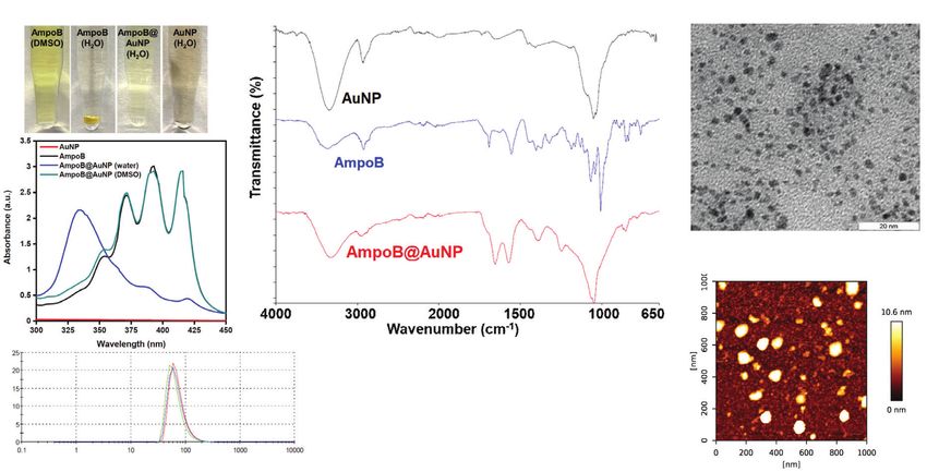
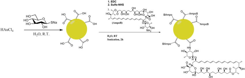
www.advancedsciencenews.com www.advtherap.com

Therapy against biofilms and intracellular forms of C. neoformans undergoes partial oxidation during the process to yield COOH
is ineffective or drastically reduced.[8,7b] The complications as- groups that could be used for further functionalization.
sociated with C. neoformans infections are a challenging unmet Modification on the mycosamine nitrogen leads to less
medical need that is being addressed via different paths.[9] toxic amphotericin B derivatives without adversely affect-
 Leishmaniasis, a parasitic tropical disease with high mortality ing the activity.[26] Thus, it was reasoned that conjuga-
in its most severe form is caused by parasites that exist in two tion of amphothericin B to glycogold nanoparticles via the
different forms within their host.[10] Extracellular forms called amine group may reduce the toxicity of the drug. The car-
promastigotes are transferred by a sand-fly bite to the host upon boxylic acid moieties on the nanoparticles will impart charge
a blood meal. These promastigotes are engulfed by phagocytes, to the conjugate and allow for more water molecules to
in particular, macrophages where they transform to amastig- interact with the nanoparticle for better aqueous disper-
otes and multiply by division. As the disease develops, amastig- sion. The carboxylic acid groups of the naked glycogold
otes released from host cells are propagated as more and more nanoparticles were activated by sonicating AuNP solution with
macrophages become infected. Alternatively, amastigote infected 1-ethyl-3-(3-dimethylaminopropyl)carbodiimide (EDC) and N-
cells are taken up by the sand fly upon biting an infected host, to hydroxysulfosuccinimide (Sulfo-NHS) for five minutes before a
enter into a new life cycle. solution of amphotericin B in dimethyl sulfoxide (DMSO) was
 Amphotericin B, the drug used to treat both cryptococcosis added and sonicated for 2 h (Scheme 1). The resultant solution
and in particular, the severe mucocutaneous and visceral forms was subjected to dialysis in milliQ water overnight to obtain the
of leishmaniasis is poorly water-soluble and quite toxic.[10b] Li- amphotericin B-conjugated nanoparticles (AmpoB@AuNP) that
posomal formulations of amphotericin B such as AmBisome were further purified by passing them through a 0.45 µm fil-
have reduced its toxicity, but the storage of liposomal formula- ter. This solution was characterized using ultraviolet absorption
tions is more difficult and costly.[11] To overcome some of these (UV), infrared spectroscopy (IR), DLS, TEM, and atomic force
shortcomings, amphotericin B, has been chemically modified, microscopy (AFM).
conjugated to polymers,[12] dendrimers,[13] carbon nanotubes,[14] Amphotericin B is soluble in DMSO and not in water
and other nanoparticles.[15] Attempts to improve the solubility (Figure 1A). In comparison, the dispersion of AmpoB@AuNP in
of amphotericin B have been made but the toxicity has not water is clear (Figure 1A). UV spectroscopy is an important tool
been addressed.[16] Recent attempts toward understanding tox- to characterize amphotericin B as it absorbs sharply at 365, 384,
icity caused by amphotericin B has led to it being used for other and 408 nm in DMSO (Figure 1B). Amphotericin B is not water-
diseases as well.[17] Nevertheless, orally available nanoformula- soluble, so its UV absorption spectrum in water was not recorded.
tions of amphotericin B remain elusive.[18] More importantly, the The suspension of naked gold nanoparticles in water absorbs very
drug fails to reduce intracellular infections of C. neoformans and little (Figure 1B). The AmpoB@AuNP dispersion in water ab-
their biofilms effectively. sorbs at around 325 nm. Both Ambiosome and Fungizone are
 The optical properties of inert and biocompatible[19] gold known to absorb very strongly at 322 nm, which is indicative of
nanoparticles[20] can be exploited for diagnostic and other the dimeric aggregates of amphotericin B (Figure S1, Supporting
applications.[21] Nanoparticulate gold is not toxic to animals[22] Information).[27] In case of AmpoB@AuNP, although the maxi-
and glycosylated-gold nanoparticles[23] are non-toxic and water- mum is at 325 nm, other small humps (from 375 to 425 nm) indi-
soluble.[24] The presence of the sugar not only improves the dis- cated presence of other aggregates (Figure S1, Supporting Infor-
persibility in water but also facilitates further functionalization. mation). Similar observations were made with super-aggregates
Gold nanoparticles can bind to cysteine-rich surface proteins of of amphotericin B.[28] However, lyophilization of the water dis-
pathogens due to the affinity for gold to thiols for targeted drug persion followed by resuspension in DMSO shows the charac-
delivery.[25] Here, we show that conjugation of amphotericin B to teristic peaks of amphotericin B allowing for quantification of
glycogold nanoparticles renders the drug water dispersible and the drug conjugated to the nanoparticles (explained below). IR
lowers its toxicity. A significant improvement in drug activity was of AmpoB@AuNP reveals distinct peaks at 1654 and 1574 cm−1
observed against L. mexicana and C. neoformans. Unlike ampho- that represent the carbonyl stretch and N─H bending respec-
tericin B, the conjugate was effective in reducing biofilms and tively, convincingly proving the existence of the amide bonds ex-
intracellular burden of C. neoformans. pected from the reaction (Figure 1C). A shoulder at around 1700
 cm−1 may reflect the free carboxylic acid group of amphotericin
 B. DLS was used to determine the hydrodynamic radii of the Am-
2. Results and Discussion poB@AuNP suspension. (Figure 1D). It was observed that the hy-
 drodynamic radius of the particles increases by 20–30 to ≈45 nm
2.1. Synthesis and Characterization in comparison to the naked gold nanoparticles (≈15 nm, Figure
 S2B, Supporting Information). The nanoparticles have a poten-
The parent gold nanoparticles were prepared by modifying a pub- tial of the nanoparticles remain similar both before (−30 mV) and
lished protocol.[24] Aurochloric acid was reduced and capped with after conjugation (−28 mV) confirming their stability in water
thioglucose to yield glyco-gold nanoparticles
www.advancedsciencenews.com www.advtherap.com

Scheme 1. Synthesis of the amphotericin B—nanoparticle conjugates. (EDC: 1-Ethyl-3-(3-dimethylaminopropyl)carbodiimide; Sulfo-NHS: N-
hydroxysulfosuccinamide; AmpoB: Amphotericin B. Only one AmpoB in the conjugated form is shown for clarity.)

Figure 1. Characterization of AmpoB@AuNP. A) Amphotericin B is soluble in DMSO and insoluble in water, while Ampob@AuNP result in a clear
solution. B) UV absorption of the different materials used in the study. AuNP absorbs slightly. For AmpoB in DMSO three distinct peaks are observed at
365, 384, and 408 nm. AmpoB@AuNP do not show signature peaks but have strong absorbance around 340 nm while the same sample in DMSO shows
distinct features. C) IR spectra of AmpoB@AuNP shows prominent amide bond peaks at 1654 and 1574 cm−1 that are absent in case of Amphotericin
B and AuNP. D) DLS measurements indicate that the hydrodynamic radii AmpoB@AuNP are mostly ≈45 nm. E) Zeta potential of AmpoB@AuNP is
−28 mV (colored lines represent triplicates in 1D and 1E). F) TEM images of AmpoB@AuNP show the core structure of the gold nanoparticles to be
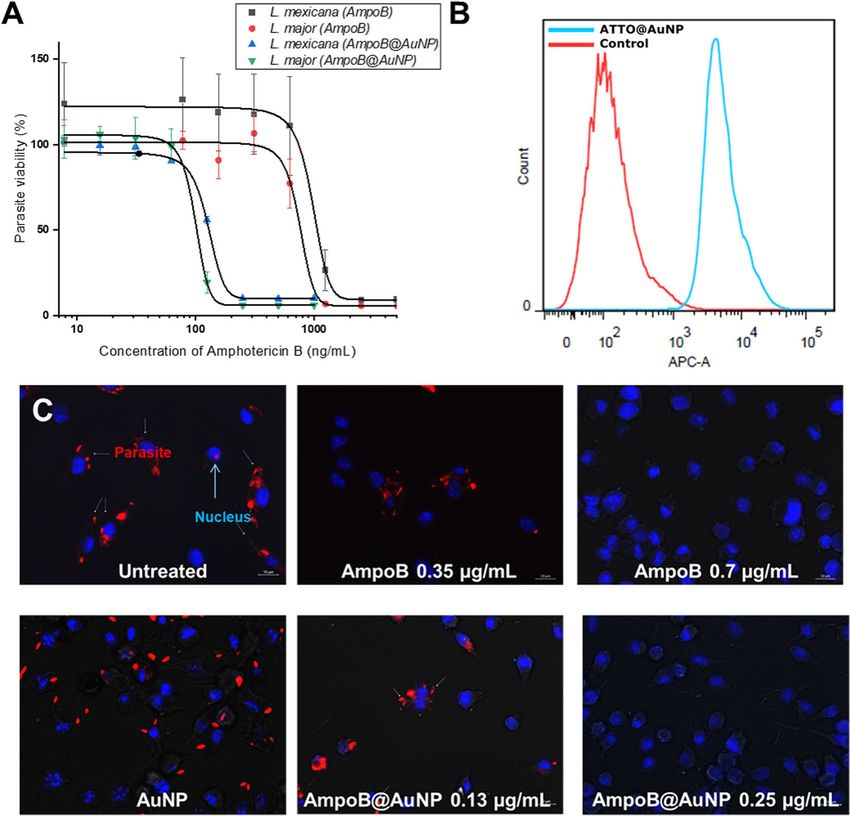
www.advancedsciencenews.com www.advtherap.com

Figure 2. A) Activity of AmpoB@AuNP against metacylcic promastigotes of Leishmania sp. Error bars represent standard deviation values. B) Uptake of
ATTO@AuNP nanoparticles by macrophages analyzed by flow cytometry. The shift in the intensity of the fluorescence signal indicates that ATTO@AuNP
cells were internalized. The same number of events was recorded. C) Activity against intramacrophage L. mexicana (DSred). Macrophage cells are stained
with DAPI and the infected cells are recognized by the red fluorescence of the parasites. Amphotericin B is partly active at 0.35 µg mL−1 but completely
active at 0.7 µg mL−1 . AmpoB@AuNP are partly active at 0.13 µg mL−1 while completely active at 0.25 µg mL−1 . AuNP do not show inhibition. Error
bars represent standard deviation values for triplicates performed using OriginPro 2020b (OriginLab, Northampton, Massachusetts, USA).

Information). The concentration of gold (Au) in the samples was measured at ≈0.1 µg mL−1 against L. major and ≈0.13 µg mL−1
measured using inductively coupled plasma-optical emission against L. mexicana while free drug displayed values between 0.7
spectroscopy. The average ratio between the concentration of am- and 1 µg mL−1 (Figure 2A).
photericin B and the concentration of gold in the Ampob@AuNP
solution (based amount present in µg mL−1 ) was determined to
 2.4. Intracellular Uptake of Nanoparticles
be 2.5. That corresponds to ≈30 amphotericin B molecules per
nanoparticle based on the calculations published previously. For
 To study intracellular uptake, AuNPs functionalized with a dye
ease of comparison, all activity values are reported in terms of
 ATTO647N was incubated with macrophages. After washing any
concentration of amphotericinAmphotericin B present in the
 surface bound nanoparticles, internalization using flow cytome-
solution of AmpoB@AuNP as opposed to the concentration of
 try was measured. From the flow cytometry analysis, a clear uni-
nanoparticles present.
 form shift in fluorescence intensity of ATTO@AuNP treated cells
 was observed in comparison to untreated cells confirming the
 internalization of the particles within macrophages (Figure 2B).
2.3. Activity against Extracellular Leishmania sp.
 Internalization was also observed previously in RBCs.[25]
The nanoparticles were checked for activity against the extracel-
lular form of L. major and L. mexicana parasites and compared 2.5. Activity against Intracellular L. mexicana
to the parent drug. Complete inhibition of cell growth was ob-
served at 250 ng mL−1 of AmpoB@AuNP against both L. mexi- The genetically modified L. mexicana parasite fluoresces red and
cana and L. major (Figure 2A). The IC50 of AmpoB@AuNP was can be easily observed under a fluorescence microscope. Upon

Adv. Therap. 2021, 4, 2000293 2000293 (4 of 8) © 2021 The Authors. Advanced Therapeutics published by Wiley-VCH GmbH
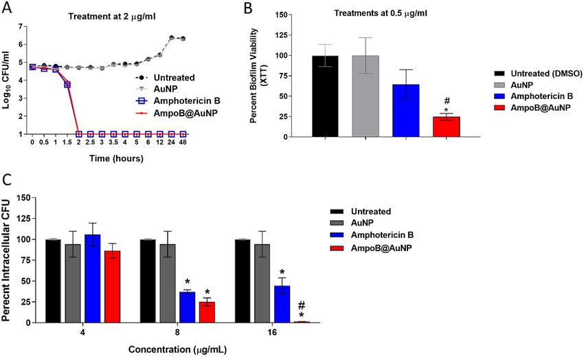
www.advancedsciencenews.com www.advtherap.com

Table 1. Summary of antifungal activity. Table 2. Antifungal activity against Cryptococcus species.

Fungal isolates MIC range [µg mL−1 ] Fungal isolate MIC [µg mL−1 ]

 Fluconazole Amphotericin B AmpoB@AuNP Fluconazole Amphotericin B AmpoB@AuNP

Aspergillus sp. ⇒128 2 1 C. gattii NR-43208 8 1 0.5
Candida sp. 0.5–>128 0.5–2 0.5–2 C. gattii NR-43209 16 1 0.5
Cryptococcus sp. 4–32 1–2 0.5 C. gattii NR-43210 16 1 0.5
 C. gattii NR-43213 8 1 0.5
Unconjugated AuNP was not active.
 C. neoformans NR-41291 16 2 0.5
 C. neoformans NR-41295 32 2 0.5
staining the macrophage nuclei with DAPI, the parasites inside C. neoformans NR-41298 4 1 0.5
the cell can be easily identified. In the negative control, most of
 C. neoformans NR-48767 4 1 0.5
the macrophages (stained blue) contain parasites (stained red)
(Figure 2C). Amphotericin B was less effective at 0.35 µg mL−1 Unconjugated AuNP was not active.
while no parasites were observed at 0.7 µg mL−1 . The IC50 of
AmpoB@AuNP against extracellular parasites was determined at
 formans NR41298 with AmpoB@AuNP was determined. The ki-
0.13 µg mL−1 , such that treatment with 0.125 µg mL−1 , resulted
 netics of the fungicidal activity of AmpoB@AuNP and free am-
in some parasite-infected macrophages. At 0.25 µg mL−1 of Am-
 photericin B were the same at 2 µg mL−1 as fungal burden was
poB@AuNP, no parasites were found within the macrophages
 reduced by five logarithmic scales within 2 h. Free AuNP had no
illustrating that passive internalization and targeted delivery en-
 effect on the cells (Figure 3A).
hances drug efficacy. Encouraged by these results, the activity of
AmpoB@AuNP against fungi was evaluated next.

 2.8. Activity against C. Neoformans NR-41298 Biofilms
2.6. Activity against Planktonic Fungi
 C. neoformans notoriously forms and survives within biofilms,
The minimum inhibitory concentrations (MIC) of amphotericin a.k.a. cryptococcomas that are recalcitrant to antifungals and
B, unconjugated gold nanoparticle (AuNP), and AmpoB@AuNP the human immune response. Proteins associated with C. neo-
were determined against Aspergillus sp., Candida sp., and Crypto- formans virulence and biofilms contain cysteine-rich regions.[29]
coccus sp to test whether the nanoparticle formulation enhanced Thus, multivalent presentation of the drug and interaction with
the antifungal activity of amphotericin B (Table 1 and Table S1, gold might be able to destroy fungal biofilms more effectively
Supporting Information). The compounds were tested against than the unbound drug. Thus, we subjected preformed biofilms
three clinical isolates of Aspergillus sp. and revealed that Am- of C. neoformans to treatment with AmpoB@AuNP and the free
poB@AuNP is twice as active (MIC = 1 µg mL−1 ) as the parent drug. Metabolic activity of the biofilms after 24 h indicated that
drug (MIC = 2 µg mL−1 ) (Table 1). AuNP were not active against even at concentrations as low as 0.25 µg mL−1 , AmpoB@AuNP
the pathogens. Activity against Candida spp. was tested using four were able to reduce the burden by 50% while free amphotericin
strains of C. albicans, five strains of the new emerging multidrug- B was not very active at this concentration (Figure S5, Support-
resistant C. auris, four strains of C. glabrata, two strains of C. kru- ing Information). At higher concentrations, the activity of both
sei, two strains of C. parapsilosis and C. tropicalis. AmpoB@AuNP AmpoB@AuNP and Amphotericin B increased. When treated
were as active as amphotericin B against all the strains of Candida with 0.5 µg mL−1 of AmpoB@AuNP, the metabolic activity of
except C. albicans ATCC 10231, where it was twice more potent the biofilms was reduced by 75% to further emphasize the po-
than the drug alone. tency of AmpoB@AuNP against biofilm infections of C. neofor-
 Next, the compounds were evaluated against C. gattii and C. mans (Figure 3B). This observation was confirmed by confocal
neoformans, the causative agents of cryptococcosis, and cryptococ- microscopy (Figure S6, Supporting Information). Amphotericin
cal meningitis (Table 2). AmpoB@AuNP were twice more potent B treated biofilms were stained green (SYTO-9) only and not red
than amphotericin B against the four clinical isolates of C. gattii. (PI), like that negative control and unconjugated gold nanoparti-
The MIC of AmpoB@AuNP was 0.5 µg mL−1 while the unconju- cles. On the contrary, significant population of AmpoB@AuNP
gated drug was active at 1 µg mL−1 . The two clinical isolates of C. treated cells were stained red indicative of death (Figure S6, Sup-
neoformans that are resistant to fluconazole require 2 µg mL−1 of porting Information).
amphotericin B for activity while AmpoB@AuNP remained ac-
tive at 0.5 µg mL−1 . AmpoB@AuNP were equally or more potent
than free amphotericin B against all the fungi that we tested. 2.9. Activity against Intracellular C. neoformans

 C. neoformans survive and replicate within immune cells. This
2.7. Kinetics of Killing/Antifungal Activity form of the pathogen is extremely difficult to treat as no dedi-
 cated therapeutic is available. Clinically used antifungals are not
To assess whether conjugation of amphotericin B to gold active against the intracellular form of the pathogen.[8,30] Since
nanoparticles alters the rate of killing, the kill kinetics of C. neo- AmpoB@AuNP were able to clear L. mexicana residing within

Adv. Therap. 2021, 4, 2000293 2000293 (5 of 8) © 2021 The Authors. Advanced Therapeutics published by Wiley-VCH GmbH
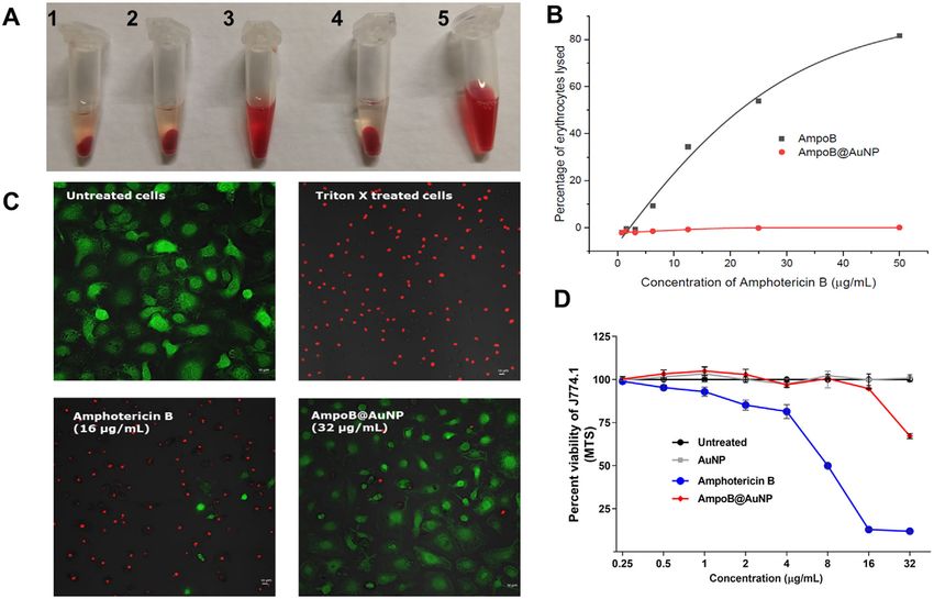
www.advancedsciencenews.com www.advtherap.com

Figure 3. A) Time-kill analysis of Amphotericin B nanoparticles (AmpoB@AuNP) and free Amphotericin B (at 2 µg mL−1 ) against C. neoformans NR-
41298 over a 48 h incubation period at 35 °C. DMSO, AuNP served as controls. B) Biofilm eradicating activity of Amphotericin B nanoparticles (Am-
poB@AuNP) against C. neoformans NR-41298 biofilm evaluated with the XTT assay over a 24-h period. The percent metabolic activity for each treatment
was calculated relative to untreated wells. Error bars represent standard deviation values. C) Activity against intracellular C. neoformans. Fungal burden of
J774 mouse macrophages infected with C. neoformans NR-41298, was reduced drastically when treated with AmpoB@AuNP from 8 µg/mL. Amphotericin
B is not active. The reduction observed here is partly because of its toxic effect on J774.1 cells. (*) denotes the statistical difference between amphotericin
nanoparticles and the untreated control group whereas hash (#) indicates a statistical significance between the amphotericin nanoparticles and the free
amphotericin. The statistical significance was assessed with one-way ANOVA, with post hoc Dunnet’s multiple comparisons test (p < 0.05), utilizing
GraphPad Prism 6.0 (GraphPad Software, La Jolla, CA).

macrophages we hypothesized that the intracellular C. neofor- staining with a mixture of fluorescein diacetate (FDA) and
mans can be eradicated by AmpoB@AuNP. At 8 µg mL−1 Am- propidium iodide (PI) was used to determine the toxicity. FDA
poB@AuNP were able to reduce 75% of the intracellular burden is a non-fluorescent, cell-permeant dye that is converted to
of C. neoformans (Figure 3C). AmpoB@AuNP (16 µg mL−1 ) re- fluorescein intracellularly by esterases expressed by viable cells.
duced the fungal burden by more than 90%. The less significant The resultant green fluorescence is an indicator of cell-viability.
reduction in case of amphotericin B at 8 and 16 µg mL−1 was In contrast, PI is non-permeant across cells and stains the nu-
attributed to its cytotoxic effect on the cells. cleus of membrane-compromised cells. When untreated murine
 macrophages were stained with a mixture of dyes, only green
 fluorescence was observed (Figure 4C). Upon treatment with
2.10. Toxicity Triton X, the cells were compromised allowing PI to stain them
 red. Similarly, at 16 µg mL−1 , amphotericin B was toxic to the
Finally, the toxicity of AmpoB@AuNP against mammalian cells cells (mostly red fluorescence is observed) while AmpoB@AuNP
was evaluated. The toxicity was evaluated against erythrocytes were not toxic even at 32 µg mL−1 (only green fluorescence
and the two different murine macrophages used for leishmanial was observed). Then, viability of J774.1 cells (the hosts for C.
studies and fungal studies (J774.1) respectively (Figure 4A–C). neoformans), when treated with different nanoparticles at varying
Incubation with freshly isolated erythrocytes for 1h revealed that concentrations, was quantified using MTS assay (Figure 4D).
AmpoB@AuNP do not induce visible haemolysis (Figure 4A) Amphotericin B was toxic at concentrations as low as 2 µg mL−1 .
even till 64 µg mL−1 . Amphotericin B, on the other hand, At 8 µg mL−1 amphotericin B treated cells, only 50% of the
induces haemolysis at concentrations as low as 6.2 µg mL−1 macrophages remain viable and at 16 µg mL−1 almost 80% of the
(Figure 4B). cells are lysed. In comparison, upon treatment with 32 µg mL−1
 Then, the toxicity of the compounds was evaluated in murine of AmpoB@AuNP, more than 70% of the cells are viable. No
macrophages that served as hosts for L. mexicana. LIVE/DEAD toxicity is observed at 16 µg mL−1 of AmpoB@AuNP. Thus,

Adv. Therap. 2021, 4, 2000293 2000293 (6 of 8) © 2021 The Authors. Advanced Therapeutics published by Wiley-VCH GmbH
www.advancedsciencenews.com www.advtherap.com

Figure 4. Toxicity of different Amphotericin B formulations. A) Visual representation of haemolysis. 1) Negative control (not haemolytic) 2) Am-
poB@AuNP are not haemolytic at concentrations of 64 µg mL−1 . 3) Unbound Amphotericin B shows haemolysis even at 12.5 µg mL−1 4) Naked
AuNP show no hemolysis and 5) Triton X (positive control) 100% haemolysis. B) Percentage of haemolysis of Amphotericin B and AmpoB@AuNP
at different concentrations. C). LIVE/DEAD staining to determine toxicity of AmpoB@AuNP against murine macrophages. Fluorescence microscopy
images of macrophage cells after treatment with or without Triton X, Amphotericin B, and AmpoB@AuNP. Staining was performed with fluorescein
diacetate (stains intact cells green) and propidium iodide (stains dead cells red). Scale bar 10 µm. D) Toxicity analysis of murine macrophage cells
(J774.1) exposed to AmpoB@AuNP, free Amphotericin B, and AuNP for 24 h. Data represent percent viable cells after exposure to the tested treatments
at a concentration range 0.25 to 32 µg mL−1 using the MTS assay. Dimethyl sulfoxide (DMSO) was used as a negative control. Error bars represent
standard deviation values for triplicates performed using GraphPad Prism 6.0 (GraphPad Software, La Jolla, CA).

AmpoB@AuNP have no associated toxicity against mammalian Acknowledgements
cells at therapeutically relevant concentrations.
 The authors thank the Max-Planck Society for generous financial support.
 The authors would also like to thank Eva Settels, Reinhild Dünnebacke,
3. Conclusion and Rona Pitschke for technical support. The authors would also like to
 thank Bruna Mara Silva Seco and Dr. Michael Downey for their meticulous
Multivalent presentation of amphotericin B on ultrasmall scrutiny of the manuscript.
nanoparticles renders the formulation dispersible in aqueous Open access funding enabled and organized by Projekt DEAL.
media and no loss of activity due to aggregation was observed.
Macrophage entry was facilitated due to improved cellular up-
 Conflict of Interest
take, and less amphotericin B was required for activity while the
toxicity was reduced. Although amphotericin B is used as a drug The authors declare no conflict of interest.
to treat cryptococcosis, it cannot clear biofilms and intracellular
forms of C. neoformans. The nanogold formulation inhibits all
forms of cryptococcal infections at concentrations that are not
 Data Availability Statement
toxic to the mammalian cells. These glycogold conjugates may The data that supports the findings of this study are available in the sup-
be useful for the treatment of intracellular C. neoformans. plementary material of this article.

Supporting Information Keywords
Supporting Information is available from the Wiley Online Library or from amphotericin B, Cryptococcus neoformans, glycogold nanoparticles, intra-
the author. cellular fungi, Leishmania mexicana, nanomedicine

Adv. Therap. 2021, 4, 2000293 2000293 (7 of 8) © 2021 The Authors. Advanced Therapeutics published by Wiley-VCH GmbH
www.advancedsciencenews.com www.advtherap.com

 Received: December 29, 2020 [15] N. Bruni, B. Stella, L. Giraudo, C. Della Pepa, D. Gastaldi, F. Dosio,
 Revised: February 14, 2021 Int. J. Nanomed. 2017, 12, 5289.
 Published online: March 18, 2021 [16] A. A. Volmer, A. M. Szpilman, E. M. Carreira, Nat. Prod. Rep. 2010,
 27, 1329.
 [17] a) T. M. Anderson, M. C. Clay, A. G. Cioffi, K. A. Diaz, G. S. Hisao,
 M. D. Tuttle, A. J. Nieuwkoop, G. Comellas, N. Maryum, S. Wang, B.
 [1] The top 10 causes of death, https://www.who.int/news-room/fact- E. Uno, E. L. Wildeman, T. Gonen, C. M. Rienstra, M. D. Burke, Nat.
 sheets/detail/the-top-10-causes-of-death (accessed: July 2019). Chem. Biol. 2014, 10, 400; b) K. A. Muraglia, R. S. Chorghade, B. R.
 [2] D. E. Bloom, S. Black, D. Salisbury, R. Rappuoli, Proc. Natl. Acad. Sci. Kim, X. X. Tang, V. S. Shah, A. S. Grillo, P. N. Daniels, A. G. Cioffi, P.
 USA 2018, 115, 12868. H. Karp, L. Zhu, M. J. Welsh, M. D. Burke, Nature 2019, 567, 405.
 [3] A. L. Armstead, B. Li, Int. J. Nanomed. 2011, 6, 3281. [18] T. T. Pham, P. M. Loiseau, G. Barratt, Int. J. Pharm. 2013, 454, 539.
 [4] a) N. Abed, P. Couvreur, Int. J. Antimicrob. Agents 2014, 43, 485; b) [19] R. Shukla, V. Bansal, M. Chaudhary, A. Basu, R. R. Bhonde, M. Sastry,
 C. Coelho, A. L. Bocca, A. Casadevall, Annu. Rev. Pathol. 2014, 9, Langmuir 2005, 21, 10644.
 219. [20] M. Shah, V. D. Badwaik, R. Dakshinamurthy, J. Nanosci. Nanotechnol.
 [5] N. F. Kamaruzzaman, S. Kendall, L. Good, Br. J. Pharmacol. 2017, 174, 2014, 14, 344.
 2225. [21] K. Saha, S. S. Agasti, C. Kim, X. Li, V. M. Rotello, Chem. Rev. 2012,
 [6] a) L. Aslanyan, D. A. Sanchez, S. Valdebenito, E. A. Eugenin, R. L. 112, 2739.
 Ramos, L. R. Martinez, J. Fungi 2017, 3, 10; b) R. Rajasingham, R. [22] J. Xu, M. Yu, C. Peng, P. Carter, J. Tian, X. Ning, Q. Zhou, Q. Tu, G.
 M. Smith, B. J. Park, J. N. Jarvis, N. P. Govender, T. M. Chiller, D. W. Zhang, A. Dao, X. Jiang, P. Kapur, J. T. Hsieh, X. Zhao, P. Liu, J. Zheng,
 Denning, A. Loyse, D. R. Boulware, Lancet Infect. Dis. 2017, 17, 873; c) Angew. Chem., Int. Ed. Engl. 2018, 57, 266.
 H. Mohammad, N. H. Elghazawy, H. E. Eldesouky, Y. A. Hegazy, W. [23] F. Compostella, O. Pitirollo, A. Silvestri, L. Polito, Beilstein J. Org.
 Younis, L. Avrimova, T. Hazbun, R. K. Arafa, M. N. Seleem, ACS Infect. Chem. 2017, 13, 1008.
 Dis. 2018, 4, 403; d) B. J. Park, K. A. Wannemuehler, B. J. Marston, N. [24] S. Varela-Aramburu, R. Wirth, C. H. Lai, G. Orts-Gil, P. H. Seeberger,
 Govender, P. G. Pappas, T. A. Chiller, Aids 2009, 23, 525. Beilstein J. Nanotechnol. 2016, 7, 1278.
 [7] a) L. R. Martinez, A. Casadevall, Infect. Immun. 2006, 74, 6118; b) L. [25] S. Varela-Aramburu, C. Ghosh, F. Goerdeler, P. Priegue, O. Moscovitz,
 R. Martinez, A. Casadevall, Antimicrob. Agents Chemother. 2006, 50, P. H. Seeberger, ACS Appl. Mater. Interfaces 2020, 12, 43380.
 1021. [26] E. Borowski, N. Salewska, J. Boros-Majewska, M. Serocki, I.
 [8] J. L. Herrmann, N. Dubois, M. Fourgeaud, D. Basset, P. H. Lagrange, Chabowska, M. J. Milewska, D. Zietkowski, S. Milewski, Med. Chem.
 J. Antimicrob. Chemother. 1994, 34, 1051. 2020, 16, 128.
 [9] C. Coelho, A. Casadevall, Cell. Microbiol. 2016, 18, 792. [27] R. Espada, S. Valdespina, C. Alfonso, G. Rivas, M. P. Ballesteros, J. J.
[10] a) Y. Yeshaw, A. T. Tsegaye, S. G. Nigatu, Infect. Drug Resist. 2020, Torrado, Int. J. Pharm. 2008, 361, 64.
 13, 881; b) S. Burza, S. L. Croft, M. Boelaert, Lancet 2018, 392, [28] a) J. Barwicz, S. Christian, I. Gruda, Antimicrob. Agents Chemother.
 951. 1992, 36, 2310; b) Q. Zia, O. Mohammad, M. A. Rauf, W. Khan, S.
[11] J. P. Adler-Moore, J.-P. Gangneux, P. G. Pappas, Med. Mycol. 2016, 54, Zubair, Sci. Rep. 2017, 7, 11873; c) Q. Zia, O. Mohammad, M. A.
 223. Rauf, W. Khan, S. Zubair, Sci. Rep. 2017, 7, 11873.
[12] a) E. Palma, A. Pasqua, A. Gagliardi, D. Britti, M. Fresta, D. Cosco, [29] R. Gyawali, S. Upadhyay, J. Way, X. R. Lin, Appl. Environ. Microb. 2017,
 Materials 2018, 11, 1167; b) S. Nicoletti, K. Seifert, I. H. Gilbert, Int. 83, e02967.
 J. Antimicrob. Agents 2009, 33, 441. [30] a) A. Butts, L. DiDone, K. Koselny, B. K. Baxter, Y. Chabrier-Rosello,
[13] K. Jain, A. K. Verma, P. R. Mishra, N. K. Jain, Nanomedicine 2015, 11, M. Wellington, D. J. Krysan, Eukaryotic Cell 2013, 12, 278; b) L. S.
 705. Joffe, R. Schneider, W. Lopes, R. Azevedo, C. C. Staats, L. Kmetzsch,
[14] V. K. Prajapati, K. Awasthi, S. Gautam, T. P. Yadav, M. Rai, O. N. Sri- A. Schrank, M. Del Poeta, M. H. Vainstein, M. L. Rodrigues, 2017, 8,
 vastava, S. Sundar, J. Antimicrob. Chemother. 2011, 66, 874. 535.

Adv. Therap. 2021, 4, 2000293 2000293 (8 of 8) © 2021 The Authors. Advanced Therapeutics published by Wiley-VCH GmbH
You can also read