Sago Palm Genome Size Estimation via Real-Time Quantitative PCR - ThaiJO
←
→
Page content transcription
If your browser does not render page correctly, please read the page content below
Current Applied Science and Technology Vol. 20 No. 2 (May-August 2020)
Sago Palm Genome Size Estimation via Real-Time
Quantitative PCR
Hairul Azman Roslan1*, Md Anowar Hossain2, Ngieng Ngui Sing1 and Ahmad Husaini1
1
Faculty of Resource Science and Technology, Universiti Malaysia Sarawak, Sarawak,
Malaysia
2
Department of Biochemistry and Molecular Biology, University of Rajshahi, Rajshahi,
Bangladesh
Received: 9 December 2019, Revised: 23 February 2020, Accepted: 27 February 2020
Abstract
Sago palm, Metroxylon sagu Rottb., is an underutilized indigenous food crop that can be found
mainly in the South East Asia and Pacific regions. It is a main starch producer and
socioeconomically important crop in the South East Asia region including Malaysia. The sago
starch provides for considerable potential to food security in the places where it is grown.
However, not many molecular works have been reported thus far. In the post genomic era, sago
plant genome sequencing is very important for sustainable starch development in these regions.
Therefore, determination of the genome size is prerequisite to full genome sequencing and
assembly. Here we report on the use of real-time quantitative polymerase chain reaction (qPCR) in
determining the genome size. For this work, we calculated the genome size, (bp) of M. sagu
based on qPCR-derived copy number of two single copy genes. Pichia pastoris, with a known
genome size, was used as a control to estimate sago palm genome size. With this technique, the
genome size of M. sagu was calculated to be 1.87 Gbp. This genome size information would be
beneficial for subsequent molecular work including genome sequencing and analysis on this
economically important crop plant.
Keywords: Genome size, Metroxylon sagu, real-time PCR, copy number, Pichia pastoris
DOI 10.14456/cast.2020.10
1. Introduction
Sago palm (Metroxylon sagu Rottb.) is a palm that is widely distributed in the South East Asia
region. It is a starch-producing crop and economically important to the state of Sarawak in
Malaysia. Sarawak has the largest acreage planted with sago palm and exports sago starch to
countries such as Taiwan, Japan, Singapore and others, generating incomes of up to US$10.8
million/year [1]. Despite its importance, little research in molecular biology has been completed
on M. sagu molecular biology, and it has not been sequenced, because genome size is not known
for this emerging crop. The genome size is a prerequisite in genome sequencing project where it is
needed to calculate the number of clones needed to be generated in shotgun sequencing and library
_______________________
*Corresponding author: Tel.: +60 82 58 3038 Fax: +60 82 58 3160
E-mail: hairulroslan@hotmail.com
208Current Applied Science and Technology Vol. 20 No. 2 (May-August 2020)
screening. The information would also be beneficial to estimate read coverage in next generation
sequencing of the genome and its subsequent analyses [2].
An organism genome size is defined as the amount of its DNA and commonly denoted as
“C” value or “”. The content represents the amount of DNA per haploid genome and is
calculated in picograms. Several techniques can be used to estimate genome size such as pulse
field gel electrophoresis (PFGE) where the genome was cut into smaller pieces and directly
measured [3]. PFGE have been successfully used to determine smaller genomes typically
prokaryotes and unicellular eukaryotes [3]. Earlier methods used to estimate genome size are
based on either determining the phosphate contents in DNA strands of a defined number of cells
or on re-association kinetics of high molecular weight of genomic DNA (C ot assay) [3]. More
recently, D’Hondt and co-workers reported the use of flow and absorption cytometry that
determined genome size characterization via DNA-tagged fluorescent dyes [4].
In this work, to estimate the genome size of sago palm, we followed a real-time
quantitative polymerase chain reaction (qPCR) technique described by Mounsey and co-workers
in 2012 [5] where the amount of genetic materials can be determined based on a known amount of
DNA. The real time qPCR combines the nucleic acid amplification and identification in one step
and finally quantify the product in a real time by fluorescent detection. This method uses a known
DNA mass that was determined by UV spectrophotometry and relatively pure DNA content, that
is then used to calculate the C-value by dividing the mass of the DNA sample by the copy number
of single copy genes [5]. Using this simple strategy, the genome sizes of various eukaryotic
organisms have been estimated such as fungus [6], lichen [7], house-fly [8], fruit fly [9], mites
[10], wasp [11], human liver fluke [12], Venus flytrap [13] and three different eukaryotes (yeast,
swordtail fish and human) [14]. Here we report on the work to determine the genome size of sago
palm using the real-time quantitative PCR technique.
2. Material and Methods
2.1 Collection of plant materials
Fresh young leaf samples (5 months old) of M. sagu were collected from the Faculty of Resource
Science and Technology garden located at GPS coordinates of 1.469852°N, 110.428175°E. The
midribs and stem were removed and then washed with distilled water to remove any debris. The
leaves were then surface sterilized using 70% ethanol, blotted dry and cut into small pieces. The
processed samples were either used immediately or stored at -80oC.
2.2 Metroxylon sagu DNA isolation and purification
Total genomic DNA isolation was carried out using the CTAB method according to Wee and
Roslan [1]. Approximately 0.1 g of the M. sagu leaves were ground to powder in liquid nitrogen
and added into 1 ml of cetyl trimethylammonium bromide (CTAB) buffer (2% CTAB; 1.4 M
NaCl; 20 mM EDTA; 100 mM Tris-HCl, pH 8.0) preheated together with 2 μl of β-
mercaptoethanol at 65°C. The mixture was incubated for 1 h at 65°C before gently mixed with
400 μl of 24:1 ratio of chloroform: isoamyl alcohol and centrifuged at 13,000 rpm for 5 min.
The upper aqueous layer containing DNA was transferred to a new tube and treated with
0.2 μl of RNase A (10 μg/μl) (Qiagen, USA) at 37ºC for 1 h, to digest any possible RNA.
Genomic DNA was then precipitated overnight at -20°C with equal volume of ice-cold propan-2-
ol. The mixture was then centrifuged at 13,000 rpm for 2 min and the resulting pellet was washed
209Current Applied Science and Technology Vol. 20 No. 2 (May-August 2020)
with 1 ml of 70% ethanol, air-dried and re-suspended in 50 μl of 15 mM TE buffer. The DNA
samples were stored at -80°C for use in quantification and quality checking.
2.3 DNA isolation from Pichia pastoris
Pichia pastoris (strain GS115) was used as the positive control for the method. Three milliliter of
P. pastoris was cultured overnight in yeast peptone dextrose (YPD) medium. Yeast genomic DNA
was isolated using the method described by Mounsey and co-workers [5]. Genomic DNA was
extracted by homogenizing P. pastoris with a conical grinder in a microcentrifuge frozen with
liquid nitrogen. Two milliliter of digestion buffer (800 mM guanidine HCl; 30 mM Tris-Cl, pH
8.0; 30 mM EDTA, pH 8.0; 5% Tween-20; 0.5% Triton X-100) was added to the homogenate
containing 2 μl RNase A (10 μg/μl) (Fermentas, Lithuania) and the mixture was incubated at 37°C
for 30 min. To further digest all proteins, 100 μl of proteinase K (Fermentas, Lithuania) was added
and incubated at 50°C for 1 h. The lysate was then centrifuged at 4000 x g for 10 min at 4°C. The
resulting supernatant was transferred into a Genomic-tip 20/G (Qiagen, USA) and DNA extraction
was performed according to the manufacturer’s instructions. The DNA pellet was then re-
suspended in 50 μl of 15 mM Tris-EDTA buffer and stored at -80°C prior to further quantification
and quality checking.
2.4 DNA quality control
The concentration and purity of all extracted DNA samples were determined by measuring
absorbance at 260 nm using U2900 UV-VIS spectrophotometer (Hitachi, Japan). The DNA purity
with A260:A280 ratio of between 1.8 and 1.9 was regarded as having good quality, where one
A260 unit correspond to 50 μg/ml DNA.
2.5 PCR primer design and preparation of qPCR standards
Sago palm primers were designed based on sago palm EST sequence available in the National
Center for Biotechnology Information database (GenBank). Two sago palm single-copy nuclear
genes were selected for this study, the beta-actin (GI: JK731311.1) and elongation factor (EF3)
(GI: JK731265.1) genes. For the positive control using P. pastoris (genome size 9.43 Mbp), the
primers selected were the genes actin [AF216956] and elongation factor 3 (EF3) [FN392322] [5].
Detailed information for the primers used is shown in Table 1. The PCR conditions were
optimized to determine that the optimum annealing temperatures were between 55-65°C. The PCR
reactions contained 0.2 mM dNTPs, 0.4 μM of each primer, 2.5 mM MgCl 2, 0.2 U Taq
Polymerase, and 1 μl template gDNA. PCR cycle conditions include an initial denaturation at
95°C for 2 min, followed by 35 cycles of a denaturation-step of 94°C for 30 s, annealing-step of
55-65°C for 30 s, and elongation-step of 72°C for 1 min, and a final extension for 10 min at 72°C.
The PCR products were visualized on 1.5% agarose gels, purified and ligated into pGEM-T vector
(Promega, USA). The PCR products were then sequenced to confirm the genes had been amplified
as intended. The plasmid was linearized with NcoI restriction enzyme and purified. The DNA was
then quantified using NanoDrop 2000/2000c spectrophotometer (Thermo Scientific, USA).
210Current Applied Science and Technology Vol. 20 No. 2 (May-August 2020)
Table 1. Primers used for qPCR in this study.
PCR
Gene Primers Sequence (5'→3') Tm
Size
Beta-actin (P. PpAct-F GAT GCG ATC TCC GTT TGA TT 246 bp 60.04
pastoris) PpAct-R GGA AGC GTA CAG GGA CAA AA 60.11
Elongation Factor 3 PpEF3-F CCA TTT GGA CAC TGT CAA CG 246 bp 60.0
(P. pastoris) PpEF3-R CCT GGT TCT GGG AAC TTG AA 60.08
Beta-actin (M. sagu) MsAct-F GCA CGA TTG AAG GAC CAC TT 185 bp 60.12
MsAct-R TGC TGA TCG TAT GAG CAA GG 59.97
Elongation Factor MsEF-F CTC TCA CAG CAA AAC GAC CA 216 bp 60.02
(M. sagu)
MsEF-R GTT ATG CCC CTG TGC TTG AT 59.96
2.6 Real-time quantitative PCR
Real-time qPCR was utilized to quantify the amount of a targeted genomic DNA according to
modified method described by Mounsey and co-workers [5]. The quantity of amplified product
monitored in qPCR was achieved via calculating the fluorescence signal intensities upon
integration into DNA strands and are proportional to the amount of amplified PCR. The signal
curves that are produced by the standards and target samples, in the same run, were then used to
quantify the target DNA.
Real-time qPCR experiment was conducted using Rotor-Gene Q Cycler (Qiagen, USA).
A PCR total volume of 25 μl reaction mixture was prepared containing 12.5 μl of 2X SYBR green
master mix (Applied Biosystems, USA), 1.0 μl 10 μM of each primer, and 1.0 μl template DNA.
The PCR cycle conditions include an initial denaturation-step at 95°C for 3 min followed by 40
cycles of denaturation-step at 95°C for 15 s, annealing-step at 60°C for 20 s and extension-step at
72°C for 30 s. Two reaction runs were conducted with each run consisted of a series of linearized
plasmid standards (six, 10-fold dilutions of standard DNA as template), genomic DNA and a no-
template control. After the PCR extension step, SYBR Green I fluorescence was calculated and a
melting curve analysis was measured. The melting curve confirms the amplification of specific
product and was achieved with a temperature gradient of 0.2 Ks-1 from 62°C to 95°C.
A DNA weight-to-moles calculator was used to calculate the copy number (Practical
Molecular Biology, http://molbiol.edu.ru/eng/scripts/h01_07.html). This calculation determined
the number of template concentration and size. To calculate the copy number of targeted gDNA, a
standard curve was generated using the CT values for plasmid amplifications. In order to estimate
the genome size, only qPCR runs with standard curve efficiency of more than 90% and duplicates
of less than 0.5 CT standard deviation were used. The C value of a single copy gene of a nuclear
genome (picograms) was calculated by dividing the input of the template concentration by the
qPCR-derived copy number [5]. The genome size of the unknown gDNA was estimated using the
genome size formula described by Mounsey et al. [5] and Dolezel et al. [15]:
Genome size (bp), = (0.978 X10⁹) X C (pg)
Where, the mean weight of one nucleotide base pair is 1.023X10⁻⁹ pg.
211Current Applied Science and Technology Vol. 20 No. 2 (May-August 2020)
3. Results and Discussion
The technique used in this work requires the calculation of the absolute amount of single-copy
genes present in a DNA sample that is then used to estimate the C values and . To achieve this
aim, DNA dilution with a known concentration of a standard (single-copy and gene specific) was
required. Two pairs of primers for M. sagu single copy genes of beta-actin and translation
elongation factor (EF3) were designed and used to amplify the single-copy genes. The PCR
products were cloned into a cloning vector, products were confirmed via sequencing and the
plasmids were linearized by restriction enzyme NcoI. The linearized DNAs were used as standard
DNAs in real-time PCR to determine the copy number of genomic DNA present in genomic DNA
samples. To validate the protocol, positive control real-time PCR using the beta-actin and
elongation factor (EF3) primers was carried out to estimate P. pastoris genome size.
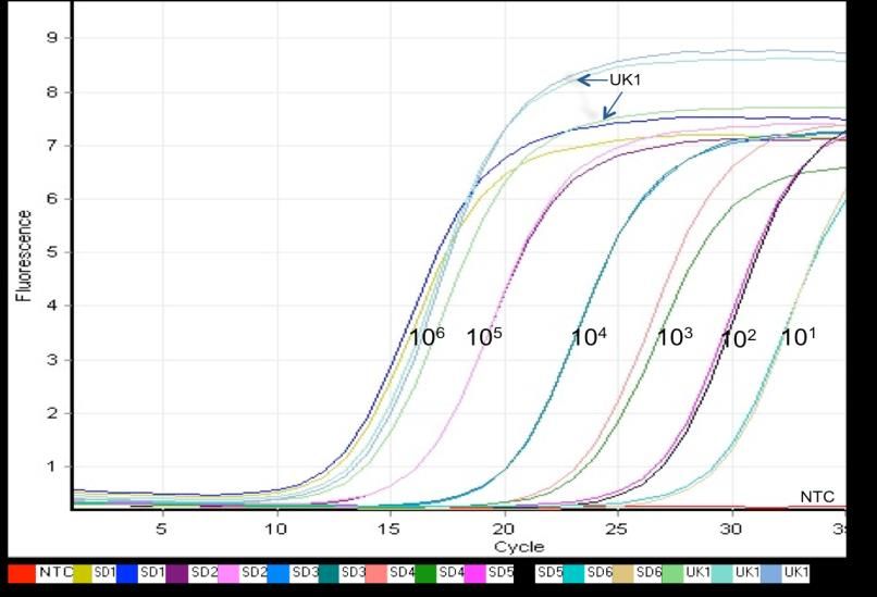
An amplification curve generated for elongation factor gene sequence quantification of P.
pastoris is shown in Figure 1, where fluorescence signals from SYBR Green I were measured at
520 nm. The different color curves are the standard PCR amplified from 106 to 101 copies of the
genes, while the curves for the targeted genomic sample, measured six times, are indicated by
arrows. The red curve represents the amplification profile of the no-template control (NTC). A CT
value calibration curve of the standard versus the concentrations in copies per microliter, was
plotted using Q-Rex software and used to determine the target copy numbers based on their
fractional cycle number or CT values (Figure 2), where the values are proportional to the log of
initial target concentrations. To determine the concentrations of targeted gDNA, a calibration
curve of the CT values of the standard dilution series versus the concentrations was then calculated
[5, 14].
Melting curve analysis was done by slowly increasing the PCR amplification temperature
from 72° to 90°C. The signal was continuously recorded to ensure that non-specific products were
not amplified before the signal curves reached the threshold. The cooperative melting process of
the dsDNA causes an abrupt decrease in fluorescence signal that can be seen as a clear peak in the
negative derivative (±dF/dT) of the melting curves. The melting curve analysis of the specific
PCR product showed that the standard melted at approximate 85°C (Figure 3). The same melting
temperature of 85°C was also observed for the PCR product of the targeted genomic DNA,
therefore confirming the specificity of the amplification that gave rise to the same PCR product.
The methanotrophic yeast P. pastoris strain GS115 was chosen as the positive control for
the method due to the availability of its complete genome sequence which was reported to be 9.43
Mbp [16] and 8.7 Mbp using similar technique [5]. Comparatively via real-time qPCR, the
genome size of P. pastoris was estimated to be 8.52 Mbp (Table 2) which was within 10% of the
size reported by De Schutter et al. [16] and closer to the size by Mounsey et al. [5]. Quantitative
real-time PCR of two single copy genes of M. sagu gave a mean genome estimate of 1.87 Gbp for
M. sagu (Table 2) with no large differences in genome size estimated using either of the two
specific primers. Assuming the 10% upper error limit for the genome size of P. pastoris, the actual
genome size of M. sagu is therefore estimated to be 2.06 Gbp.
This estimated genome size of M. sagu is very much larger than that of any other
economically important plant of known genome size such as agarwood (Aquilaria malaccensis)
which range from 894.65 to 938.88 Mbp [17]. The genome size for the Para rubber tree (Hevea
brasiliensis) has been estimated to range from 1.37 to 1.47 Gbp [18] and the genome size for oil
palm (Elaeis guineensis) closely related to M. sagu and from the same family Arecaceae was
estimated at the 1.8 Gbp [19]. However, genome size across angiosperms has been revealed to
be
212Current Applied Science and Technology Vol. 20 No. 2 (May-August 2020)
Figure 1. Signal curves obtained from real-time PCR. An amplification curve used for
quantification of elongation factor gene from P. pastoris. Fluorescence signals from SYBR Green
1 were measured at 520 nm. Curves obtained for the standards with 106, 105, 104, 103, 102 copies
of the standards PCR products as templates are shown in different colors (SD1 to SD6, each
duplicate samples). Samples curves are shown by black arrows (UK1, triplicate samples); NTC
indicates the no-template control.
Figure 2. CT values of serially 10-fold diluted standard of known concentrations (copy number) of
elongation factors (EF3) gene sequence and unknown genomic samples of P. pastoris. Standard
and unknown samples CT values are indicated by blue and red colors, respectively.
213Current Applied Science and Technology Vol. 20 No. 2 (May-August 2020)
Figure 3. Melting curve analysis of standard and samples after amplification with the PpEF3
specific primers (Amplification curve is shown in Figure 1). Melting curve analysis was carried
directly after the PCR by slowly increasing the temperature at 0.2 Ks-1 from 72 to 90C while the
signal was taken continuously. Cooperative melting process of the dsDNA causes a steep decrease
in the fluorescence signal around the melting temperature of the PCR products. The temperature
where the top value is reached is considered as the melting temperature: the specific PCR product
obtained from the amplification of the standard PCR product melts at 85C. Same melting
temperature was also determined for the PCR product of the genomic DNA sample.
Table 2. Quantitative real time PCR based genome size estimates of P. pastoris and M. sagu
Species Sample Gene qPCR Target C Genome Average
conc. products copies/l (Mean, Size, Genome
(ng/l) size (bp) SD, (Mean, size
pg) SD) ()
0.0086 8.45
134 Beta-actin 246 15.5x10⁶ ( 0.5)
8.52
P. 0.0006 Mbp
( 0.42)
pastoris 0.0088 8.62
Elongation Mbp
254 246 28.81x10⁶ ( 0.4)
factor 3
0.0004 Mbp
1.89 1.85
151 Beta-actin 185 7.95x10⁴ 1.87
0.11 0.09 Gbp
M. sagu ( 0.08)
Elongation 1.95 1.90 Gbp
160 209 8.20x10⁴
factor 0.12 0.08 Gbp
diverse with differences in C value ranging up to 2000-fold [20]. The genome size estimated here
for M. sagu of 1.87 Gbp, is therefore still considered small compared to the monocot Trillium
hagae (Melanthiaceae) that is known to have the largest genome size (129.54 Gbp) among all
angiosperms analyzed to date [21]. The relatively small size of M. sagu genome nevertheless
214Current Applied Science and Technology Vol. 20 No. 2 (May-August 2020)
allows a fair challenge to its genome sequence project. Although flow cytometry remains the most
ideal method for determination of genome size, this method is not available to many investigators,
and the PCR method we use here is demonstrated to be quite reliable. Up until now, there has
been no reported analyses of M. sagu genome size. Therefore, this is the first report for genome
size estimation for M. sagu, which will be helpful for future M. sagu genome sequencing and
subsequent molecular analyses projects.
4. Conclusions
Quantitative real time PCR is an invaluable tool that could be used in many applications including
in evaluating the genome size of an underutilized crop plant, Metroxylon sagu. The qPCR
technique indicated that the genome size of M. sagu is determined to be 1.87 Gbp. With the
determination of genome size of M. sagu, this would enable other molecular research of the
species, such as genome sequencing, and further drive molecular analysis of this important but
underutilized crop plant.
5. Acknowledgements
This study was funded by grants derived from UNIMAS (F07/DPD01/1119/2014(01)) and partially
funded by the Malaysia Ministry of Higher Education (FRGS/2/2013/ST03/UNIMAS/02/2). The
authors would also like to thank the Faculty of Resource Science and Technology (UNIMAS) for
providing the research facilities.
References
[1] Wee, C.C. and Roslan, H.A., 2012. Expressed sequence tags (ESTs) from young leaves of
Metroxylon sagu. 3 Biotech, 2, 211-218.
[2] Hare, E.E. and Johnston, J.S., 2011. Genome size determination using flow cytometry of
propidium iodide-stained nuclei. Methods in Molecular Biology, 772, 3-12.
[3] Zhang, J.Z. and Fan M.Y., 2002. Determination of genome size and restriction fragment
length polymorphism of four Chinese rickettsial isolates by pulsed-field gel electrophoresis.
Acta Virologica, 46, 25-30.
[4] D’Hondt, L., Hofte, M., Van Bockstaele, E. and Leus, L., 2011. Applications of flow
cytometry in plant pathology for genome size determination, detection and physiological
status. Molecular Plant Pathology, 12, 815-828.
[5] Mounsey, K.E., Willis, C., Burgess, S.T.G., Holt, D.C., McCarthy, J. and Fischer, K., 2012.
Quantitative PCR-based genome size estimation of the astigmatid mites Sarcoptes scabiei,
Psoroptes ovis and Dermatophagoides pteronyssinus. Parasites & Vectors, 5, 3.
https://doi.org/10.1186/1756-3305-5-3.
[6] Henk, D.A. and Fisher, M.C., 2012. The gut fungus Basidiobolus ranarum has a large
genome and different copy numbers of putatively functionally redundant elongation factor
genes. Plos One; 7 (2), e31268. https://doi.org/10.1371/journal.pone.0031268
[7] Armaleo, D. and May, S., 2009. Sizing the fungal and algal genomes of the lichen Cladonia
grayi through quantitative PCR. Symbiosis, 49, 43, https://doi.org/10.1007/s13199-009-0012-
3
215Current Applied Science and Technology Vol. 20 No. 2 (May-August 2020)
[8] Gao, J. and Scott, J.G., 2006. Use of quantitative real-time polymerase chain reaction to estimate
the size of the house-fly Musca domestica genome. Insect Molecular Biology, 15, 835-837.
[9] Tsoumani, K.T. and Mathiopoulos, K.D., 2012. Genome size estimation with quantitative real-
time PCR in two Tephritida species: Ceratitis capitata and Bactrocera oleae. Journal of Applied
Entomology, 136, 626-631.
[10] Kim, J.H., Roh, J.Y., Kwon, D.H., Kim, Y.H., Yoon, K.A., Yoo, S., Noh, S.J., Park, J.H.,
Shin, E.H., Park, M.Y. and Lee, S.H., 2014. Estimation of the genome sizes of the chigger
mites Leptotrombidium pallidum and Leptotrombidium scutellare based on quantitative PCR
and k-mer analysis. Parasites & Vectors, 7, 279. https://doi.org/10.1186/1756-3305-7-279.
[11] Park, B. and Kim, Y., 2012. Genome size estimation of an endoparasitoid wasp, Cotesia plutellae,
using quantitative real-time polymerase chain reaction. Journal of Asia-Pacific Entomology, 15,
349-353.
[12] Kaewkong, W., Imtawil, K., Maleewong, W., Intapan, P.M., Sri-Aroon, P., Wongkham, S.
and Wongkham, C., 2012. Genome size estimation of liver fluke Opisthorchis viverrini by
real-time polymerase chain reaction based method. Parasitology International, 61, 77-80.
[13] Jensen, M.K., Vogt, J.K., Bressendorff, S., Seguin-Orlando, A., Petersen, M., Sicheritz-
Pontén, T. and Mundy, J., 2015. Transcriptome and genome size analysis of the Venus
flytrap. Plos One, 10: e0123887. https://doi.org/10.1371/journal.pone.0123887
[14] Wilhelm, J., Pingoud, A. and Hahn, M., 2003. Real-time PCR-based method for the estimation of
genome sizes. Nucleic Acids Research, 31: e56. https://doi.org/10.1093/ nar/gng056
[15] Dolezel, J., Bartos, J., Voglmayr, H. and Greilhuber, J., 2003. Nuclear DNA content and genome
size of trout and human. Cytometry. Part A , 51, 127-128.
[16] De Schutter, K., Lin, Y-C., Tiels, P., Van Hecke, A., Glinka, S., Weber-Lehmann, J., Rouzé,
P., Van de Peer, Y. and Callewaert, N., 2009. Genome sequence of the recombinant protein
production host Pichia pastoris. Nature Biotechnology, 27, 561-566.
[17] Siti Suhaila, A.R., Mohd Saleh, N., Norwati, M., Mahani, M.C., Namasivayam, P. and
Kandasamy, K.I., 2018. DNA content and genome size of highly valued malaysian agarwood,
Aquilaria malaccensis LAMK. Malaysian Applied Biology, 47, 13-21.
[18] Tang, C., Yang, M., Fang, Y., Luo, Y., Gao, S., Xiao, X., An, Z., Zhou, B., Zhang, B., Tan,
X., Yeang, H.Y., Qin, Y., Yang, J., Lin, Q., Mei, H., Montoro, P., Long, X., Qi, J., Hua, Y.,
He, Z., Sun, M., Li, W., Zeng, X., Cheng, H., Liu, Y., Yang, J., Tian, W., Zhuang, N., Zeng,
R., Li, D., He, P., Li, Z., Zou, Z., Li, S., Li, C., Wang, J., Wei, D., Lai, Z.Q., Luo, W., Yu, J.,
Hu, S. and Huang, H., 2016. The rubber tree genome reveals new insights into rubber
production and species adaptation. Nature plants, 2, 16073 https://doi:
10.1038/nplants.2016.73.
[19] Singh, R., Ong-Abdullah, M., Low, E-L., Manaf, M.A., Rosli, R., Nookiah, R., Ooi, L.C.,
Ooi, S.E., Chan, K.L., Halim, M.A., Azizi, N., Nagappan, J., Bacher, B., Lakey, N., Smith,
S.W., He, D., Hogan, M., Budiman, M.A., Lee, E.K., DeSalle, R., Kudrna, D., Goicoechea,
J.L., Wing, R.A., Wilson, R.K., Fulton, R.S., Ordway, J.M., Martienssen, R.A.,
Sambanthamurthi, R., 2013. Oil palm genome sequence reveals divergence of interfertile
species in Old and New worlds. Nature, 500, 335-339.
[20] Pellicer, J., Fay, M.F. and Leitch, I.J., 2010. The largest eukaryotic genome of them all? Botanical
Journal of the Linnean Society 164, 10-15.
[21] Zonneveld, B.J.M., 2010. New record holders for maximum genome sizes for monocots and
eudicots. Journal of Botany, 2010, article ID 527357. https://doi.org/ 10.1155/2010/527357.
216You can also read