Digestive system - Sher-e-Bangla Agricultural University
←
→
Page content transcription
If your browser does not render page correctly, please read the page content below
Mohammad Saiful Islam
                                                                                Phd (Japan) Post doc (Australia)
Digestive system                                                                Dept. of Anatomy, Histology & Physiology
                                                                                Sher-e-Bangla Agricultural University, Dhaka

Functions of the digestive tract:

The digestive tract has following functions
   • Ingestion: This is the active process of bringing material into the oral cavity.
   • Propulsion: Ingested materials are moved through the digestive tract by swallowing and peristalsis.
   • Mechanical processing: Material entering the digestive tract is physically reduced in size. This begins in the
       oral cavity where food is crushed and sheared before being propelled along the digestive tract
   • Digestion: Following reduction in size, ingested nutrients are chemically broken down into particles small
       enough for absorption. Although simple molecules such as monosaccharides and amino acids can be absorbed
       without further reduction in size, macromolecules such as protein, DNA, polysaccharides, and triglycerides
       must first be reduced into smaller molecules. Specific enzymes complete such reduction.
   • Secretion: Water, mucus, acids, enzymes, buffers, and salts are released into the lumen of the digestive tract
       along its length. Secretions come from epithelial cells and glandular organs.
   • Absorption: Along the length of the digestive tract, nutrients including organic substrates, electrolytes,
       vitamins, and water pass from the lumen into the body. In addition to absorbing ingested nutrients, the
       digestive tract must absorb secreted water, salts, and other secreted material. Failure of such absorption will
       result in dehydration.
   • Excretion: The digestive tract is a site of elimination of waste products.
   • Immunity. The digestive tract provides a substantial barrier to prevent the entry of pathogens into the body.
       The digestive tract acts not only as a physical barrier, but also has an innate immune system.

Classification of animals according to their diet:

        Animals can be classified into four groups as follows. The first group includes animals with a simple stomach,
such as humans, pigs, dogs and cats. The second group is foregut fermenters, which includes cattle, sheep, and goats.
These animals have a ruminant stomach in which they can ferment nondigestible carbohydrates. The third group
consists of hindgut fermenters such as horses, rabbits, guinea pigs, etc. These animals rely on fermentation that
occurs in the cecum and colon. The final group consists of birds in which various adaptations have occurred to both
store and grind various foodstuffs.

Pregastric Physiology

    •   The stomach is the first major organ associated with digestion. Before food can be received by the stomach,
        important pregastric functions are performed to receive, prepare, and deliver a bolus to the stomach.
        Performance of these functions varies among the animals and depends mostly on adaptations associated with
        their diet.

Prehension

        The first mechanical function necessary for the digestive process is prehension. The seizing and conveying of
food into the mouth is called prehension. Lips, teeth, and tongue are the principal prehensile structures in domestic
animals. The lips of the horse, the tongue of the cow and sheep, and the snout of the pig are used extensively in
obtaining food.

Mastication

        Mastication refers to the mechanical breakdown of food in the mouth. It is commonly called chewing. It is
carried out to varying degrees by different animals. The fibrous nature of the diet of herbivores requires more chewing
than the meat diet of carnivores. A bolus (rounded ball) of food is formed by the mastication process.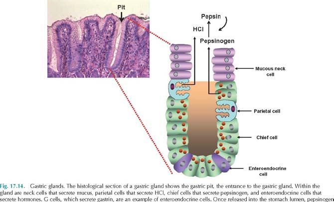
Deglutition
          Deglutition is the act of swallowing or conveying the food mass from the mouth to the stomach. This complex
process involves a number of reflexes that are coordinated by a swallowing center in the brain. There are three stages
of swallowing: 1) through the mouth (voluntary), 2) through the pharynx (reflex) and 3) through the esophagus
(reflex).

Saliva

         Saliva consists of water (97–99.5%), and it is therefore hypoosmotic. Electrolytes in the saliva include
sodium, potassium, chloride, bicarbonate, and phosphate. It tends to be slightly acidic (pH 6.75–7.00). Saliva has
several functions:
1. Solubilizes food. Dissolves foods so they can be tasted and digestive reactions can occur.
2. Provides alkaline buffering and fluid. Bicarbonate and phosphate in the saliva can neutralize acidic
feedstuffs. Alkaline fluid via the saliva is particularly important in ruminants.
3. Removes wastes. Metabolic waste products such as urea and uric acid are excreted in the saliva.
4. Lubricates and binds. The mucus in the saliva helps bind masticated food so that it can be formed into a bolus. In
addition, saliva coats the oral cavity and esophagus, thus protecting the mucosa of the oral cavity and esophagus.
5. Initiates starch digestion. The starch-digesting enzyme amylase is present in the saliva of omnivores (pig) and to
a limited degree in horses but absent in ruminants and carnivores (dog).
6. Assists oral hygiene. Lysozyme, found in saliva, is a bacteriostatic enzyme that lyses bacteria, thus protecting the
mouth.
7. Enables evaporative cooling. This is particularly important in dogs, which have very poorly developed sweat
glands.

Enteric nervous system
The digestive tract has its own nervous system called the enteric nervous system. It is composed mostly of two large
plexuses:
    • Submucosal plexus/Meissner’s plexus: located within the submucosal layer
    • Myenteric plexus/ Auerbach plexus: located between the two layers of smooth muscle fibers in the
        muscularis externa

Segmentation and Peristalsis

Peristalsis (peri = around + stalsis = constriction) can be defined as alternating waves of contraction and relaxation of
muscles along the digestive tract wall.
During peristalsis, the circular smooth muscle layer behind the bolus contracts while that in front of the bolus relaxes.
Conversely, the longitudinal smooth muscle layer behind the bolus relaxes while that in front of the bolus contracts.
This increases the diameter of the lumen in front of the bolus while constricting the diameter of the lumen behind the
bolus. This results in propulsion of the bolus down the digestive tract.

Mechanical digestion and motility in the
small intestine
Small intestine motility is regulated mainly
by the enteric reflex responding to the
presence of materials in the intestinal
lumen. There are two types of movements
within the small intestine: segmentation
and peristalsis.
Segmental contractions characterized by
ring like contractions separated by relaxed
areas containing a bolus of chyme. The
constant formation and then relaxation of
these contractual rings along the length of
the small intestine result in a mixing
action.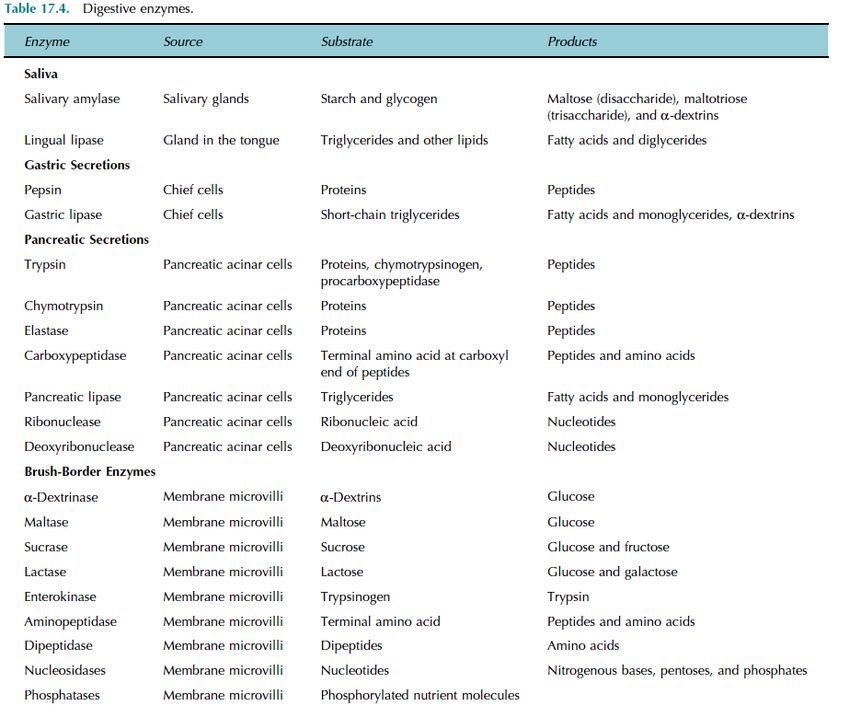
Gastric juice: means secretion of gastric gland into the stomach lumen by gastric glands.
Gastric juice contains water, hydrochloric acid, mucus, intrinsic factor, pepsinogen, and rennin. There are four cell
types in gastric glands.
1. Mucous neck cells. Found in the upper, or neck, region of a gastric gland, they produce a more acidic mucus than
goblet cells.
2. Parietal cells. Found in the middle region, they secrete hydrochloric acid (HCl) and intrinsic factor. Intrinsic factor
is a glycoprotein necessary for absorption of vitamin B12 in the small intestine. The HCl decreases the pH of the
stomach to 1.5– 2. This low pH has several functions: (1) It is necessary for the function of pepsin; (2) it provides a
harsh environment for bacteria ingested with food; (3) it denatures proteins and inactivates enzymes in food; and (4) it
breaks down cell walls of plant material and connective tissue in meat.
3. Chief cells. These cells
produce pepsinogen, the
inactive form of pepsin,
and an enzyme that
digests proteins. When
pepsinogen is first
released, it interacts with
HCl and converts it to its
active form, pepsin. Once
activated, pepsin can
convert other molecules
of pepsinogen to pepsin.
Regulation of gastric
secretions and emptying
Gastric secretions are
controlled by neural and
hormonal mechanisms.
Gastric secretions are
controlled at three levels
and can be classified into
three phases: cephalic
phase, gastric phase, and
intestinal phase of gastric
secretion. These control
mechanisms can either
increase or decrease
gastric secretions.
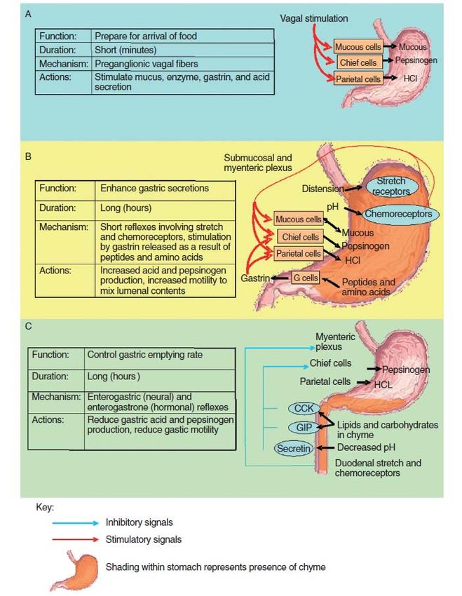
Cephalic phase
The cephalic
phase causes an increase
in gastric secretions prior
to the arrival of food.
This stage is controlled
Fig. 1 Phases of Gastric secrions; A: Cephalic phase, B: Gastric phase C: intestinal phase
by the central nervous
system, and it prepares the stomach for the arrival of food. The sight, smell, and taste of food stimulate the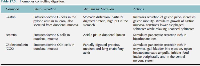
parasympathetic nervous system to send signals via the vagus nerve that synapse on the submucosal plexus located in
the wall of the stomach. This stimulates the postganglionic parasympathetic fibers innervating mucous cells, chief
cells, parietal cells, and G cells in the stomach, thus increasing gastric secretions. This phase is short, lasting minutes.
Emotional responses associated with activation of the fight-or-flight response decrease gastric secretions and gastric
motility.

Gastric phase

Beginning with the arrival of food in the stomach, this phase further stimulates gastric secretion and motility. This
phase accounts for about two-thirds of gastric secretions.

Stimuli for the gastric phase include distention of the stomach, an increase in gastric pH, and the presence of
undigested food, especially proteins and peptides.

Intestinal phase

        This phase functions to decrease gastric motility. Stimulation of chemoreceptors and stretch receptors triggers
the enterogastric reflex. This reflex inhibits gastrin production and gastric motility, and stimulates contraction of the
pyloric sphincter, thus slowing gastric emptying into the duodenum.

Pepsin secretions regulations

    •    Pepsin is not produced within the chief cells because this would cause the self-digestion of the cells. Instead,
         chief cells produce the zymogen (i.e., the precursor form of an enzyme) pepsinogen, which is activated when
         it enters the stomach lumen and comes in contact with HCl.
    • Protein digestion is initiated in the stomach via the action of pepsin, the only enzyme found in the stomach of
         adult animals. Dietary proteins are denatured by HCl secreted by the parietal cells.
HCl secretions and regulations
         HCl is not produced within the parietal
cells because it would destroy the cell. Both H+
and Cl− are independently transported from the
parietal cells into the stomach lumen. Hydrogen
ions are generated from the dissociation of
carbonic acid that is produced by the enzyme
carbonic anhydrase acting upon CO2 and H2O.
Hydrogen ions are then transported into the
stomach lumen in exchange for K+. Chloride
ions enter the parietal cell in exchange for
bicarbonate ions. The chloride ions then travel
down their concentration gradient and enter the
stomach lumen. Once in the lumen, hydrogen
and chloride ions combine, producing HCl.
When parietal cells are producing considerable
HCl, a significant amount of bicarbonate enters
the blood, thus increasing the pH (called the
alkaline tide). Parietal cells respond to many
signals. Located on their surface are receptors
for histamine, acetylcholine (ACh), and gastrin.
Histamine comes from mast cells located in the
lamina propria, ACh from postganglionic parasympathetic fibers, and gastrin from G cells. Histamine acts at H2
receptors, whereas ACh acts at muscarinic receptors. Stimulation of these receptors results in stimulation of protein
kinase, other neural and hormonal stimuli such as ACh and histamine.

Emesis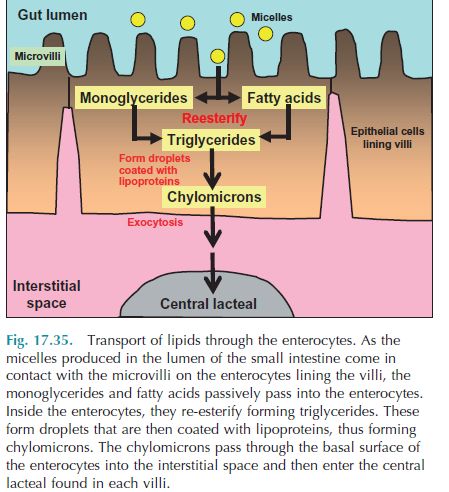
Emesis (vomiting) is an emptying of the cranial part of the duodenum and stomach in an orad (toward the
mouth) direction. A series of reflexes is involved to initiate antiperistalsis and closure of the epiglottis and nasal
cavity. Swine, dogs, and cats vomit easily. Vomiting is a protective mechanism that helps prevent absorption of
noxious substances. Vomiting in the horse is rare because of the difficulty in opening the cardia from a reverse
direction. The reflexes of vomiting are controlled by a vomiting center in the brain.

Defecation
Defecation is a complex reflex act in which feces are evacuated from the terminal colon and rectum.The frequency of
defecation varies among animals but can occur 5 to 10 times daily in vigorous horses, 10 to 20 times daily in cattle,
and 2 to 3 times daily in carnivores. The reflex can be assisted or inhibited by certain voluntary muscles.

•   Animals that regurgitate and remasticate their food are called ruminants.
•   There are two suborders of ruminant animals: 1) Ruminantia- includes the bison, cow, sheep, goat deer, reindeer,
    antelope, giraffe, musk ox, and 2) Tylopoda-which includes the camel, llama, alpaca, and vicuna.
•   The main difference between the two suborders is Tylopoda do not have an omasum. Another difference is that
    Tylopoda have cardiac glands that open into ventral surfaces of the reticulum and rumen.

Rumination
   • The process of bringing food material back from the ruminant stomach to the mouth for further mastication is
      known as rumination. Rumination is a cycle of activity composed of four phases: 1) regurgitation (returning it
      to the mouth), 2) remastication, 3) resalivation, and 4) redeglutition. It is a reflex initiated by mechanical
      stimulation of receptors in the mucosa of the reticulum and rumen in the area of the cardia.
   • The time spent in rumination each day varies with species and diet. Generally, the coarseness of the ration
      influences the time for rumination—cattle on a hay diet average about 8 hr/day. When only concentrates are
      fed, rumination time can be reduced to about 2-1/2 hr/day.

Chemical digestion in the small intestine Some/little starch digestion occurs in the mouth by the action of salivary
amylase. In the stomach, some chemical protein digestion occurs as pepsin converts proteins to peptides.
However, most chemical digestion occurs within the small intestine.

Carbohydrate digestion
Since food remains in the mouth only a short time, little starch is totally digested. Instead, pancreatic amylase
completes this process. Starch and glycogen are acted upon by salivary and pancreatic amylase to form maltose,
maltotriose, and α-dextrins. This is the luminal phase of carbohydrate digestion since it occurs within the lumen. The
smaller di- and trisaccharides then move
into contact with the brush border where
mucosal digestion by brush-border
enzymes digests these sugars to
monosaccharides.

Protein digestion
Chemical digestion of protein begins in
the stomach by the action of pepsin.
Pepsin, which works optimally at a pH of
1.5–2.5, cleaves bonds involving tyrosine
and phenylalanine. Pepsin digests
approximately 10–15% of dietary protein
before being inactivated in the lumen of
the small intestine. Once in the small
intestine, trypsin and chymotrypsin
secreted by the pancreas break down
proteins into peptides. Carboxypeptidase,
another pancreatic enzyme, cleaves one
amino acid at a time from the carboxyl
and amino end of a polypeptide,
respectively, while the brush-border
enzymes aminopeptidase and dipeptidase
further cleave the proteins.

Lipid digestion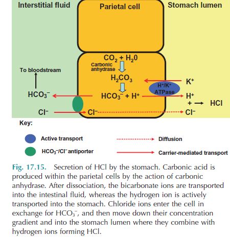
Triglycerides are the most abundant lipid in the diet. Triglycerides and phospholipids are digested by lipases. Bile salts
assist in emulsifying dietary lipids within the aqueous environment found in the small intestine lumen. During this
emulsification process, large lipid masses are converted into small droplets (Micelle) which allows water-soluble
pancreatic lipase to act more efficiently. Pancreatic lipase cleaves off two fatty acids from triglycerides producing two
free fatty acids and monoglyceride.

Absorption in the small intestine
         Absorption is the process whereby compounds and ions move through the epithelial cells lining the mucosa
and pass into the bloodstream or lymphatic system. About 90% of absorption occurs within the small intestine with the
rest occurring in the stomach and large intestine. Absorption occurs via diffusion, facilitated diffusion, osmosis, and
active transport.

Absorption of amino acids, dipeptides, and tripeptides

         Although it was once believed that only amino acids are
absorbed, it is now well established that di- and tripeptides are also
actively absorbed in the small intestine. Some amino acids enter the
epithelial cells by a secondary active transport system similar to
that described for glucose and galactose. There are some amino
acids that utilize a sodium-independent cotransporter in which the
amino acids enter along with H+ instead of Na+. In this case, H+ is
pumped into the intestinal lumen in exchange for Na+. The Na+ is
then pumped out of the cell by the Na+-K+-ATPase on the
basolateral membrane. This creates a concentration gradient for H+,
which is high concentrations within the lumen. As H+ enters the
epithelial cells, selected amino acids are cotransported. Peptides are
absorbed via this sodium-independent co-transporter. Once inside
the epithelial cell, the peptides are hydrolyzed to single amino acids,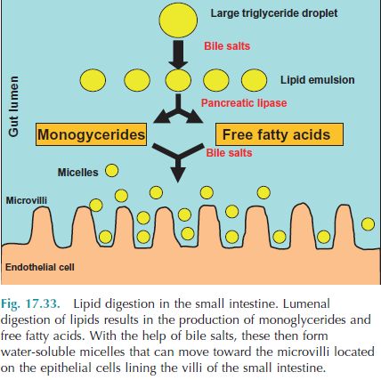
which then move by diffusion into the hepatic portal vein
Absorption of lipids
   • Since lipids are not water soluble so lipid absorption and transport within the body face unique challenges
        compared to carbohydrate and protein absorption.
   • Within the small intestine lumen, triglycerides are broken down into fatty acids and monoglycerides. The bile
        salts within the gut lumen help emulsify the lipids by forming water-soluble particles (micelles), which helps
        the lipids migrate within the aqueous chyme found in the gut. Since lipids are fat soluble; once the micelles
        come in contact with the lumen wall, the monoglycerides and free fatty acids can cross the epithelial
        membrane by simple diffusion. The bile salts help in the formation of micelles.
   • Once inside the epithelial cells lining the gut pass into the hepatic portal system similar to amino acids and
        monosaccharides. Within the enterocytes, absorbed monoglycerides and fatty acids are used to resynthesize
        triglycerides. These triglycerides combine with cholesterol and intracellular proteins and form a particle which
        is called chylomicrons. The chylomicrons are secreted by a process of exocytosis into the interstitial fluid of
        the villus. In mammals, these chylomicrons then enter the lacteal (smallest lymphatic vessels) and are carried
        to thoracic ducts and ultimately added to the blood.

ACCESSORY ORGANS

Pancreas
Chyme passes from the stomach to the small intestine. The chemical digestive processes that occur within the small
intestine depend upon accessory organs, including the pancreas, liver, and gallbladder.

Composition and function of pancreatic juice
Pancreatic juice is a clear, colorless liquid. Containing mostly water, it also has salts, sodium bicarbonate,
and enzymes. Sodium bicarbonate serves as a buffer to neutralize stomach acid within the small intestine, thus
stopping the action of gastric pepsin. Neutralizing gastric acid also allows pancreatic enzymes to function. Pancreatic
enzymes include the carbohydrate-digesting enzyme pancreatic amylase; several protein-digesting enzymes, including
trypsin, chymotrypsin, carboxypeptidase, and elastase; the triglyceride-digesting enzyme pancreatic lipase; and the
nucleic acid-digesting enzymes ribonuclease and deoxyribonuclease.

Protein-digesting enzymes are produced within the pancreas in an inactive form so that they do not digest the
pancreas. Pancreatic acinar cells secrete a protein called trypsin inhibitor that prevents the activation of trypsinogen..
Upon entering the duodenal lumen, trypsinogen is activated by the brush-border enzyme enterokinase. Trypsin then
activates the remaining zymogens chymotrypsinogen, procarboxypeptidase,
and proelastase producing the respective active enzymes.

Regulation of pancreatic secretions
Similar to gastric secretions, pancreatic secretions are controlled by both neural and hormonal mechanisms:

    1. During the cephalic and gastric phases of gastric secretion, parasympathetic signals carried via the vagus
       nerve (cranial nerve X) increase secretion of pancreatic enzymes.

    2. Partially digested lipids and proteins within the duodenal lumen stimulate the secretion of CCK from
       enteroendocrine cells in the duodenal wall. CCK stimulates the secretion of pancreatic enzymes.

    3. Decreased pH in the duodenal lumen stimulates the release of secretin from enteroendocrine cells in the
       duonenal wall. Secretin stimulates release of bicarbonate ions from the pancreas.

Bile composition and function
Bile consists of water, bile salts, bile acids, cholesterol, the phospholipid (lecithin), bile pigments (bilirubin), and ions.
The bile salts include sodium and potassium salts of bile acids, (glycocholic and taurocholic acids). Bile salts assist in
emulsification of lipids within the small intestine.

Regulation of bile secretion
        In those animals that have a gallbladder, bile is stored between meals. The sphincter of the hepatopancreatic
ampulla (sphincter of Oddi) restricts the entrance to the duodenum. There are neural and hormonal stimuli that can
stimulate bile secretion:
1. Parasympathetic signals traveling along the vagus nerve can stimulate bile production by the liver.
2. Fatty acids, particularly medium- and long-chain, and amino acids in the chyme cause duodenal enteroendocrine
cells to secrete CCK. CCK causes the smooth muscle cells of the gallbladder to contract, and bile is squeezed into the
cystic duct and through the common bile duct. CCK also relaxes the sphincter of the hepatopancreatic ampulla.
Functions of the liver: The liver is a vital organ. It performs many functions:
1. Carbohydrate metabolism. The liver plays a vital role in maintaining blood glucose levels. When blood
glucose levels are high, the liver converts glucose to glycogen (glycogenesis) and triglycerides so that energy
can be stored until needed. When blood glucose levels drop, the liver can break down glycogen to glucose
(glycogenolysis) and release the glucose into the bloodstream. In addition, the liver can convert certain amino
acids to glucose (gluconeogenesis), as well as lactic acid to glucose.
2. Lipid metabolism. Hepatocytes can store triglycerides as well as use fatty acids to synthesize ATP. In
addition, hepatocytes synthesize lipoproteins that carry fatty acids, triglycerides, and cholesterol throughout
the body. Cholesterol can be synthesized in the liver, and cholesterol is used to make bile salts.
3. Protein metabolism. Hepatocytes remove the amino group (deamination) of amino acids so they can be used
for ATP synthesis. Hepatocytes also synthesize carbohydrates and fats from certain amino acids. Hepatocytes
can synthesize various plasma proteins such as albumin, prothrombin, fibrinogen, and alpha and beta
globulins (needed for hemoglobin synthesis).
4. Removal of waste products. The liver detoxifies substances such as alcohol and antibiotics, and can alter and
excrete steroid hormones. The liver is an important site of detoxification of ammonia, which is converted to
the less toxic urea, which is excreted in the urine. The waste product of red blood cell destruction, bilirubin is
eliminated via the bile.
5. Synthesis of bile salts. The bile salts necessary for lipid emulsification within the small intestine are
synthesized in the liver.
6. Storage. The liver is the primary storage site of fat-soluble vitamins (A, D, E, and K), as well as vitamin B12.
Glycogen and certain minerals (Fe and Cu) are also stored in the liver.
7. Phagocytosis. Kupffer cells destroy aged blood cells and microbes that may have entered via the hepatic
portal blood.
8. Activation of vitamin D. The liver combines with the skin and kidneys to synthesize the active form of
vitamin D.
Large intestine
The terminal portion of the digestive tract is the large intestine. It extends from the end of the ilium to the anus. The
primary functions of the large intestine are electrolyte and water absorption, microbial digestion and vitamin
production, formation of feces, and expulsion of feces. These functions require considerable time, so transit time
through the large intestine is slow
Chemical digestion in the large intestine
Although no chemical digestion occurs in
the large intestine, considerable
fermentation takes place, especially in
those animals that are hindgut fermenters.
Microbes ferment any remaining
carbohydrates releasing hydrogen, carbon dioxide, and methane. These gases lead to flatulence in the large intestine.
Amino acids are converted to indole, skatole, hydrogen sulfide, and fatty acids. Indoles and skatole are eliminated in
the feces, contributing to its odor, while some of the remaining products are absorbed and
transported to the liver. The microbes synthesize B vitamins and vitamin K, which are also absorbed by
the large intestine.
Micelle: small droplets formed in the intestinal chymes which contain lipids, bile salts, and products of lipid digestion.
Emulsification: The process of breakdown of fat globules into smaller globules by the bile salts.You can also read