Changes in the interstitial cells of Cajal in the gallbladder of guinea pigs fed a lithogenic diet
←
→
Page content transcription
If your browser does not render page correctly, please read the page content below

EXPERIMENTAL AND THERAPEUTIC MEDICINE 22: 823, 2021

            Changes in the interstitial cells of Cajal in the gallbladder
                       of guinea pigs fed a lithogenic diet
                                          LEI HUANG1*, CHAO DING2* and XINMIN SI2

                    1
                     Department of General Surgery, Children's Hospital of Nanjing Medical University, Nanjing,
                         Jiangsu 210008; 2Department of Gastroenterology, The First Affiliated Hospital of
                                 Nanjing Medical University, Nanjing, Jiangsu 210029, P.R. China

                                           Received June 11, 2020; Accepted March 12, 2021

                                                        DOI: 10.3892/etm.2021.10255

Abstract. Cholesterol cholelithiasis is a common disease                 Introduction
and gallbladder hypomotility may underlie its pathogenesis.
Interstitial cells of Cajal (ICCs) in the gallbladder serve              Cholelithiasis is a highly prevalent gastroenterological disease
vital roles in regulating gallbladder motility. The aim of               and one of the leading causes of hospital admissions world‑
the present study was to investigate changes in gallbladder              wide (1). In China, the incidence of cholelithiasis is 8‑10%,
ICCs during the development of cholesterol cholelithiasis. A             and the incidence rate has been gradually increasing in recent
total of 40 male guinea pigs were randomly assigned to four              years (2). It was reported that females of childbearing age
groups and fed a standard diet (SD) or lithogenic diet (LD)              are approximately twice more likely than males to develop
for 2 or 8 weeks. The LD significantly increased the total               cholelithiasis (3). Furthermore, among the subtypes of chole‑
cholesterol levels in the serum and bile, as well as the serum           lithiasis, cholecystolithiasis accounts for ~85% of cases (4).
levels of high‑density lipoprotein‑cholesterol and low‑density           Cholelithiasis is composed of >90% cholesterol, and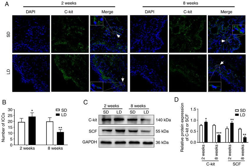
2                           HUANG et al: LITHOGENIC DIET INDUCED CHANGES IN GALLBLADDER ICCs

contraction (15). However, to the best of our knowledge, no        at room temperature. Subsequently, sections were incubated
studies have shown whether CCK1R expression in the gall‑           overnight at 4˚C with the following primary antibodies:
bladder, particularly in gallbladder ICCs, is altered during the   Anti‑C‑kit (1:200; cat. no. ab25022; Abcam) and anti‑CCK1R
development of cholesterol cholelithiasis.                         (5 µg/ml; cat. no. ab77269; Abcam). Following three washes
    The lithogenic diet (LD), which contains a higher propor‑      with PBS, sections were incubated with rabbit anti‑rat
tion of cholesterol, is widely used to induce cholesterol          IgM/Alexa Fluor 488 antibody (1:500; cat. no. bs‑0346R;
cholelithiasis in animals (16,17). The aim of the present          BIOSS) and rabbit anti‑goat IgG/Alexa Fluor 647 antibody
study was to detect the changes in the density, ultrastructure     (1:500; cat. no. bs‑0294R; BIOSS) for 2 h at room tempera‑
and CCK1R expression levels in gallbladder ICCs in LD‑fed          ture, and then incubated with DAPI (Beyotime Institute of
guinea pigs.                                                       Biotechnology) for 10 min at room temperature. Finally,
                                                                   sections were mounted using antifade mounting medium
Materials and methods                                              (Beyotime Institute of Biotechnology). For the negative control,
                                                                   the primary antibodies were omitted. Images were captured
Animal models. A total of 40 male guinea pigs (4 weeks             on a laser scanning confocal microscope (magnification, x200
old; weight, 250‑280 g) were purchased from Charles River          or x400). C‑kit‑positive cells excluding those with a round
Laboratories, Inc. and randomly assigned to four groups            shape (mast cells) were considered as gallbladder ICCs. To
(n=10): Standard diet (SD) for 2 weeks; SD for 8 weeks; LD for     quantify the number of gallbladder ICCs, three gallblad‑
2 weeks and LD for 8 weeks. Guinea pigs in the LD group were       ders were obtained from each group, three sections were
fed with LD (chow diet supplemented with ~1.25% cholesterol,       prepared from each gallbladder and three random fields of
0.5% bile salt and 15% fat) and animals in the SD group were       view (magnification, x200) in each section were selected. The
fed standard chow. All guinea pigs were maintained in cages        number of gallbladder ICCs in the lamina propria was counted
with a 12‑h light/dark cycle with free access to food and water.   by two researchers in a blinded manner.
All animal experiments were performed according to the
National Institutes of Health Guide for the Care and Use of        Western blot analysis. The resected gallbladders were washed
Laboratory Animals and approved by the Institutional Animal        with PBS and lysed in RIPA lysis buffer (Thermo Fisher
Care and Use Committee of the First Affiliated Hospital of         Scientific, Inc.) to extract total protein. Protein concentration
Nanjing Medical University (Nanjing, China).                       was measured using a BCA protein assay kit (Beyotime Institute
                                                                   of Biotechnology) according to the manufacturer's protocol.
Measurement of total cholesterol (T‑CHOL), lipoproteins            Subsequently, 50 µg of protein was loaded on an 8% SDS gel,
and CCK octapeptide (CCK8) levels in the serum and bile.           resolved using SDS‑PAGE and subsequently transferred to a
After feeding for 2 or 8 weeks, guinea pigs in the SD group        PVDF membrane (EMD Millipore). PVDF membranes were
(weighing 340‑800 g) and the LD group (weighing 420‑1200 g)        blocked in TBS (Boster Biological Technology) containing
were anesthetized by 1.5‑3.0% isoflurane, and blood samples        5% skimmed milk powder for 2 h at room temperature.
were obtained from the eyes of guinea pigs (~1 ml per guinea       Subsequently, membranes were washed three times with TBS
pig) and collected in tubes containing heparin. Plasma was         supplemented with 0.05% Tween‑20 and incubated overnight
obtained following centrifugation at 1,789 x g for 15 min          at 4˚C with one of the following antibodies: Anti‑C‑kit
at 4˚C. Moreover, after resecting the gallbladder, bile was        (1:1,000; cat. no. ab256345; Abcam), anti‑stem cell factor
collected and diluted with saline water (1:10), followed by        (SCF; 1:1,000; cat. no. ab64677; Abcam), anti‑Connexin
centrifugation at 2,795 x g for 10 min at 4˚C to remove the        (Cx) 43 (1:2,000; cat. no. ab11370; Abcam), anti‑CCK1R
granular components. Following which, all guinea pigs were         (1:500; cat. no. ab75153; Abcam) or anti‑GAPDH (1:1,000;
euthanized by an overdose of isoflurane. To degrade the            cat. no. AF0006; Beyotime Institute of Biotechnology),
bile pigment, bile was illuminated under a daylight lamp           followed by incubation with horseradish peroxidase‑
for 12 h at 4˚C. The serum levels of T‑CHOL, high‑density          conjugated goat anti‑rabbit IgG (1:5,000; cat. no. bs‑0295G;
lipoprotein cholesterol (HDL‑C) and low‑density lipoprotein        BIOSS) for 2 h at room temperature. Protein signals were
cholesterol (LDL‑C), as well as the T‑CHOL levels in bile          detected using an enhanced chemiluminescence detection kit
were measured using an automatic biochemical analyser              (Thermo Fisher Scientific Inc.) on the ChemiDoc XRS+
(Hitachi, Ltd.) with the corresponding reagents (T‑CHOL,           chemiluminescence imaging system (Bio‑Rad Laboratories.
cat. no. SNM226; HDL‑C, cat. no. SNM224; LDL‑C,                    Inc.). Densitometric analysis was performed using
cat. no. SNM222; all purchased from Beijing Biolab Science         ImageJ v1.8.0 software (National Institutes of Health), and
& Technology Co., Ltd.). The concentration of CCK8 in              expression of each protein was normalized to the respective
serum was measured using an ELISA kit (cat. no. abx150409;         GAPDH loading control band.
Abbexa Ltd.) according to the manufacturer's protocol.
                                                                   Transmission electron microscopy. Gallbladder specimens
Immunofluorescence staining. After snap‑freezing, the               were fixed in 2.5% glutaraldehyde/0.1 M cacodylate buffer for
resected gallbladders were cut into 5‑µm sections using a          2 h at 4˚C and 1% perosmic acid for 2 h at room temperature.
freezing microtome. Subsequently, all sections were fixed          Specimens were then dehydrated using a series of ethanol
in 4% paraformaldehyde for 20 min at room temperature              solutions of increasing concentration and embedded in Epon
and washed three times with PBS. To prevent non‑specific           ethoxyline resin at 60˚C. Ultrathin sections with 70‑80‑nm
binding, sections were incubated with immunostaining               thickness were obtained using a Leica EM UC7 ultramicro‑
blocking buffer (Beyotime Institute of Biotechnology) for 1 h      tome (Leica Microsystems, Inc.) and subsequently placed on
EXPERIMENTAL AND THERAPEUTIC MEDICINE 22: 823, 2021 3 Figure 1. LD significantly increases T‑CHOL levels in the serum and bile and the serum levels of HDL‑C and LDL‑C. Serum levels of T‑CHOL, HDL‑C and LDL‑C were measured after (A) 2 and (B) 8 weeks in guinea pigs fed with SD or LD. The bile levels of T‑CHOL were measured after (C) 2 and (D) 8 weeks in guinea pigs fed with SD or LD. **P
4                                HUANG et al: LITHOGENIC DIET INDUCED CHANGES IN GALLBLADDER ICCs

Figure 2. LD alters the number of ICCs and protein expression levels of C‑kit and SCF in the gallbladders of guinea pigs. (A) Immunofluorescence staining
of ICCs in the gallbladders of guinea pigs fed a SD or LD. ICCs were labelled using a primary antibody targeting C‑kit (green), and nuclei were labelled
using DAPI (blue). White arrowheads indicate C‑kit‑positive gallbladder ICCs, which are shown in the inset. Magnification, x200. (B) The mean number
of ICCs in a field of view of guinea pig gallbladder. (C and D) Western blotting of C‑kit and SCF protein expression levels in guinea pig gallbladders of the
SD and LD groups. *P
EXPERIMENTAL AND THERAPEUTIC MEDICINE 22: 823, 2021                                                    5

                                                                                   results, it was hypothesized that guinea pigs may enhance their
                                                                                   gallbladder emptying capacity via a compensatory increase in
                                                                                   the density of ICCs during the early stages of high cholesterol
                                                                                   stimulation, and their gallbladder motility is considerably
                                                                                   impaired at the later stages of cholesterol cholelithiasis devel‑
                                                                                   opment due to decreases in the density of ICCs.
                                                                                       Subsequently, the mechanisms underlying the decrease
                                                                                   in the density of gallbladder ICCs were investigated. The
                                                                                   SCF/C‑kit pathway is well established as a key regulator of
                                                                                   the differentiation and proliferation of ICCs (26). The present
                                                                                   study detected for the first time that the protein expression
                                                                                   levels of SCF and C‑kit in guinea pig gallbladders were signifi‑
                                                                                   cantly increased after feeding a LD for 2 weeks, indicating
                                                                                   increased activation of the SCF/C‑kit pathway. Furthermore,
                                                                                   consistent with previous studies in which the mRNA and
                                                                                   protein expression levels of SCF and C‑kit were found to be
                                                                                   significantly attenuated in the gallbladders of animal models
                                                                                   fed a high cholesterol diet for 8 weeks (12,27), the present
                                                                                   study also demonstrated that activity of the SCF/C‑kit pathway
                                                                                   was significantly suppressed after being fed a LD for 8 weeks.
                                                                                   Taken together, it was hypothesized that differential activity
                                                                                   of the SCF/C‑kit pathway is observed at various stages of
                                                                                   cholesterol cholelithiasis development, and this may lead to the
                                                                                   corresponding alterations in the density of gallbladder ICCs at
                                                                                   each stage.
                                                                                       A previous study reported that feeding a high cholesterol
                                                                                   diet for 8 weeks did not significantly result in ultrastructural
                                                                                   changes in the gallbladder ICCs in mice (12). However, in
Figure 3. LD alters the ultrastructure of gallbladder ICCs in SD‑fed guinea        the present study, although the ultrastructure of gallbladder
pigs after 8 weeks and the protein expression levels of Cx43 in guinea pig         ICCs in guinea pigs was not notably altered after feeding a
gallbladders. (A) Ultrastructure of gallbladder ICCs in the guinea pigs fed        LD for 2 weeks, they were markedly altered after 8 weeks.
with SD or LD was visualized using a transmission electron microscope. The         It was hypothesized that the differences in outcomes may be
white arrowhead indicated enlarged mitochondria and the white triangle indi‑
cates the vacuole in the cytoplasm. Magnification, x10,000. Scale bar, 2 µm.       attributed to the different susceptibilities of various species to
(B and C) Western blotting of Cx43 expression in guinea pig gallbladders fed       high cholesterol diets. It was reported that gallbladder ICCs
with SD or LD. *P
6 HUANG et al: LITHOGENIC DIET INDUCED CHANGES IN GALLBLADDER ICCs Figure 4. CCK1R expression in gallbladder ICCs is altered in guinea pigs fed a LD. (A) Serum CCK8 levels were measured after 2 and 8 weeks in guinea pigs fed a SD or LD. (B and C) Western blotting for CCK1R protein expression in guinea pig gallbladders fed a SD or LD. (D) Immunofluorescence labelling of CCK1R (red) expression in C‑kit (green)‑positive gallbladder ICCs of guinea pigs fed a SD or LD. Magnification, x400. Cell nuclei were labelled using DAPI (blue). White arrowheads indicate C‑kit‑positive gallbladder ICCs. *P
EXPERIMENTAL AND THERAPEUTIC MEDICINE 22: 823, 2021                                                       7

Patient consent for publication                                           16. Tharp KM, Khalifeh‑Soltani A, Park HM, Yurek DA, Falcon A,
                                                                              Wong L, Feng R, Atabai K and Stahl A: Prevention of gallbladder
                                                                              hypomotility via FATP2 inhibition protects from lithogenic
Not applicable.                                                               diet‑induced cholelithiasis. Am J Physiol Gastrointest Liver
                                                                              Physiol 310: G855‑G864, 2016.
                                                                          17. Liu M, Liu C, Chen H, Huang X, Zeng X, Zhou J and Mi S:
Competing interests                                                           Prevention of cholesterol gallstone disease by schafto‑
                                                                              side in lithogenic diet‑induced C57BL/6 mouse model.
The authors declare that they have no competing interests.                    Eur J Pharmacol 815: 1‑9, 2017.
                                                                          18. Zheng Y, Xu M, Heianza Y, Ma W, Wang T, Sun D,
                                                                              Albert CM, Hu FB, Rexrode KM, Manson JE and Qi L:
References                                                                    Gallstone disease and increased risk of mortality: Two large
                                                                              prospective studies in US men and women. J Gastroenterol
 1. Munoz LE, Boeltz S, Bilyy R, Schauer C, Mahajan A, Widulin N,             Hepatol 33: 1925‑1931, 2018.
    Gruneboom A, Herrmann I, Boada E, Rauh M, et al: Neutrophil           19. Lee S, Kweon OK and Kim WH: Associations between serum
    extracellular traps initiate gallstone formation. Immunity 51:            leptin levels, hyperlipidemia, and cholelithiasis in dogs.
    443‑450.e4, 2019.                                                         PLoS One 12: e0187315, 2017.
 2. Cai JS, Qiang S and Bao‑Bing Y: Advances of recur‑                    20. Di Ciaula A, Garruti G, Fruhbeck G, De Angelis M, de Bari O,
    rent risk factors and management of choledocholithiasis.                  Wang DQ, Lammert F and Portincasa P: The role of diet in
    Scand J Gastroenterol 52: 34‑43, 2017.                                    the pathogenesis of cholesterol gallstones. Curr Med Chem 26:
 3. Figueiredo JC, Haiman C, Porcel J, Buxbaum J, Stram D,                    3620‑3638, 2019.
    Tambe N, Cozen W, Wilkens L, Le Marchand L and                        21. Wu X, Liang X, Du Y, Zhang Y, Yang M, Gong W,
    Setiawan VW: Sex and ethnic/racial‑specific risk factors for              Liu B, Dong J, Zhang N and Zhang H: Prevention of gall‑
    gallbladder disease. BMC Gastroenterol 17: 153, 2017.                     stones by Lidan Granule: Insight into underlying mechanisms using a
 4. Chen CH, Lin CL and Kao CH: Association between Hashimoto's               guinea pig model. Biomed Rep 5: 50‑56, 2016.
    thyroiditis and cholelithiasis: A retrospective cohort study in       22. Rong ZH, Chen HY, Wang XX, Wang ZY, Xian GZ, Ma BZ,
    Taiwan. BMJ Open 8: e020798, 2018.                                        Qin CK and Zhang ZH: Effects of sphincter of Oddi motility on
 5. Lammert F, Gurusamy K, Ko CW, Miquel JF, Mendez‑Sanchez N,                the formation of cholesterol gallstones. World J Gastroenterol 22:
    Portincasa P, van Erpecum KJ, van Laarhoven CJ and Wang DQ:               5540‑5547, 2016.
    Gallstones. Nat Rev Dis Primers 2: 16024, 2016.                       23. Zheng H, Drumm BT, Zhu MH, Xie Y, O'Driscoll KE, Baker SA,
 6. Wang HH, Portincasa P, Liu M, Tso P, Samuelson LC and                     Perrino BA, Koh SD and Sanders KM: Na+/Ca2+ exchange
    Wang DQ: Effect of gallbladder hypomotility on cholesterol                and pacemaker activity of interstitial cells of Cajal. Front
    crystallization and growth in CCK‑deficient mice. Biochim                 Physiol 11: 230, 2020.
    Biophys Acta 1801: 138‑146, 2010.                                     24. Sanders KM, Ward SM and Koh SD: Interstitial cells: Regulators
 7. Pasternak A, Szura M, Gil K and Matyja A: Interstitial cells of           of smooth muscle function. Physiol Rev 94: 859‑907, 2014.
    Cajal‑systematic review. Folia Morphol 75: 281‑286, 2016.             25. Huang ZP, Qiu H and Yu BP: Distribution changes of inter‑
 8. Radenkovic G, Radenkovic D and Velickov A: Development                    stitial cells of Cajal during cholesterol gallstone formation in
    of interstitial cells of Cajal in the human digestive tract as the        guinea pigs fed a high cholesterol diet. Int J Clin Exp Pathol 11:
    result of reciprocal induction of mesenchymal and neural crest            1653‑1659, 2018.
    cells. J Cell Mol Med 22: 778‑785, 2018.                              26. Chai Y, Huang Y, Tang H, Tu X, He J, Wang T, Zhang Q, Xiong F,
 9. Chen L and Yu B: Telocytes and interstitial cells of Cajal in the         Li D and Qiu Z: Role of stem cell growth factor/c‑Kit in the
    biliary system. J Cell Mol Med 22: 3323‑3329, 2018.                       pathogenesis of irritable bowel syndrome. Exp Ther Med 13:
10. Pasternak A, Gil K, Matyja A, Gajda M, Sztefko K, Walocha JA,             1187‑1193, 2017.
    Kulig J and Thor P: Loss of gallbladder interstitial Cajal‑like       27. Fan Y, Wu S, Fu B, Yan X, Wang X and Zhang W: Decreased
    cells in patients with cholelithiasis. Neurogastroenterol Motil 25:       expression of stem cell factor mRNA and protein in the gallblad‑
    e17‑e24, 2013.                                                            ders of guinea pigs fed on high cholesterol diet. Int J Clin Exp
11. Spetana J, Lipinski A and Jelen M: Comparison of the ICC loca‑            Med 8: 6379‑6383, 2015.
    tion in the gallbladder wall in patients with cholelithiasis and      28. Wang L, Liang Y, Chen Q, Ahmed N, Wang F, Hu B and Yang P:
    patients with non‑calculous changes. Pol J Pathol 70: 205‑209,            Identification and distribution of the interstitial cells of Cajal in
    2019.                                                                     the abomasum of goats. Cell Transplant 27: 335‑344, 2018.
12. Fan Y, Wu S, Fu B, Weng C and Wang X: The role of interstitial        29. Wang HH, Portincasa P and Wang DQ: Update on the molecular
    Cajal‑like cells in the formation of cholesterol stones in guinea         mechanisms underlying the effect of cholecystokinin and
    pig gallbladder. Hepatol Int 9: 612‑620, 2015.                            cholecystokinin‑1 receptor on the formation of cholesterol
13. Konno K, Takahashi‑Iwanaga H, Uchigashima M, Miyasaka K,                  gallstones. Curr Med Chem 26: 3407‑3423, 2019.
    Funakoshi A, Watanabe M and Iwanaga T: Cellular and subcel‑           30. Xu GQ, Xu CF, Chen HT, Liu S, Teng XD, Xu GY and Yu CH:
    lular localization of cholecystokinin (CCK)‑1 receptors in the            Association of caveolin‑3 and cholecystokinin A receptor with
    pancreas, gallbladder, and stomach of mice. Histochem Cell                cholesterol gallstone disease in mice. World J Gastroenterol 20:
    Biol 143: 301‑312, 2015.                                                  9513‑9518, 2014.
14. Rathore RM, Angotzi AR, Jordal AE and Ronnestad I:
    Cholecystokinin receptors in Atlantic salmon: Molecular                                This work is licensed under a Creative Commons
    cloning, gene expression, and structural basis. Physiol Rep 1:                         Attribution-NonCommercial-NoDerivatives 4.0
    e00069, 2013.                                                                          International (CC BY-NC-ND 4.0) License.
15. Xu D, Yu BP, Luo HS a nd Chen LD: Cont rol of
    gallbladder contractions by cholecystokinin through cholecys‑
    tokinin‑A receptors on gallbladder interstitial cells of Cajal.
    World J Gastroenterol 14: 2882‑2887, 2008.
You can also read