XPD inhibits cell growth and invasion and enhances chemosensitivity in esophageal squamous cell carcinoma by regulating the PI3K/AKT signaling pathway
←
→
Page content transcription
If your browser does not render page correctly, please read the page content below
INTERNATIONAL JOURNAL OF MOlecular medicine

               XPD inhibits cell growth and invasion and enhances
             chemosensitivity in esophageal squamous cell carcinoma
                 by regulating the PI3K/AKT signaling pathway
                             JIE JIAN1*, SHUANG LI2*, LI‑ZHEN LIU3, LI ZHEN1, LING YAO1,
                                   LI‑HONG GAN1, YA‑QING HUANG1 and NIAN FANG1

     Departments of 1Gastroenterology and 2Geriatrics and General Medicine, Third Affiliated Hospital of Nanchang University,
                 Nanchang, Jiangxi 330008; 3Department of Oncology, Jiading District Central Hospital Affiliated
                       Shanghai University of Medicine and Health Sciences, Shanghai 201800, P.R. China

                                       Received January 20, 2020; Accepted April 13, 2020

                                                  DOI: 10.3892/ijmm.2020.4593

Abstract. Esophageal squamous cell carcinoma (ESCC) is a           and increased the apoptotic rate of EC9706 and EC109
lethal disease due to its high aggressiveness. The aim of the      cells. Furthermore, the overexpression of XPD significantly
present study was to investigate the role of xeroderma pigmen‑     increased the chemosensitivity of EC9706 and EC109 cells to
tosum complementation group D (XPD) in the growth and              cisplatin or fluorouracil. Following XPD overexpression, the
invasion of ESCC and to elucidate the potential underlying         expression levels of PI3K, p‑AKT, c‑Myc, Cyclin D1, Bcl‑2,
molecular mechanisms. Western blot analysis and RT‑qPCR            vascular endothelial growth factor (VEGF) and matrix metal‑
were used to detect the expression level of XPD in ESCC tissue     loproteinase (MMP)‑9 were markedly downregulated, while
samples and adjacent normal esophageal tissue samples. The         the expression level of p21 was markedly upregulated. On
pEGFP‑N2/XPD plasmid was transfected into human ESCC               the whole, the findings of the present study demonstrate that
cell lines (EC9706 and EC109). The proliferation, apoptosis,       XPD inhibits the growth and invasion of EC9706 and EC109
migration and invasion of EC9706 or EC109 cells were assessed      cells, whilst also enhancing the chemosensitivity of EC9706
following transfection with the XPD overexpression plasmid.        and EC109 cells to cisplatin or fluorouracil by regulating the
The chemosensitivity of EC9706 or EC109 cells to cisplatin         PI3K/AKT signaling pathway. XPD may thus be an underlying
or fluorouracil was evaluated by CCK‑8 assay. The expres‑          target for ESCC treatment and drug resistance.
sion levels of phosphoinositide 3‑kinase (PI3K)/AKT, nuclear
factor (NF)‑κ B, Janus kinase 2 (JAK2)/signal transducer and       Introduction
activator of transcription 3 (STAT3) and mitogen‑activated
protein kinase (MAPK) signaling pathway‑related genes were         Esophageal squamous cell carcinoma (ESCC), a major histo‑
detected by RT‑qPCR and western blot analysis. The results         logical type of esophageal cancer, is one of the most lethal
demonstrated that the expression level of XPD was mark‑            malignant cancers. In 2018, there were an estimated 455,800
edly lower in ESCC tissue samples than in adjacent normal          new cases and 400,200 deaths related to ESCC worldwide (1).
esophageal tissue samples. The pEGFP‑N2/XPD plasmid                The 5‑year survival rate of patients with ESCC is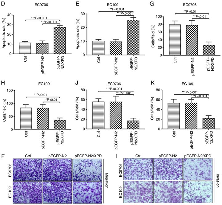
2             JIAN et al: XPD INHIBITS CELL GROWTH AND INVASION AND ENHANCES CHEMOSENSITIVITY IN ESCC

altered. Genetic mutation is the most common cause of DNA         Hospital of Nanchang University (Nanchang, China) between
damage. Xeroderma pigmentosum complementation group D             September, 2018 and December, 2018. The 20 ESCC cases
(XPD) can regulate the transcription initiation and cleavage      were obtained from 14 males and 6 females aged 41‑77 years.
repair of damaged nucleotide sequences and maintain the           No patient had received radiotherapy or chemotherapy prior
biological process in a normal and orderly fashion (6). XPD       to the endoscopic biopsy. The present study was approved by
plays an important role in the repair of damage caused by         the Human Ethics Committee of Third Affiliated Hospital of
oxidative stress (7). A previous study found that XPD gene        Nanchang University and prior written consent was obtained
polymorphism increases the risk of lung cancer in residents       from all patients.
of coal mines (8). XPD polymorphisms are associated with
the development of pre oral cancer as well as oral cancer and     Cells and cell culture. The human ESCC cell lines, EC9706
its clinical course (9). It has been found that XPD can inhibit   and EC109, were obtained from the American Type Culture
the proliferation and migration of hepatocellular carcinoma       Collection (ATCC). All cells were cultured in DMEM supple‑
cells (10). However, the biological roles of XPD in ESCC          mented with 10% FBS, 100 U/ml penicillin and 100 µg/ml
remain unclear.                                                   streptomycin and incubated in a humidified incubator at 37˚C,
    In the present study, the mechanisms through which XPD        5% CO2 and 95% air.
participates in the tumorigenesis and progression of ESCC
were investigated. An XPD gene‑encoding plasmid was trans‑        Cell transfection. The EC9706 or EC109 Cells were divided
fected into ESCC cell lines (EC9706 and EC109 cells), and         into 3 groups as follows: i) The untransfected control group
changes in the molecular biological behavior of EC9706 or         (Ctrl); ii) pEGFP‑N2 empty plasmid transfection group
EC109 cells were observed. In addition, the molecular mecha‑      (pEGFP‑N2); and iii) the pEGFP‑N2/XPD plasmid transfection
nisms underlying the XPD‑mediated regulation of ESCC cell         group (pEGFP‑N2/XPD). The detailed procedures for trans‑
growth and invasion were investigated.                            fection were described in a previous study by the authors (12).
                                                                  At 48 h following the transfection of the pEGFP‑N2 or
Materials and methods                                             pEGFP‑N2/XPD plasmids, green fluorescence was observed
                                                                  under a fluorescence microscope (Olympus Corp.). Western
Reagents. Dulbecco's modified Eagle's medium (DMEM)               blot analysis and RT‑qPCR were used to detect the protein and
and fetal bovine serum (FBS) were purchased from                  mRNA expression levels of XPD.
Gibco; Thermo Fisher Scientific, Inc. The vacant vector
plasm id pEGF P‑N2 a nd the recombina nt plasm id                 Cell proliferation assay. At 0, 24, 48, 72 or 96 h following trans‑
pEGFP‑N2/XPD were generously provided by Jiangxi                  fection with XPD plasmid, the EC9706 or EC109 cells were
Provincial Key Laboratory of Molecular Medicine and these         seeded into 96‑well culture plates at a density of 4x10³ cells/well.
plasmids have been described in previous studies (10,11).         In addition, for the pEGFP‑N2/XPD + LY294002 group,
Lipofectamine™ 2000 and TRIzol reagent were purchased             the EC9706 or EC109 cells were treated with 10 µmol/l of
from Invitrogen; Thermo Fisher Scientific, Inc. PCR primers       LY294002 for 0, 24, 48, 72 or 96 h following transfection with
were synthesized by Sangon Biotech. The total protein             XPD plasmid. The cells in each group were washed with PBS
extraction kit was purchased from AmyJet Scientific Inc.          and incubated with 100 µl CCK‑8 solution for 1 h at 37˚C.
The reverse transcription kit was purchased from Fermentas;       The absorbance at 450 nm was measured using a microplate
Thermo Fisher Scientific, Inc. The Annexin V‑FITC/PI kit          reader (Thermo Fisher Scientific, Inc.). Each independent
was purchased from Vazyme Biotech. The Cell Counting              experiment was performed 3 times. Data were calculated as
kit‑8 was purchased from Solarbio Science Technology.             the means ± SD.
Transwell chambers were purchased from BD Biosciences.
Anti‑XPD (ab54676) primary antibody was purchased                 Cell apoptosis assay. The EC9706 or EC109 cells in each
from Abcam. Primary antibodies against phosphoinositide           group were trypsinized and collected by centrifugation
3‑kinase (PI3K; #4249), AKT (#4685), p‑AKT (Ser473)               at 37˚C for 5 min at a speed of 1,000 x g. A total of 1x10⁵ cells
(#4060), Bcl‑2 (#15071), p21 (#2947), p‑p65 (Ser536) (#3033),     were then resuspended in 500 µl of buffer and incubated with
p65 (#8242), p‑signal transducer and activator of transcrip‑      Annexin V‑FITC/PI kit for 15 min at room temperature. The
tion 3 (STAT3; Tyr705) (#9145), STAT3 (#12640), p‑p38             apoptotic rate of EC9706 or EC109 cells was detected using
mitogen‑activated protein kinase (MAPK; Thr180/Tyr182)            a flow cytometer (FACSCalibur, BD Biosciences). Each
(#4511), p38 MAPK (#8690) and β ‑actin (#4970) were               experiment was performed in triplicate independently.
purchased from Cell Signaling Technology, Inc. Horseradish
peroxidase‑conjugated secondary antibodies (ZB‑2305 and           Cell Transwell migration and invasion assays. Cell migration
ZB‑2306) were purchased from Beijing Zhongshan Golden             and invasion assays were performed using Transwell cham‑
Bridge Biotechnology Co. Ltd. Cisplatin, fluorouracil and         bers without or with Matrigel according to the manufacturer's
LY294002 were purchased from MedChemExpress (MCE).                instructions and as previously described (12). A total of 2x10⁵
                                                                  EC9706 or EC109 cells were seeded into the upper chamber
Clinical specimens. A total of 20 ESCC tissue samples and         of the insert in serum‑free DMEM. The lower chamber of
adjacent normal esophageal tissue samples (>5 cm away             the insert contained DMEM supplemented with 10% FBS
from the tumor) were collected from patients who underwent        as a chemoattractant. Following incubation in a humidified
gastroscopy, endoscopic biopsy and pathological diagnosis         incubator at 37˚C 5% CO2 and 95% air for 48 h, EC9706 or
at the Department of Gastroenterology of Third Affiliated         EC109 cells remaining on the insert's top layer were wiped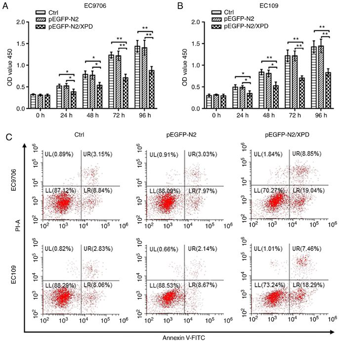
INTERNATIONAL JOURNAL OF MOlecular medicine                                                  3

off with a cotton swab. Cells that migrated or invaded to the      Table I. Primer sequences used for RT‑qPCR.
lower surface of the membrane were stained with crystal violet
(Beijing Solarbio Science & Technology Co., Ltd.) for 20 min       Gene                         Primer sequences
at room temperature and imaged under an inverted light
microscope (x50 magnification). Cells in 5 fields were counted     XPD            F: 5'‑TCTGCCTCTGCCCTATGAT‑3'
to calculate the cell migration or invasion. Each experiment                      R: 5'‑CGATTCCCTCGGACACTTT‑3'
was performed in triplicate.                                       PI3K           F: 5'‑TGGCCTTAGCTCTTAGCCAAACAC‑3'
                                                                                  R: 5'‑ATTGGAACACGGCCTTTGACA‑3'
Chemosensitivity assay. At 24 h following transfection with        AKT            F: 5'‑CTGTGCCTATGCTGCCCAT‑3'
the XPD plasmid, EC9706 or EC109 cells were seeded into                           R: 5'‑CAGTGCGATGTCGTGGAGG‑3'
96‑well plates at a density of 4x10³ cells/well. The medium        Bcl‑2          F: 5'‑GGATAACGGAGGCTGGGATGC‑3'
was then discarded and the cells were incubated at 37˚C in                        R: 5'‑GACTTCACTTGTGGCCCAGAT‑3'
the presence of 0, 20, 40, 80 and 100 µg/ml cisplatin or fluoro‑   c‑Myc          F: 5'‑TGTGTTACGGTCGCGTCTTT‑3'
uracil for 72 h. EC9706 or EC109 cell sensitivity to cisplatin                    R: 5'‑AACAGCTCGGTCACCATCTC‑3'
or fluorouracil in each group was detected by CCK‑8 assay as
                                                                   p21            F: 5'‑GACCTGTCACTGTCTTGTAC‑3'
described above. Cell viability (%) was calculated by using the
                                                                                  R: 5'‑CTCTCATTCAACCGCCTAG‑3'
following formula: OD value (0, 20, 40, 80, or 100 µg/ml)/OD
value (0 µg/ml) x100%.                                             Cyclin D1      F: 5'‑CCAGACCCACGTTTCTTTGC‑3'
                                                                                  R: 5'‑ATCCCTAGAAACACCACGGC‑3'
RT‑qPCR. Total RNA was isolated from all tissue samples,           VEGF           F: 5'‑ ACCCACTCACTGGCTGTTT‑3'
EC9706, or EC109 cells according to the standard TRIzol                           R: 5'‑CGGGCTCTGAGATGTTCAG‑3'
method. A total of 1 µg RNA was used as a template for cDNA        MMP‑9          F: 5'‑ TAGACGCTGCTCCCCTCA‑3'
synthesis using a reverse transcription kit at 37˚C for 15 min                    R: 5'‑GCGGGTGTAACCATAGCG‑3'
and 85˚C for 30 sec. XPD, PI3K, AKT, Bcl‑2, c‑Myc, p21,            β‑actin        F: 5'‑AAGGTGACAGCAGTCGGTT‑3'
Cyclin D1, vascular endothelial growth factor (VEGF), matrix                      R: 5'‑TGTGTGGACTTGGGAGAGG‑3'
metalloproteinase (MMP)‑9 and β‑actin primers were designed
using Primer Premier 5.0 software. The primer sequences            XPD, xeroderma pigmentosum complementation group D; PI3K,
are presented in Table I. Quantitative PCR was performed           phosphoinositide 3‑kinase; VEGF, vascular endothelial growth
using TB Green Premix Ex Taq (Takara Bio, Inc.) under the          factor; MMP‑9, matrix metalloproteinase 9, F, forward, R, reverse.
following thermal cycling conditions: Initial denaturation
at 95˚C for 5 min; subsequent 40 cycles of 95˚C for 30 sec and
60˚C for 30 sec. The relative expression mRNA levels of XPD,
PI3K, AKT, Bcl‑2, c‑Myc, p21, Cyclin D1, VEGF and MMP‑9            among groups and the LSD post hoc test. The t‑test was used
were calculated using the 2‑ΔΔCq (13) method. β‑actin was used     to determine differences between 2 groups. Differences in the
as an endogenous control. All reactions were repeated 3 times.     expression levels of XPD in ESCC tissue samples were evalu‑
                                                                   ated using a Fisher's exact test or Pearson's χ2 test. Differences
Western blot analysis. Total protein of all tissue samples,        at P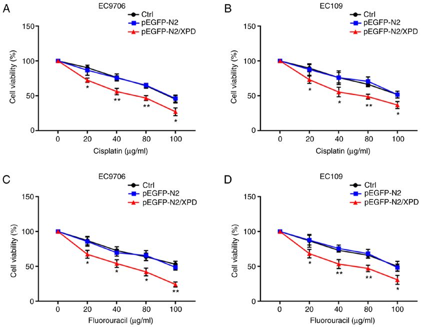
4 JIAN et al: XPD INHIBITS CELL GROWTH AND INVASION AND ENHANCES CHEMOSENSITIVITY IN ESCC
Figure 1. XPD expression in ESCC and adjacent normal esophageal tissues. The expression levels of XPD (A) mRNA and (B and C) protein in ESCC tissue
samples and adjacent normal esophageal tissue samples were detected by RT‑qPCR and western blot analysis, respectively. Data represent the means ± standard
deviation. ESCC, esophageal squamous cell carcinoma; XPD, xeroderma pigmentosum complementation group D; Adjacent NE, adjacent normal esophagus.
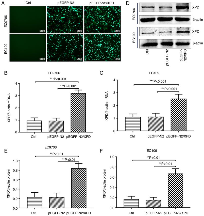
Table II. Association between XPD expression and clinico‑ XPD in EC9706 cells or EC109 cells of the pEGFP‑N2/XPD
pathological factors of ESCC patients. group were markedly upregulated.
XPD XPD overexpression suppresses the proliferation of EC9706
expression cells and EC109 cells. Cell viability was used to investi‑
No. of ‑‑‑‑‑‑‑‑‑‑‑‑‑‑‑‑‑‑‑‑‑‑ gate whether XPD overexpression affects the proliferation
Factors patients Low High P‑value of EC9706 or EC109 cells. At 24, 48, 72 or 96 h following
transfection with XPD plasmid, the optical density (OD)
Sex 0.354 value of the pEGFP‑N2/XPD group was significantly reduced
Male 14 8 6 compared to the that of the control (Ctrl) group and pEGFP‑N2
Female 6 5 1 group (PINTERNATIONAL JOURNAL OF MOlecular medicine 5 Figure 2. XPD expression is upregulated following transfection with pEGFP‑N2/XPD plasmid. (A) Green fluorescent protein was observed after pEGFP‑N2 or pEGFP‑N2/XPD plasmid transfection (x100 magnification). The expression levels of XPD (B and C) mRNA and (D‑F) protein in EC9706 or EC109 cells were detected by RT‑qPCR and western blot analysis, respectively. **P
6                JIAN et al: XPD INHIBITS CELL GROWTH AND INVASION AND ENHANCES CHEMOSENSITIVITY IN ESCC

Figure 3. Effects of XPD overexpression on the proliferation, apoptosis, migration and invasion of EC9706 and EC109 cells. (A and B) CCK‑8 assay was
used to detect the proliferation of EC9706 or EC109 cells. (C‑E) Flow cytometry was used to detect the apoptosis of EC9706 or EC109 cells. The effects
of XPD overexpression on the migration (F‑H) and the invasion (I-K) of EC9706 or EC109 cells were examined by Transwell assay. Data represent the
means ± standard deviation. *P
INTERNATIONAL JOURNAL OF MOlecular medicine                                                             7

Figure 4. Upregulation of XPD enhances the chemosensitivity of EC9706 and EC109 cells to cisplatin or fluorouracil. The effects of XPD upregulation
on the chemosensitivity of EC9706 and EC109 cells to (A and B) cisplatin or (C and D) fluorouracil were determined by CCK‑8 assay. Date represent the
means ± standard deviation. *P
8                JIAN et al: XPD INHIBITS CELL GROWTH AND INVASION AND ENHANCES CHEMOSENSITIVITY IN ESCC

Figure 5. Effects of XPD overexpression on multiple signaling pathways in EC9706 cells and EC109 cells. (A and B) The mRNA expression levels of
PI3K, AKT, Bcl‑2, c‑Myc, Cyclin D1, VEGF, MMP‑9 and p21 in EC9706 or EC109 cells were detected by RT‑qPCR. *P
INTERNATIONAL JOURNAL OF MOlecular medicine                                                     9

as surgical resection, chemotherapy, and radiotherapy have             In conclusion, the findings of the present study demon‑
been applied for ESCC treatment, the prognosis of ESCC             strate that the upregulation of XPD inhibits the proliferation,
remains poor (22‑24). Therefore, it is of utmost importance to     abrogates the migration and invasion, and promotes the apop‑
better understand the molecular mechanisms responsible for         tosis of EC9706 and EC109 cells by inhibiting the PI3K/AKT
the development of ESCC and to explore novel therapies with        signaling pathway. XPD overexpression also enhanced the
which to improve the survival of patients with ESCC.               chemosensitivity of EC9706 and EC109 cells to cisplatin or
    XPD is located on 19q13.2‑q13.3 and encodes an                 fluorouracil. Based on the results of the present study, XPD
ATP‑dependent DNA helicase (25). XPD751 polymorphism               may thus become a potential target for ESCC treatment and
has been shown to be associated with the occurrence and devel‑     drug resistance in the future.
opment of a wide range of malignancies, such as esophageal
cancer, gastric cancer, and colorectal cancer (26‑28). Previous    Acknowledgements
studies have demonstrated that XPD expression serves as a
tumor suppressor in HCC (10,11,29). In a present study by the      The authors would like to thank Dr Bin Li, Dr Jian‑Bin Qin
authors, it was found that the mRNA and protein expression         and Dr Feng Deng (Department of Gastroenterology, Third
levels of XPD in ESCC tissue samples were significantly lower      Affiliated Hospital of Nanchang University) for providing
than those in the adjacent normal esophageal tissue samples.       their valuable assistance with this research.
To determine the role of XPD in ESCC, the in vitro cellular
effects of XPD overexpression in ESCC were investigated            Funding
through XPD transfection into EC9706 and EC109 cells. In
the present study, it was demonstrated that XPD gene overex‑       The present study was supported by the National Natural
pression significantly reduced the proliferation and inhibited     Science Foundation of China (grant. no. 81660408) and
the migration and invasion of EC9706 or EC109 cells, whilst        the Health and Family Planning Commission Science and
increasing cell apoptosis. Additionally, the upregulation of       Technology Plan of Jiangxi Province (grant. no. 20184002).
XPD gene enhanced the chemosensitivity of EC9706 and
EC109 cells to cisplatin or fluorouracil.                          Availability of data and materials
    Previous studies have indicated that the PI3K/AKT
signaling pathway plays an important role in the occurrence,       The datasets used in the present study can be obtained from
development and invasion of malignant tumors, such as              the corresponding author upon reasonable request.
esophageal cancer, colon cancer and gastric cancer (30‑32). A
previous study by the authors demonstrated that the inhibition     Authors' contributions
of the activation of AKT and the promotion of the expression of
p21 inhibited cell proliferation and promotes cell apoptosis in    JJ, SL and NF designed the experiments. JJ, SL, LZL, LZ, LY,
hepatocellular carcinoma (12). In the present study, XPD was       LHG and YQH performed the experiments. SL contributed to
shown to be involved in the phosphorylation of AKT. Following      the data analysis. JJ wrote the manuscript and conducted the
XPD upregulation, the protein expression level of p‑AKT was        revision of the manuscript. NF was responsible for the final
significantly decreased, indicating that XPD overexpression        modification of the manuscript. All authors read and approved
may inhibit the activation of AKT and suppress PI3K/AKT            the final manuscript.
signal transduction. p21 has been demonstrated to be involved
in cell cycle progression and apoptosis, as well as in behaviors   Ethics approval and consent to participate
essential for tumorigenesis and tumor progression (33). The
present study also demonstrated that the mRNA and protein          The present study was approved by the Human Ethics
expression levels of p21 were significantly upregulated            Committee of Third Affiliated Hospital of Nanchang University
following the overexpression of XPD. Previous studies have         and prior written consent was obtained from all patients.
demonstrated that c‑Myc, Cyclin D1 and Bcl‑2 play crucial
roles in regulating tumorigenesis and are significantly upregu‑    Patient consent for publication
lated during tumor progression (34‑36). The present study also
revealed that the expression levels of c‑Myc, Cyclin D1 and        Not applicable.
Bcl‑2 were significantly downregulated following the over‑
expression of XPD. Previous studies have demonstrated that         Competing interests
the functions of VEGF and MMP‑9 are essential for tumor
invasion (37,38). The present study also demonstrated that the     The authors declare that they have no competing interests.
mRNA expression levels of VEGF and MMP‑9 were both
significantly decreased following the overexpression of XPD.       References
As shown by the results of the present study, the expression
level of XPD was low in ESCC tissues, EC9706 cells, or EC109        1. Bray F, Ferlay J, Soerjomataram I, Siegel RL, Torre LA and
cells; thus, XPD overexpression experiments were conducted             Jemal A: Global cancer statistics 2018: GLOBOCAN estimates
                                                                       of incidence and mortality worldwide for 36 cancers in 185 coun‑
and this is the reason that XPD knockdown experiments were             tries. CA Cancer J Clin 68: 394‑424, 2018.
not conducted in the EC9706 or EC109 cells. As such, the fact       2. Yutong H, Xiaoli X, Shumei L, Shan S, Di L and Baoen S:
that there no XPD knockdown experiments were performed is              Increased neutrophil‑lymphocyte ratio is a poor prognostic factor
                                                                       in patients with esophageal cancer in a high incidence area in
a limitation of the present study.                                     China. Arch Med Res 46: 557‑563, 2015.
10 JIAN et al: XPD INHIBITS CELL GROWTH AND INVASION AND ENHANCES CHEMOSENSITIVITY IN ESCC
3. Toh Y, Oki E, Ohgaki K, Sakamoto Y, Ito S, Egashira A, Saeki H, 22. Hong H, Jie H, Liyu R, Zerui C, Borong S and Hongwei L:
Kakeji Y, Morita M, Sakaguchi Y, et al: Alcohol drinking, ciga‑ Prognostic significance of middle paraesophageal lymph node
rette smoking, and the development of squamous cell carcinoma metastasis in resectable esophageal squamous cell carcinoma: A
of the esophagus: Molecular mechanisms of carcinogenesis. Int STROBE‑compliant retrospective study. Medicine (Baltimore) 98:
J Clin Oncol 15: 135‑144, 2010. e17531, 2019.
4. Moon DH, Jeon JH, Yang HC, Kim YI, Lee JY, Kim MS, Lee JM 23. Zhao Y, Han L, Zhang W, Shan L, Wang Y, Song P, Peng C and
and Lee GK: Intramural metastasis as a risk factor for recurrence Zhao X: Preoperative chemotherapy compared to postoperative
in esophageal squamous cell carcinoma. Ann Thorac Surg 106: adjuvant chemotherapy for squamous cell carcinoma of the
249‑256, 2018. thoracic oesophagus with the detection of circulation tumour
5. Wu H, Chen S, Yu J, Li Y, Zhang XY, Yang L, Zhang H, Hou Q, cells randomized controlled trial. Int J Surg 73: 1‑8, 2020.
Jiang M, Brunicardi FC, et al: Single‑cell transcriptome analyses 24. Zhang Z, Xu L, Di X, Zhang C, Ge X and Sun X: A retrospective
reveal molecular signals to intrinsic and acquired paclitaxel study of postoperative radiotherapy for locally advanced esopha‑
resistance in esophageal squamous cancer cells. Cancer Lett 420: geal squamous cell carcinoma. Ann Palliat Med 8: 708‑716, 2019.
156‑167, 2018. 25. Guan Q, Chen Z, Chen Q and Zhi X: XRCC1 and XPD
6. Oksenych V and Coin F: The long unwinding road: XPB and polymorphisms and their relation to the clinical course in hepa‑
XPD helicases in damaged DNA opening. Cell Cycle 9: 90‑96, tocarcinoma patients. Oncol Lett 14: 2783‑2788, 2017.
2010. 26. Yoon HH, Catalano PJ, Murphy KM, Skaar TC, Philips S,
7. Lerner LK, Moreno NC, Rocha CRR, Munford V, Santos V, Powell M, Montgomery EA, Hafez MJ, Offer SM, Liu G, et al:
Soltys DT, Garcia CCM, Sarasin A and Menck CFM: Genetic variation in DNA‑repair pathways and response to radio‑
XPD/ERCC2 mutations interfere in cellular responses to oxida‑ chemotherapy in esophageal adenocarcinoma: A retrospective
tive stress. Mutagenesis 34: 341‑354, 2019. cohort study of the eastern cooperative oncology group. BMC
8. Minina VI, Bakanova ML, Soboleva OA, Ryzhkova AV, Cancer 11: 176, 2011.
Titov RA, Savchenko YA, Sinitsky MY, Voronina EN, Titov VA 27. Engin AB, Karahalil B, Engin A and Karakaya AE: DNA repair
and Glushkov AN: Polymorphisms in DNA repair genes in lung enzyme polymorphisms and oxidative stress in a Turkish popula‑
cancer patients living in a coal‑mining region. Eur J Cancer tion with gastric carcinoma. Mol Biol Rep 38: 5379‑5386, 2011.
Prev 28: 522‑528, 2019. 28. Huang MY, Wang JY, Huang ML, Chang HJ and Lin SR:
9. Nigam K, Yadav SK, Samadi FM, Bhatt ML, Gupta S and Polymorphisms in XPD and ERCC1 associated with colorectal
Sanyal S: Risk modulation of oral pre cancer and cancer cancer outcome. Int J Mol Sci 14: 4121‑4134, 2013.
with polymorphisms in XPD and XPG genes in North 29. Zheng JF, Li LL, Lu J, Yan K, Guo WH and Zhang JX: XPD
Indian population. Asian Pac J Cancer Prev 20: 2397‑2403, 2019. functions as a tumor suppressor and dysregulates autophagy in
10. Xiao Z, Wang Y and Ding H: XPD suppresses cell proliferation cultures HepG2 cells. Med Sci Monit 21: 1562‑1568, 2015.
and migration via miR‑29a‑3p‑Mdm2/PDGF‑B axis in HCC. 30. Javadinia SA, Shahidsales S, Fanipakdel A, Mostafapour A,
Cell Biosci 9: 6, 2019. Joudi‑Mashhad M, Ferns GA and Avan A: The esophageal cancer
11. Ding H, Xu JJ, Huang Y, Du FT and Zhang JX: XPD could and the PI3K/AKT/mTOR signaling regulatory microRNAs:
suppress growth of HepG2.2.15 and down‑regulate the expres‑ A novel marker for prognosis, and a possible target for immuno‑
sion of hepatitis B virus x protein through P53 pathway. Biochem therapy. Curr Pharm Des 24: 4646‑4651, 2018.
Biophys Res Commun 419: 761‑767, 2012. 31. Han B, Jiang P, Li Z, Yu Y, Huang T, Ye X and Li X:
12. Huang DH, Jian J, Li S, Zhang Y and Liu LZ: TPX2 silencing Coptisine‑induced apoptosis in human colon cancer cells
exerts anti‑tumor effects on hepatocellular carcinoma by regu‑ (HCT‑116) is mediated by PI3K/Akt and mitochondrial‑associ‑
lating the PI3K/AKT signaling pathway. Int J Mol Med 44: ated apoptotic pathway. Phytomedicine 48: 152‑160, 2018.
2113‑2122, 2019. 32. Huang Y, Zhang J, Hou L, Wang G, Liu H, Zhang R, Chen X and
13. Livak KJ and Schmittgen TD: Analysis of relative gene expres‑ Zhu J: LncRNA AK023391 promotes tumorigenesis and invasion
sion data using real‑time quantitative PCR and the 2(‑Delta Delta of gastric cancer through activation of the PI3K/Akt signaling
C(T)) method. Methods 25: 402‑408, 2001. pathway. J Exp Clin Cancer Res 36: 194, 2017.
14. Wang L, Zhang Z, Yu X, Li Q, Wang Q, Chang A, Huang X, 33. Parveen A, Akash MS, Rehman K and Kyunn WW: Dual role
Han X, Song Y, Hu J, et al: SOX9/miR‑203a axis drives of p21 in the progression of cancer and its treatment. Crit Rev
PI3K/AKT signaling to promote esophageal cancer progression. Eukaryot Gene Expr 26: 49‑62, 2016.
Cancer Lett 468: 14‑26, 2020. 34. Zhang D, Qi J, Liu R, Dai B, Ma W, Zhan Y and Zhang Y: c‑Myc
15. Zheng TL, Li DP, He ZF and Zhao S: MiR‑145 sensitizes esopha‑ plays a key role in TADs‑induced apoptosis and cell cycle arrest
geal squamous cell carcinoma to cisplatin through directly in human hepatocellular carcinoma cells. Am J Cancer Res 5:
inhibiting PI3K/AKT signaling pathway. Cancer Cell Int 19: 250, 1076‑1088, 2015.
2019. 35. Cheng G, Zhang L, Lv W, Dong C, Wang Y and Zhang J:
16. Zhou J, Zheng S, Liu T, Liu Q, Chen Y, Tan D, Ma R and Lu X: Overexpression of cyclin D1 in meningioma is associated with
MCP2 activates NF‑κ B signaling pathway promoting the migra‑ malignancy grade and causes abnormalities in apoptosis, inva‑
tion and invasion of ESCC cells. Cell Biol Int 42: 365‑372, 2018. sion and cell cycle progression. Med Oncol 32: 439, 2015.
17. Lu Z, Lu C, Li C, Jiao Y, Li Y and Zhang G: Dracorhodin 36. Alam M, Kashyap T, Pramanik KK, Singh AK, Nagini S and
perchlorate induces apoptosis and G2/M cell cycle arrest in Mishra R: The elevated activation of NFκ B and AP‑1 is corre‑
human esophageal squamous cell carcinoma through inhibition lated with differential regulation of Bcl‑2 and associated with
of the JAK2/STAT3 and AKT/FOXO3a pathways. Mol Med oral squamous cell carcinoma progression and resistance. Clin
Rep 20: 2091‑2100, 2019. Oral Investig 21: 2721‑2731, 2017.
18. Wang WW, Zhao ZH, Wang L, Li P, Chen KS, Zhang JY, Li WC, 37. Su F, Geng J, Li X, Qiao C, Luo L, Feng J, Dong X and Lv M: SP1
Jiang GZ and Li XN: MicroRNA‑134 prevents the progression of promotes tumor angiogenesis and invasion by activating VEGF
esophageal squamous cell carcinoma via the PLXNA1‑mediated expression in an acquired trastuzumab‑resistant ovarian cancer
MAPK signalling pathway. EBioMedicine 46: 66‑78, 2019. model. Oncol Rep 38: 2677‑2684, 2017.
19. Ferlay J, Soerjomataram I, Dikshit R, Eser S, Mathers C, 38. Zhao G, Zhang H, Huang Z, Lv L and Yan F: Cortactin and Exo70
Rebelo M, Parkin DM, Forman D and Bray F: Cancer incidence mediated invasion of hepatoma carcinoma cells by MMP‑9
and mortality worldwide: Sources, methods and major patterns secretion. Mol Biol Rep 43: 407‑414, 2016.
in GLOBOCAN 2012. Int J Cancer 136: E359‑E386, 2015.
20. Wiedmann MW and Mössner J: New and emerging combination
therapies for esophageal cancer. Cancer Manag Res 5: 133‑146, This work is licensed under a Creative Commons
2013. Attribution-NonCommercial-NoDerivatives 4.0
21. Ohashi S, Miyamoto S, Kikuchi O, Goto T, Amanuma Y and International (CC BY-NC-ND 4.0) License.
Muto M: Recent advances from basic and clinical studies of esoph‑
ageal squamous cell carcinoma. Gastroenterology 149: 1700‑1715,
2015.You can also read