Ulinastatin alleviates traumatic brain injury by reducing endothelin-1
←
→
Page content transcription
If your browser does not render page correctly, please read the page content below
Translational Neuroscience 2021; 12: 1–8
Research Article
Ting Liu, Xing-Zhi Liao*, Mai-Tao Zhou*
Ulinastatin alleviates traumatic brain injury by
reducing endothelin-1
https://doi.org/10.1515/tnsci-2021-0001
received June 22, 2020; accepted November 3, 2020
1 Introduction
Abstract Traumatic brain injury (TBI) can lead to serious debili-
Background ‒ Brain edema is one of the major causes tating complications and fatality [1]. Brain edema is one
of fatality and disability associated with injury and neuro- of the major secondary reactions in TBI resulting in water
surgical procedures. The goal of this study was to eval- accumulation in extracellular or intracellular spaces within
uate the effect of ulinastatin (UTI), a protease inhibitor, the brain, in turn resulting in elevated intracranial pressure.
on astrocytes in a rat model of traumatic brain injury (TBI). Surgical interventions to cure brain edema have limited
Methodology ‒ A rat model of TBI was established. benefit, hence necessitating other methods of treating brain
Animals were randomly divided into 2 groups – one group edema and TBI.
was treated with normal saline and the second group was Cellular edema in the astrocytes and/or vasogenic
treated with UTI (50,000 U/kg). The brain water content and edema related to disruption of the blood–brain barrier
permeability of the blood–brain barrier were assessed in the (BBB) comprises brain edema and can ultimately lead
two groups along with a sham group (no TBI). Expression of to neuronal cell death [2,3]. Disruption of the BBB due
the glial fibrillary acidic protein, endthelin-1 (ET-1), vascular to brain edema has been shown to result in the develop-
endothelial growth factor (VEGF), and matrix metalloprotei- ment of neurodegenerative diseases such as traumatic
nase 9 (MMP-9) were measured by immunohistochemistry
epilepsy and Parkinson’s disease [4,5].
and western blot. Effect of UTI on ERK and PI3K/AKT sig-
Ulinastatin (UTI) has been increasingly used as a
naling pathways was measured by western blot.
protease inhibitor for organ protection [6]. UTI mainly
Results ‒ UTI significantly decreased the brain water
functions by scavenging oxygen free radicals, inhibiting
content and extravasation of the Evans blue dye. This
inflammatory reactions, and immune regulation [7]. How-
attenuation was associated with decreased activation of
ever, there are few studies on the role and mechanism
the astrocytes and ET-1. UTI treatment decreased ERK
of UTI in the central nervous system, especially in TBI.
and Akt activation and inhibited the expression of pro-
UTI was shown to significantly reduce plasma C reactive
inflammatory VEGF and MMP-9.
protein and alleviate brain tissue damage in patients
Conclusion ‒ UTI can alleviate brain edema resulting from
with severe craniocerebral injury [8]. UTI was shown to
TBI by inhibiting astrocyte activation and ET-1 production.
exert protective effect against brain injury and edema by
Keywords: ulinastatin, traumatic brain injury, brain decreasing the expression and secretion of the pro-inflam-
edema, astrocyte, endothelin-1 matory cytokines, IL-1β and TNF-α, in brain tissue and
serum of rats with TBI. TBI-associated brain edema is con-
nected with increased expression of aquaporin-4 at the site
of injury [5]; however, how aquaporin-4 contributes to the
pathology is not known. However, UTI has been shown to
* Corresponding author: Xing-Zhi Liao, The 904th Hospital of the
Joint Logistic Support Force of PLA, Wuxi, China, inhibit the expression of aquaporin 4 (AQP4) and alleviate
e-mail: liaoxingzhi@aliyun.com brain edema [10,11]. These studies also indicate that UTI is
* Corresponding author: Mai-Tao Zhou, The 904th Hospital of the BBB-permeable.
Joint Logistic Support Force of PLA, Wuxi, China, Cerebral vasospasm caused by traumatic subarach-
e-mail: zhoumaitao_904h@126.com
noid hemorrhage and facilitated by the vasoconstrictor
Ting Liu: Jiangsu Province Key Laboratory of Anesthesiology, Xuzhou
Medical University, Xuzhou, China; The 904th Hospital of the Joint
endothelin-1 (ET-1) results in secondary cerebral ischemic
Logistic Support Force of PLA, Wuxi, China, edema after brain injury [12]. Intervention with UTI in cra-
e-mail: 617396382@qq.com niocerebral injury was shown to plasma levels of ET-1,
Open Access. © 2021 Ting Liu et al., published by De Gruyter. This work is licensed under the Creative Commons Attribution 4.0 International
License.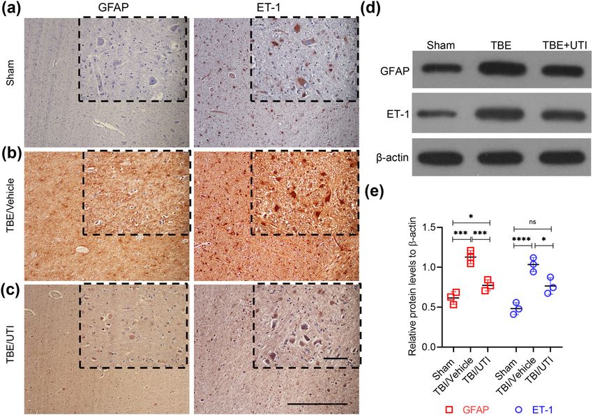
2 Ting Liu et al.
resulting in improvement of hypoperfusion status, cere- 2.3 UTI injection and experimental grouping
bral ischemia and hypoxia, and vascular endothelial func-
tion [13]. Astrocytes are the main type of glial cells, Twenty-eight rats undergoing TBI were randomly divided
accounting for more than 50% of the total volume of brain into 2 groups (n = 14 each). The sham group also had
cells. ET-1 produced by activated astrocytes binds to its another 14 rats. The TBI rats were either injected intra-
cognate receptor expressed on astrocytes and causes neu- peritoneally with UTI (Tianpu Biochemical Pharmaceutical
roinflammation [14], potentiated by increased expression Co., Ltd, Guangdong, China) at a dose of 50,000 U/kg
of matrix metalloproteinase-9 (MMP-9) and vascular (TBI/UTI) [15] or normal saline (TBI/vehicle) immediately
endothelial growth factor-A (VEGF-A) in astrocytes, ulti- after induction of TBI. The animals were sacrificed after
mately resulting in brain edema [15]. In this study, we 24 h and brain tissue was resected.
investigated whether UTI alleviates the severity of TBI by
suppressing the astrocyte activity and limiting the pro-
duction of ET-1 and inflammatory mediators.
2.4 Determination of water content in brain
tissue
2 Methods After 24 h, the resected brain tissues (n = 3 per experi-
mental group) were immediately weighed (wet weight)
2.1 Establishment of rat model of TBI and then dried in an oven at 105°C for 3 days and weighed
again (dry weight). The water content of brain tissue was
calculated as (wet weight − dry weight)/wet weight ×
Male Sprague-Dawley rats (250–300 g) were housed in 100%.
humidity- and temperature-controlled housing facility
with a 12 h light/dark cycle and free access to rodent
chow and water supply. The TBI model was generated
using previously described protocol [9]. Rats were 2.5 Assessment of permeability of BBB
anesthetized with 2% pentobarbital sodium (30 mg/kg). using Evans blue dye
After fixing in the prone position, a midline skin incision
was made. A trephine bit was then used to generate a Two percent Evans blue dye (Sigma, LA, USA) was
4 mm craniotomy in the right parietal bone between injected into the right femoral vein of rats (n = 5 per
the lambda and bregma, 2 mm dextrolateral to the experiment group) at a dose of 25 mg/kg, after 24 h, and
sagittal suture. The bumper was placed outside the allowed to circulate for 1 h. Phosphate buffered saline
dura mater, the bumper protruded 3 mm from the spiral (0.1 mol/L) was then perfused transcardially. The resected
cylinder, and the brain was contused and lacerated brain tissues were subsequently homogenized in PBS and
with a 100 g weight from a height of 30 cm, approxi- centrifuged for 30 min at 15,000g. An equal volume of
mately equivalent to a moderate TBI in human. The trichloroacetic acid was added to the supernatant and
craniotomy was closed after the blood was stopped. incubated at 48°C overnight, followed by centrifugation
Animals were only returned to their cage after they at 15,000g for 30 min. Extravasated Evans blue dye in the
were bright, alert, and responsive. Then 2% Evans supernatant was quantified by measuring absorbance at
blue liquid was injected into the right femoral vein 615 nm using a spectrophotometer.
at a dose of 25 mg/kg, and rats were sacrificed 24 h
later. The sham group went through the same protocol
except the injury procedure.
2.6 Immunohistochemistry (IHC)
Resected brain tissues (n = 3 per experiment group) were
2.2 Ethical approval fixed by immersion in 4% formaldehyde (Beyotime,
Zhejiang, China) for 60 min at 37°C before paraffin
The research related to animals use has been complied embedding. These samples were then sectioned at 5 µm
with all the relevant national regulations and institu- thickness via microtome (Model HM310, Microm Inc.,
tional policies for the care and use of animals. USA), dewaxed with xylene, and cleared with a series
Ulinastatin reduces ET-1 in astrocyte 3
of changing ethanol concentrations. Blocking was done (1:1,000; Cell Signaling Technology), P-Akt (T308) (1:1,000;
by incubation in 5% bovine serum albumin (Thermo Cell Signaling Technology), Akt (1:1,000; Cell Signaling
Fisher Scientific, Carlsbad, CA, USA) and 0.3% Triton- Technology), and β-Actin (1:6,000; Abcam). The β-actin
X-100 (Thermo Fisher Scientific) for 1 hour at room tem- protein was used as a loading control for data normaliza-
perature (RT). Antibodies used for IHC were glial fibrillary tion. Densitometry analysis was done using the NIH Image J
acidic protein (GFAP) (1:100, Abcam, Cambridge, USA), software.
ET-1 (1:50, Abcam), VEGF-A (1:100, Cell Signaling
Technology, Cambridge, MA, USA), or MMP-9 (1:100,
Cell Signaling Technology). All incubation with primary
antibodies were done in 1 BSA overnight at 4°C. Samples 2.8 Statistical analysis
were viewed at 100× magnification by light microscopy
(BX61, Olympus Optical GmbH, Hamburg, Germany) and Data were presented as mean ± standard error (SD). Data
imaged (Color View II; Soft Imaging System, Olympus were analyzed using one-way analysis of variance (ANOVA)
Optical GmbH, Hamburg, Germany). followed by the Tukey post hoc test. P < 0.05 was considered
as significant.
2.7 Western blot analysis
3 Results
Tissue specimens (n = 3 per experiment group) were
snap-frozen in liquid nitrogen and homogenized using
3.1 UTI alleviates brain edema and prevents
a mortar and pestle. Lysis was done in RIPA buffer
(Beyotime, Zhejiang, China). Protein quantification was disruption of BBB in rat model of TBI
done using the BCA assay kit (Thermo Fisher Scientific,
CA, USA). Proteins were separated on 12% SDS PAGE. Brain water content was significantly increased in rats
Antibodies used to probe the blots were: GFAP (1:1,000; subjected to UTI compared to the sham control (Figure 1a).
Abcam), ET-1 (1:2,000; Abcam), VEGF (1:1,000; Cell Intraperitoneal injection of UTI, administrated immedi-
Signaling Technology), MMP-9 (1:1,000; Cell Signaling ately after TBI induction, significantly attenuated the
Technology), P-ERK1/2 (1:1,000; Cell Signaling Technology), increase in brain water content to levels observed in
ERK1/2 (1:1,000; Cell Signaling Technology), P-Akt (S473) the sham control (Figure 1a). We next evaluated BBB
Figure 1: Administration of UTI immediately after induction of TBI alleviates brain edema and prevents BBB disruption. TBI-induced rats were
randomly divided into two groups – vehicle control (TBI/vehicle) and UTI (TBI/UTI). The latter group was intraperitoneally injected with
50,000 U/kg of UTI. No TBI was induced in the third sham control group. (a) Brain water content was determined in resected brain tissues
after 24 h; (b) the integrity of the BBB was evaluated by Evans blue dye staining; (c) quantification of Evans blue dye extravasation in the
different experimental groups. Error bars in (a) and (c) are standard deviation; **p < 0.01, ***p < 0.001, ****p < 0.0001, ns, not sig-
nificant – one-way ANOVA with Tukey’s post hoc test (n = 3 in (a) and n = 5 in (c)).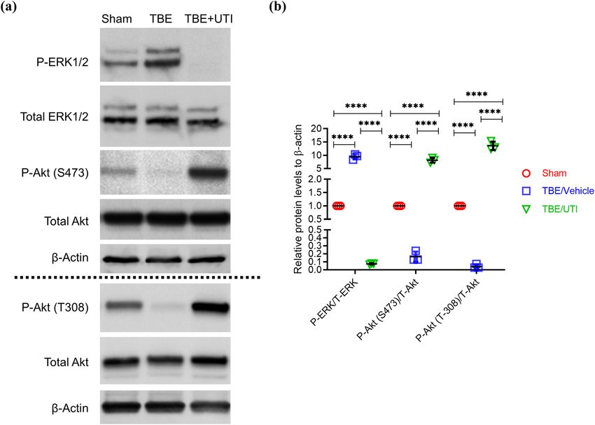
4 Ting Liu et al.
disruption after 24 h using Evans blue dye. Vehicle- western blot analyses were performed. Based on the GFAP
treated rats had significantly increased Evans blue dye level, we found that the activity of astrocytes was signifi-
extravasation in right hemisphere compared with sham cantly higher in TBI/vehicle rats when compared with the
rats (Figure 1b and c). In contrast, TBI rats treated with sham group (Figure 2a, b, d and e). Similarly, the expression
UTI had significantly reduced Evans blue dye extravasa- of ET-1 was also significantly high in the TBI/vehicle group
tion compared with the vehicle-treated TBI rats (Figure 1b compared with the sham group (Figure 2a, b, d and e).
and c). However, Evans blue dye extravasation was still However, UTI treatment resulted in significant inhibition
significantly higher in the UTI-treated TBI rats compared of both activation of astrocytes and ET-1 expression
with the sham control (Figure 1b and c). (Figure 2c–e). Overall, these results indicate that inhibi-
tory effect of UTI on astrocyte activation in TBI rats is
potentially correlated to ET-1 expression.
3.2 UTI inhibits the activity of astrocytes by
suppressing expression of ET-1
expression in TBI rats 3.3 UTI decreases expression of
inflammatory mediators in TBI rats
The GFAP is mainly distributed in the astrocytes. It parti-
cipates in cell cytoskeleton formation and maintains the Inflammatory mediators MMP-9 and VEGF are known to
tensile strength. GFAP is also used as a marker of astrocyte be regulated by ET-1 [15]. Therefore, to verify the fact that
activation [16]. Activated astrocyte exhibit increased expres- the effect of UTI treatment in TBI rats was due to the
sion of ET-1. To assess the levels of GFAP and ET-1, IHC and decreased expression of ET-1, we further assessed the
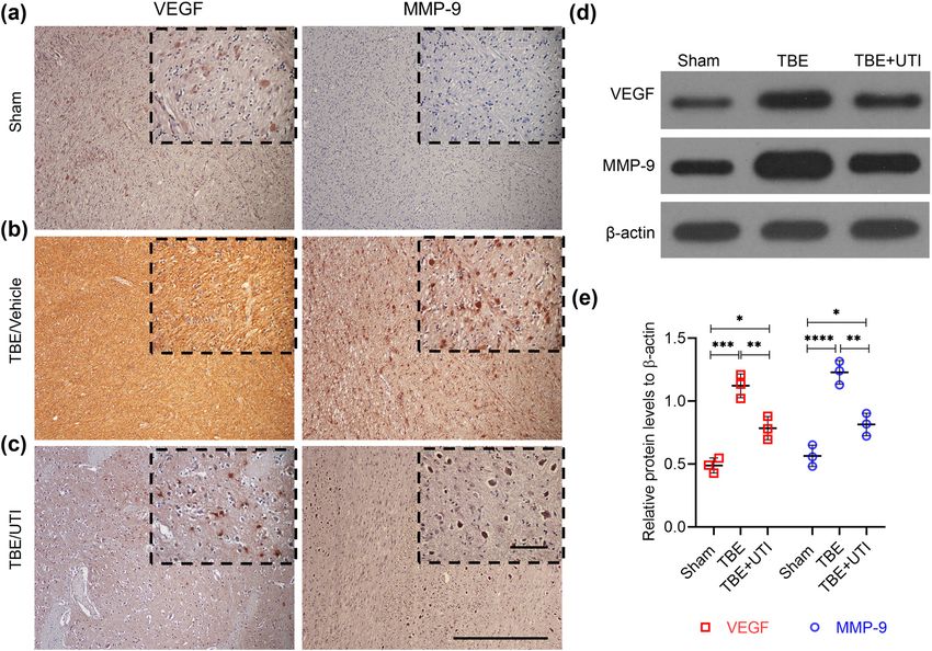
Figure 2: UTI inhibits the activity of astrocytes and decreases expression of ET-1 in the rat brain. (a–d) Representative images of IHC staining
of GFAP and ET-1 on resected brain tissues of the different experimental groups (n = 3, each group). Insets in (a–d) show images obtained under
higher magnification. Scale bar = 30 µm; (d) representative images of immunoblots showing expression of GFAP and ET-1 in the indicated
experimental groups. β-Actin was used as a loading control; (e) quantification of relative expression of GFAP and ET-1 in the indicated experi-
mental groups. Densitometry analysis of blots shown in (b) was done using Image J and data were normalized to expression of β-actin. Error bars
are standard deviation; *p < 0.05, ***p < 0.001, ****p < 0.0001, ns, not significant – one-way ANOVA with Tukey’s post hoc test (n = 3).Ulinastatin reduces ET-1 in astrocyte 5
levels of MMP-9 and VEGF. Both IHC and western blot function by the activation of ERK and inhibition of
analyses revealed that compared with the sham group, the PI3K/Akt pathway [17]. Therefore, we next evalu-
expression of VEGF and MMP-9 was significantly higher ated if induction of ET-1 in TBI rats is accompanied by
in TBI rats (Figure 3a, b, d and e). Administration of UTI the activation of ERK1/2. Results from western blot
resulted in significant decrease in MMP-9 and VEGF analysis show that compared with the sham group,
(Figure 3c–e). These results provide further evidence TBI rats had significantly higher expression of P-ERK/
that UTI-mediated alleviation of brain edema and BBB total ERK (Figure 4a and b). At the same time, using
disruption in TBI rats is mediated by a decrease in astro- levels of P-AKT (Serine 473)/total AKT and P-AKT
cyte activation and ET-1 expression. (Threonine 308)/total AKT, a significant downregula-
tion of PI3K/AKT pathway was noticed in TBI rats
(Figure 4a and b). Administration of UTI resulted in
significant decrease in ERK activation along with
3.4 UTI inhibits inflammatory mediators in an increased activation of the PI3K/AKT pathway
rat astrocytes potentially via inhibiting (Figure 4a and b). Since ERK activation is a read-out
ERK activation and stimulating the PI3K/ of the ET-1 receptor activity, these results provide con-
AKT pathway vincing evidence that UTI alleviates TBI-associated
brain edema and BBB disruption in astrocytes via
Vasoactive peptide ET-1 induces vasoconstriction and pro- downregulating expression of ET-1 and suppressing
liferation in aortic smooth muscle cells and its receptors the activity of the ET-1 receptor.
Figure 3: UTI decreases expression of inflammatory mediators in TBI rats. (a–d) Representative images of IHC staining of VEGF and MMP-9
on resected brain tissues of the different experimental groups (n = 3, each group). Insets in (a–d) show images obtained under higher
magnification. Scale bar = 30 µm; (d) representative images of immunoblots showing expression of VEGF and MMP-9 in the indicated
experimental groups. β-Actin was used as a loading control; (e) quantification of relative expression of VEGF and MMP-9 in the indicated
experimental groups. Densitometry analysis of blots shown in (d) was done using Image J and data were normalized to expression of
β-actin. Error bars are standard deviation; *p < 0.05, **p < 0.01, ***p < 0.001, ****p < 0.0001, ns, not significant – one-way ANOVA with
Tukey’s post hoc test (n = 3).6 Ting Liu et al.
4 Discussion size of lesion [24]. Additionally, matrix metalloprotei-
nases (MMPs) produced by the astrocytes function in dis-
Brain edema, increased intracranial pressure, and dis- rupting the tight junction of BBB and the basement mem-
ruption of BBB are associated to the high incidence of brane, thereby further aggravating the cerebral edema
long-term disabilities and mortality rate in cases of TBI [25]. Recently, BBB injury has also been related to neu-
[11]. Surgical intervention has limited outcomes, necessi- roinflammation [26]. In hypoxic brain tissue, vascular
tating identification of additional therapeutic options. endothelial cell growth inhibitory factor was shown to
Results from the current study indicate that administra- reduce inflammation and stabilize BBB by inhibiting the
tion of UTI might be an attractive regimen to manage TBI. TLR4/NF-κB signaling pathway [27]. Indeed, UTI admin-
BBB is mainly composed of microvascular endothe- istration has been shown to decrease expression of AQ4,
lial cells, astrocytes, microglial cells, pericytes, and base- thus restoring integrity of BBB [9].
ment membranes [18]. Incidence of TBI is directly related Therefore, to study the role and mechanism of UTI in
to the dysfunction of BBB [19]. Disruption of BBB allows TBI, we primarily focused on its effect on BBB function.
intra- and extravascular fluid accumulation, ultimately Animal experiments and morphological studies con-
leading to increased intracranial pressure and vascular firmed that UTI indeed protected the integrity of BBB
edema [20]. AQ4, which is key membrane protein pro- and alleviated brain edema. UTI decreased ET-1 expres-
duced by the astrocytes, is critical in maintaining BBB sion in astrocytes. ET-1 and nitric oxide (NO) function in
integrity and water balance in the brain. Increased AQ4 maintaining the hemodynamic stability [28]. The bio-
results in astrocyte swelling and contributes to the patho- activity of ET-1, a member of the ET family, is mediated
genesis of TBI [21–23]. In acute TBI, the absence of AQP4 by vasoconstriction and inflammation [29]. Recently, it was
could alleviate cell edema of astrocytes by reducing the suggested that ET-1 plays a crucial role in both normal
Figure 4: UTI inhibits ERK activation and induces activation of the PI3K/AKT pathway in TBI rats. (a) Representative images of immunoblots
showing expression of P-ERK1/2, total ERK1/2, P-Akt (S473), P-Akt (T308), and total Akt in the indicated experimental groups. β-Actin was
used as a loading control; (b) quantification of relative expression of P-ERK/total ERK (T-ERK), P-Akt (S473)/total Akt (T-Akt), and P-Akt
(T308)/T-Akt in the indicated experimental groups. Densitometry analysis of blots shown in (b) was done using Image J and data were
normalized to expression of β-actin. Error bars are standard deviation; ****p < 0.0001 – one-way ANOVA with Tukey’s post hoc test (n = 3).Ulinastatin reduces ET-1 in astrocyte 7
development and neurological diseases [30]. In response [4] Bhowmick S, D’Mello V, Caruso D, Wallerstein A, Abdul-
to cerebral hypoxia or ischemic injury, both ECs and Muneer PM. Impairment of pericyte-endothelium crosstalk
astrocytes activate the release of ET-1 [31]. The secreted leads to blood-brain barrier dysfunction following traumatic
brain injury. Exp Neurol. 2019;317:260–70. doi: 10.1016/
ET-1 subsequently induces the secretion of IL-1β in astro-
j.expneurol.2019.03.014.
cytes, which facilitates the disruption of BBB, cumula- [5] Prakash R, Carmichael ST. Blood-brain barrier breakdown and
tively culminating in cerebral inflammation [32]. neovascularization processes after stroke and traumatic brain
TBI stimulates ET-1 release in astrocytes via G-pro- injury. Curr Opin Neurol. 2015;28(6):556–64. doi: 10.1097/
tein coupled ET receptors [33]. The astrocytes are the wco.0000000000000248.
[6] Li WY, Li RP, Guo YZ. The effect of ulinastatin, a broad-spec-
major cell type that provides functional and structural
trum protease inhibitor, on the expression of IL-6, CRP and
support to neurons. An in vitro study showed that ET-1 NGAL in patients undergoing gynecological laparoscopic
exposure led to the overexpression of nitric oxide surgery. J Biol Regul Homeost Agents. 2019;33(3):919–23.
synthase (iNOS), production of NO, and MMP-9 in astro- [7] Li ST, Dai Q, Zhang S, Liu Y, Yu Q, Tan F, et al. Ulinastatin
cytes [34]. The interaction of ET-1 and its receptor acti- attenuates LPS-induced inflammation in mouse macrophage
RAW264.7 cells by inhibiting the JNK/NF-κB signaling pathway
vates PI3K/Akt pathway and its downstream partner,
and activating the PI3K/Akt/Nrf2 pathway. Acta Pharmacol
nuclear factor-kappa B (NF-κB) [35]. Furthermore, the
Sin. 2018;39(8):1294–304. doi: 10.1038/aps.2017.143.
vascular endothelial growth factor receptor (VEGF-R) is [8] Sakai K, Okudera H, Hongo K. Significant elevation of urinary
overexpressed in traumatic brain tissues [36]. ET-1 expo- trypsin inhibitor in patients with brain contusion – a preli-
sure was shown to induce upregulation of VEGF-R, in minary report. J Clin Neurosci. 2003;10(6):677–9.
turn activating VEGF-R1-mediated ERK1/2 signaling in doi: 10.1016/s0967-5868(03)00073-0.
[9] Cui T, Zhu G. Ulinastatin attenuates brain edema after trau-
the brain tissues [37]. Results from this study show that
matic brain injury in rats. Cell Biochem Biophys.
administration of UTI inhibited ET-1 expression, MMP-9 2015;71(2):595–600. doi: 10.1007/s12013-014-0239-3.
and VEGF production, and inactivation of ERK1/2 sig- [10] Chen HM, Huang HS, Ruan L, He YB, Li XJ. Ulinastatin attenu-
naling, while stimulating PI3K/Akt signaling. Hence, ates cerebral ischemia-reperfusion injury in rats. Int J Clin Exp
UTI is capable of disrupting the different nodes of the Med. 2014;7(5):1483–9.
cascade involved in the pathogenesis of BBB disruption [11] He W, Liu Y, Geng H, Li Y. The regulation effect of ulinastatin on
the expression of SSAT2 and AQP4 in myocardial tissue of rats
and brain edema following TBI, making it a potential
after cardiopulmonary resuscitation. Int J Clin Exp Pathol.
candidate for further preclinical evaluation in different 2015;8(9):10792–9.
models of TBI. [12] Hayman EG, Wessell A, Gerzanich V, Sheth KN, Simard JM.
Mechanisms of global cerebral edema formation in aneur-
Funding: This study was supported by the Project on ysmal subarachnoid hemorrhage. Neurocrit Care.
2017;26(2):301–10. doi: 10.1007/s12028-016-0354-7.
Scientific Research of Wuxi Municipal Health and Family
[13] He JT, Li H, Yang L, Cheng KL. Involvement of endothelin-1, H(2)
Planning Commission [MS201810]. S and Nrf2 in beneficial effects of remote ischemic precondi-
tioning in global cerebral ischemia-induced vascular dementia
Conflict of interest: Authors state no conflict of interest. in mice. Cell Mol Neurobiol. 2019;39(5):671–86. doi: 10.1007/
s10571-019-00670-y.
[14] Cheng X, Yeung PKK, Zhong K, Zilundu PLM, Zhou L, Chung SK.
Astrocytic endothelin-1 overexpression promotes neural pro-
genitor cells proliferation and differentiation into astrocytes
References via the Jak2/Stat3 pathway after stroke. J Neuroinflammat.
2019;16(1):227. doi: 10.1186/s12974-019-1597-y.
[1] Vella MA, Crandall ML, Patel MB. Acute management of trau- [15] Wang HH, Hsieh HL, Wu CY, Yang CM. Endothelin-1 enhances
matic brain injury. Surg Clin North Am. 2017;97(5):1015–30. cell migration via matrix metalloproteinase-9 up-regulation in
doi: 10.1016/j.suc.2017.06.003. brain astrocytes. J Neurochem. 2010;113(5):1133–49.
[2] Robinson BD, Isbell CL, Anasooya Shaji C, Kurek Jr S, doi: 10.1111/j.1471-4159.2010.06680.x.
Regner JL, Tharakan B. Quetiapine protects the blood– [16] Jung KT, Lee HY, Yoo MH, Lim KJ. The effect of urinary trypsin
brain barrier in traumatic brain injury. J Trauma Acute inhibitor against neuropathic pain in rat models. Korean J Pain.
Care Surg. 2018;85(5):968–76. doi: 10.1097/ 2013;26(4):335–60. doi: 10.3344/kjp.2013.26.4.356.
ta.0000000000002011. [17] Chen QW, Edvinsson L, Xu CB. Role of ERK/MAPK in endothelin
[3] Nasser M, Bejjani F, Raad M, Abou-El-Hassan H, Mantash S, receptor signaling in human aortic smooth muscle cells. BMC
Nokkari A, et al. Traumatic brain injury and blood-brain Cell Biol. 2009;10:52. doi: 10.1186/1471-2121-10-52.
barrier cross-talk. CNS Neurol Disord Drug Targets. [18] Daneman R, Prat A. The blood-brain barrier. Cold Spring
2016;15(9):1030–44. doi: 10.2174/ Harb Perspect Biol. 2015;7(1):a020412. doi: 10.1101/
1871527315666160815093525. cshperspect.a020412.8 Ting Liu et al.
[19] Zhu J, Li X, Yin J, Hu Y, Gu Y, Pan S. Glycocalyx degradation [29] Belaidi E, Morand J, Gras E, Pépin JL, Godin-Ribuot D. Targeting
leads to blood-brain barrier dysfunction and brain edema after the ROS-HIF-1-endothelin axis as a therapeutic approach for
asphyxia cardiac arrest in rats. J Cereb Blood Flow Metab. the treatment of obstructive sleep apnea-related cardio-
2018;38(11):1979–92. doi: 10.1177/0271678x17726062. vascular complications. Pharmacol Ther. 2016;168:1–11.
[20] Krueger M, Mages B, Hobusch C, Michalski D. Endothelial doi: 10.1016/j.pharmthera.2016.07.010.
edema precedes blood-brain barrier breakdown in early time [30] Lehmann LH, Stanmore DA, Backs J. The role of endothelin-1 in
points after experimental focal cerebral ischemia. Acta the sympathetic nervous system in the heart. Life Sci.
Neuropathol Commun. 2019;7(1):17. doi: 10.1186/s40478-019- 2014;118(2):165–72. doi: 10.1016/j.lfs.2014.03.005.
0671-0. [31] Kanazawa F, Nakanishi K, Osada H, Kanamaru Y, Ohrui N,
[21] Winkler EA, Minter D, Yue JK, Manley GT. Cerebral edema in Uenoyama M, et al. Expression of endothelin-1 in the brain and
traumatic brain injury: Pathophysiology and prospective lung of rats exposed to permanent hypobaric hypoxia. Brain
therapeutic targets. Neurosurg Clin N Am. 2016;27(4):473–88. Res. 2005;1036(1–2):145–54. doi: 10.1016/
doi: 10.1016/j.nec.2016.05.008. j.brainres.2004.12.019.
[22] Ikeshima-Kataoka H. Neuroimmunological implications of [32] Didier N, Romero IA, Créminon C, Wijkhuisen A, Grassi J,
AQP4 in astrocytes. Int J Mol Sci. 2016;17(8):1306. Mabondzo A. Secretion of interleukin-1beta by astrocytes
doi: 10.3390/ijms17081306. mediates endothelin-1 and tumour necrosis factor-alpha
[23] Yasui M. Aquaporin4 (AQP4) in brain disorder. Nihon effects on human brain microvascular endothelial cell perme-
Yakurigaku Zasshi. 2019;153(5):231–4. doi: 10.1254/ ability. J Neurochem. 2003;86(1):246–54. doi: 10.1046/j.1471-
fpj.153.231. 4159.2003.01829.x.
[24] Katoozi S, Skauli N, Rahmani S, Camassa LMA, Boldt HB, [33] Hostenbach S, D’Haeseleer M, Kooijman R, De Keyser J. The
Ottersen OP, et al. Targeted deletion of Aqp4 promotes the pathophysiological role of astrocytic endothelin-1. Prog
formation of astrocytic gap junctions. Brain Struct Funct. Neurobiol. 2016;144:88–102. doi: 10.1016/
2017;222(9):3959–72. doi: 10.1007/s00429-017-1448-5. j.pneurobio.2016.04.009.
[25] Rempe RG, Hartz AMS, Soldner ELB, Sokola BS, Alluri SR, [34] Wang HH, Hsieh HL, Yang CM. Nitric oxide production by
Abner EL, et al. Matrix metalloproteinase-mediated blood- endothelin-1 enhances astrocytic migration via the tyrosine
brain barrier dysfunction in epilepsy. J Neurosci. nitration of matrix metalloproteinase-9. J Cell Physiol.
2018;38(18):4301–15. doi: 10.1523/jneurosci.2751-17.2018. 2011;226(9):2244–56. doi: 10.1002/jcp.22560.
[26] Sonar SA, Lal G. Blood-brain barrier and its function during [35] D’Antoni S, Ranno E, Spatuzza M, Cavallaro S, Catania MV.
inflammation and autoimmunity. J Leukoc Biol. Endothelin-1 induces degeneration of cultured motor neurons
2018;103(5):839–53. doi: 10.1002/jlb.1ru1117-428r. through a mechanism mediated by nitric oxide and PI3K/Akt
[27] Gao W, Zhao Z, Yu G, Zhou Z, Zhou Y, Hu T, et al. VEGI pathway. Neurotox Res. 2017;32(1):58–70. doi: 10.1007/
attenuates the inflammatory injury and disruption of s12640-017-9711-3.
blood–brain barrier partly by suppressing the TLR4/NF-κB [36] Salehi A, Zhang JH, Obenaus A. Response of the cerebral
signaling pathway in experimental traumatic brain injury. vasculature following traumatic brain injury. J Cereb Blood
Brain Res. 2015;1622:230–9. doi: 10.1016/ Flow Metab. 2017;37(7):2320–39. doi: 10.1177/
j.brainres.2015.04.035. 0271678x17701460.
[28] Kansanen E, Kuosmanen SM, Ruotsalainen AK, Hynynen H, [37] Sun J, Huang W, Yang S, Zhang X, Yu Q, Zhang Z, et al. Gαi1 and
Levonen AL. Nitro-Oleic acid regulates endothelin signaling in Gαi3mediate VEGF-induced VEGFR2 endocytosis, signaling
human endothelial cells. Mol Pharmacol. 2017;92(4):481–90. and angiogenesis. Theranostics. 2018;8(17):4695–709.
doi: 10.1124/mol.117.109751. doi: 10.7150/thno.26203.You can also read