Original Article Lin28 is associated with astrocytic proliferation during intracerebral hemorrhage - ijcep
←
→
Page content transcription
If your browser does not render page correctly, please read the page content below
Int J Clin Exp Pathol 2020;13(5):1136-1145
www.ijcep.com /ISSN:1936-2625/IJCEP0110276

Original Article
Lin28 is associated with astrocytic proliferation
during intracerebral hemorrhage
Wensen Ding1*, Yuqin Wang2*, Yaqin Cheng2, Xin Chen2, Weiguan Chen3, Peng Zuo2, Weihai Chen4, Zhenguo
Qiao5, Xingjuan Fan2
1
 Department of Intensive Care Unit, Affiliated Haian Hospital of Nantong University, Nantong 226600, Jiangsu,
China; Departments of 2Neurology, 3Rehabilitation Medicine, Affiliated Hospital of Nantong University, Nantong
226001, Jiangsu, China; Departments of 4Cardiology, 5Gastroenterology, Suzhou Ninth People’s Hospital, Affili-
ated Wujiang Hospital of Nantong University, Suzhou 215200, Jiangsu, China. *Equal contributors.
Received March 4, 2020; Accepted April 14, 2020; Epub May 1, 2020; Published May 15, 2020

Abstract: As an evolutionarily conserved RNA-binding protein, LIN28 is known to be involved in the regulation of the
translation and stability of a large number of mRNAs and the biogenesis of certain miRNAs. Increasing evidence
indicates that LIN28 regulates many cellular processes, such as embryonic stem cell proliferation, cell fate succes-
sion, developmental timing, and oncogenesis. However, the expression and function of LIN28 after intracerebral
hemorrhage (ICH) are still unclear. In this study, we performed an intracranial hemorrhage model in adult rats
and western blot, immunohistochemistry, as well as immunofluorescence showed that LIN28 was obviously up-
regulation in neurons adjacent to the hematoma after ICH. Besides, the transitory increase of LIN28 expression was
paralleled with the up-regulation of proliferating cell nuclear antigen (PCNA) as well as GFAP. Hence, LIN28 might
play an important role in astrocyte proliferation after ICH.

Keywords: LIN28, intracerebral hemorrhage, astrocytes, proliferation

Introduction                                                  intracranial pressure which leads to brain
                                                              herniation and blood flow falling (ischaemia).
Intracerebral hemorrhage (ICH) is the result                  In addition, subsequent rehaemorrhagia and
of the rupture of cerebral vessels leading to                 hematoma expansion aggravate the early
bleeding into the brain parenchyma and/or                     neurological deterioration. Moreover, the sec-
subarachnoid space [1]. Although ICH contrib-                 ondary damage takes place through a series
utes to 10-15% of all strokes, it accounts for                of parallel pathological pathways including
serious morbidity and mortality worldwide. The                blood cytotoxicity, excitotoxicity, hypermeta-
30 day mortality rate is about 35-52%. The                    bolism, oxidative stress, and inflammation.
6 month functional independence is only                       Ultimately, these mechanisms lead to irrevers-
achieved in 20% of the survival individuals                   ible disruption of normal tissue structure and
[2-4]. The most notable risk factors for ICH                  neurological function through neuronal apop-
are age, hypertension, cerebral amyloid angi-                 tosis and necrosis, astrocyte proliferation,
opathy, anticoagulants, aneurysms, brain tu-                  and oligodendrocyte death [9-13]. It is to be
mours, and so on [5, 6]. Even worse, the inci-                observed that inflammatory around the clot
dence of ICH is growing and effective medical                 after ICH is regarded as one of the most im-
and surgical strategies for ICH treatment are                 portant events [12]. However, there still needs
still lacking [7, 8]. Therefore, it is essential to           researches on the exactly molecular and cellu-
develop a better understanding of the potential               lar mechanisms after ICH.
molecular mechanisms of ICH-induced brain
injury. The injury mechanisms during ICH                      As a heterochronic gene, LIN28 was first
include primary and secondary brain injury. The               identified in the nematode. Caenorhabditi-
primary brain injury is due to the formation                  selegans and the expression of LIN28 is
of hemorrhage and cerebral edema increasing                   stage- and tissue-specific [14]. In mammals,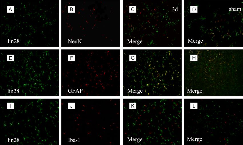
Ding et al: Lin 28 is associated with astrocyte proliferation

Lin28 is abundantly expresses in early-stage         The animals were allowed to recover at 37°C
embryos and upon induction of differentiation        room temperature and were provided sufficient
the expression is decreasing and restricted in       food and water after surgery. The animals’
several tissues such as cardiac and skeletal         brains used for our experiment were removed
muscles [15]. LIN28 is known to mediate a            at an indicated time point after the surgery.
variety of cellular processes including embryo-
genesis, skeletal myogenesis, germ cell devel-       Forelimb placing test
opment, neurogliogenesis, differentiation, lym-
phopoiesis, and glucose metabolism [16]. As          The rats were held by the dorsal torsos in order
a RNA-binding protein, LIN28 can regulate the        to make the forelimb hang freely. Independent
translation and stability of a large number of       testing of each forelimb was induced by brush-
mRNAs and the biogenesis of certain miRNAs.          ing the vibrissae on the corner edge of a
For example, Lin28 can associate with many           countertop. Intact animals rapidly placed the
complexes such as messenger ribonucleopro-           homolateral forelimb onto the countertop. In
tein particles (mRNPs), polysomes and stress         the light of the extent of damage, the ICH rats
granules. Besides, LIN28 is involved in regu-        placing of the forelimb contralateral to the
lating cell growth and differentiation in embry-     injection side indicated it was injured. Each
onic cells through interacting with let-7 family     forelimb of every rat was tested 10-15 times
microRNAs and blocking their processing into         and the percentage of placing the left fore-
mature miRNAs [16-19].                               limb was recorded.

In this study, we found LIN28 was over-expres-       Corner turn test
sion and colocalization with proliferating cell
nuclear antigen (PCNA) during an ICH mode.           The rats were permitted to proceed into a
This study indicates the insight role of LIN28       corner with an angle measuring 30°C. In order
on the cellular and molecular mechanisms             to exit the corner, the rat must turn either to
during ICH-induced astrocytes activation.            the right or to the left. Only the turns involving
                                                     full rearing along either wall was included.
Materials and methods                                According to the extent of damage, the rats
                                                     may show an inclination to turn to the homo-
Animals and intracerebral infusion                   lateral side of the injury. The test was con-
                                                     ducted 10 times, with an interval between
Male Sprague-Dawley rats (240-270 g) provid-         each test at least 30 s. The percentage of right
ed by the Department of Animal Center, Medical       turns was recorded.
College of Nantong University were used in
our study and performed in accordance with           Western blot analysis
National Institutes of Health Guidelines for the
Care and Use of Laboratory Animals published         To get the brain tissue needed for western
by The National Research Council in 1996.            blotting analysis, the rats were sacrificed at
Animals were maintained in a 12 h light/dark         different time points by intraperitoneal inject-
cycle and at a temperature controlled room           ing chloral hydrate. The brain tissue surround-
(24°C) and animals were divided into a sham          ing the hematoma (extending 2 mm to the
group and an experimental group. For ICH             hematoma) and the same part of the normal,
model, the animals were deeply anesthetized          sham-controlled, and contralateral tissue were
with 10% chloral hydrate by intraperitoneal          dissected and fleetly frozen at -80°C. To ob-
injection. 50 μL of autologous whole blood was       tain brain tissue proteins, the tissues were
collected from the tail tip of the animal and        cut and added to lyses buffer (1% Nonidet
injected into the right caudata nucleus ste-         P-40, 50 mM Tris, pH 7.5,5 mM EDTA, 1% SDS,
reotactically by use of a microinfusion pump         1% sodium deoxycholate, 1% Triton X-100, 1
through a 26-gauge needle (coordinates: 0.2          mM PMSF, 10 lg/mL aprotinin, and 1 lg/mL leu-
mm anterior, 5.5 mm ventral, and 3.5 mm              peptin) according to 0.1 g tissue/1 mL lyses
lateral to the bregma). After infusion for 10 min-   buffer. After lysed by the sonifier cell disrupter,
utes, the needle was kept in situ for over 5 min-    the solution was stewed for 40 min, centri-
utes before being withdrawn. The sham group          fuged at 14,000 rpm for 15 min in a micro-
was merely subjected to a needle insertion.          centrifuge at 4°C, and collected the superna-

1137                                                    Int J Clin Exp Pathol 2020;13(5):1136-1145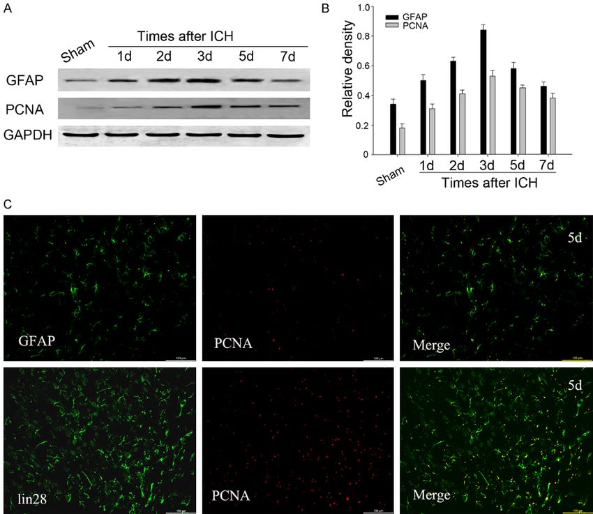
Ding et al: Lin 28 is associated with astrocyte proliferation

tant. After we measured the concentration         vine serum albumin, 0.3% Triton X-100, and
with the Bradford assay, the supernatant was      0.15% Tween-20) for 2 h at room tempera-
subjected to SDS-polyacrylamide gel electro-      ture. Then, sections were incubated with pri-
phoresis (PAGE) and transferred to a poly-        mary antibodies against LIN28 antibody (anti-
vinylidine difluoride membrane (PVDF) by a         rabbit, 1:100; abcam), NeuN (mouse; 1:100;
transfer apparatus at 300 mA. The membrane        Chemicon), GFAP (mouse; 1:100; Cell Signaling
was blocked with 5% non-fat milk for 2 h          Technology), Iba-1 (mouse; 1:100; Santa Cruz)
and incubated with primary antibody against       and PCNA (anti-mouse, 1:100; Santa Cruz) at
LIN28 (anti-rabbit, 1:800; Abcam), PCNA (anti-    4°C overnight. After washed for 15 min, sec-
mouse, 1:800; Santa Cruz), GFAP (anti-mouse,      tions were subjected to a mixture of FITC- and
1:1000; Cell Signaling Technology), and GAPDH     CY3-conjugated secondary antibodies at room
(anti-rabbit, 1:1000; Sigma) overnight at 4°C.    temperature for 2 h. After washed for 45 min,
After washing with PBST for 15 minutes, the       sections were covered with coverslips. The
membranes were incubated with secondary           sections were observed by Leica fluores-
antibodies for 2 h at room temperature and        cence microscope (Wetzlar, Germany).
after washing 45 min, the protein was visual-
ized using an enhanced chemiluminescence          Quantitative analysis
system (Pierce Company, USA).
                                                  Cells double labeled for LIN28 and other phe-
Immunohistochemistry                              notypic markers were quantified. A minimum of
                                                  200 phenotype-specific marker-positive cells
Rats used for our experiment were anesthe-        surrounding the hematoma were counted in
tized and perfused with 500 mL of 0.9% saline     each section to identify the proportion of posi-
and 4% paraformaldehyde. After perfusion, the     tive cells. The double labeled cells for LIN28
brains were removed and post-fixed in the          and phenotype-specific markers were regarded
same fixative for 1 d and the solution was         as positive. Three sections consecutive from
replaced with 20% sucrose for 2 days and          every rat were sampled.
then 30% sucrose treatment for 2 days. The
brain tissues were embedded in OCT com-           Cell culture
pound to cut 5mm frozen cross-sections. The
sections were stored at -20°C. For immuno-        Primary astrocytes were prepared from the
histochemistry, sections kept at 37°C for 2 h     cerebral cortex of newborn Sprague-Dawley
and subjected to 10 mmol/L citrate buffer (pH     rats which were provided by the Department
6.0) to retrieve the antigen. After washed by     of Animal Center, Medical College of Nantong
0.01 MPBS for 5 min, the sections were sub-       University. The meaninges were removed care-
jected to 3% H2O2 for 10 min to reduce endo-      fully to obtain the isolated neopallium which
genous peroxidase activity. After blocked by      subsequently was cut into 1 mm cubes. After
confining liquid, the sections were incubated      trypsinized by 0.125% [w/v] trypsin for 15 min
with anti-LIN28 antibody (anti-rabbit, 1:100;     at 37°C, the tissue suspension was passed
abcam) for 2 h at 37°C. The sections were         through nylon meshes of 70 mm pore size
washed for 15 min before secondary antibody       and the cells were seeded at a density of
treatment (Vector Laboratories, Burlingame,       2×10⁹ cells/flask. The cells were cultured with
CA) for 40 min at 37°C. After stained with        D/F (1:1 DMEM/F12) medium (supplemented
DAB (Vector Laboratories), the sections were      with 4-(2-hydroxyethyl)-1-piperazineethanesul-
dehydrated, cleared, and coverslipped after       fonic acid (HEPES), sodium bicarbonate, and
reaction. Slides were examined with a Leica       antibiotics) with 10% (v/v) heat-inactivated
light microscope (Germany) and strong brown       fetal bovine serum (HI-FBS) at 37°C with a
staining was regarded as positive and no stain-   humidified atmosphere of 95% air and 5% CO2
ing was regarded as negative.                     for 7-10 days. To separate the microglia from
                                                  the astrocytes, the flasks were placed on a
Immunofluorescent staining                         shaker platform and shaken for 8 h at 180
                                                  rpm at 37°C. The supernatant was discarded.
For immunofluorescent staining, sections were      The medium was changed every 1-2 days. Prior
treated with 1% Triton X-100 for 30 min and       to the experiments, the medium was switch-
blocking solution (10% donkey serum, 1% bo-       ed to serum-free DMEM/F12 culture medium.

1138                                                 Int J Clin Exp Pathol 2020;13(5):1136-1145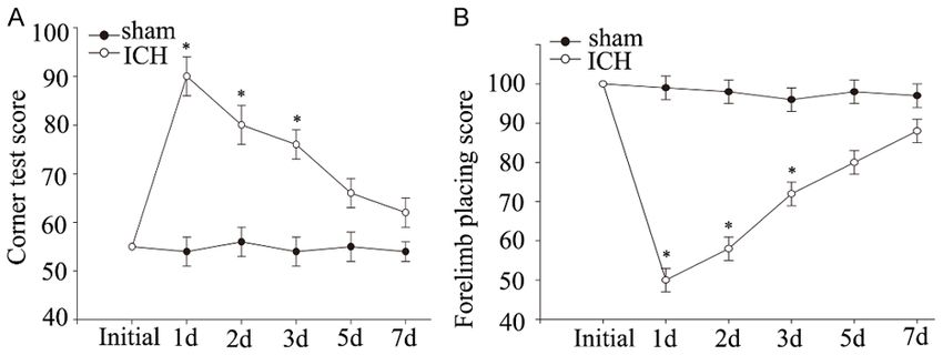
Ding et al: Lin 28 is associated with astrocyte proliferation

                                                                                cells were used for western
                                                                                blot.

                                                                                Statistical analysis

                                                                                    All statistical analyses we-
                                                                                    re conducted with a STATA
                                                                                    7.0 software package (Stata
                                                                                    Corp., College Station, TX,
                                                                                    USA). All data were expres-
Figure 1. Neurological deficits measured by Behavior Tests after ICH. Corner        sed as mean ± standard error
turn testing (A) and forelimb placing scores (B) at different time points follow-   of mean (S.E.M.). One-way
ing ICH were performed. The ICH group showed functional deficits compared           ANOVA was used to compa-
with the sham-operated group distinctly over the first 3 days (*P
Ding et al: Lin 28 is associated with astrocyte proliferation
together showed LIN28 was
up-regulation after ICH, sug-
gesting that LIN28 played a
novel biological function after
ICH.
Co-localization of LIN28 with
different cellular markers
To further address the role
of LIN28, immunofluorescent
experiment with different
marks (neuron marker NeuN,
astrocyte marker GFAP, and
microglia marker Iba1) was
conducted to determine whi-
ch cell type LIN28 expressed
after ICH. As shown in the
Figure 4, LIN28 was ex-
pressed in both neurons and
astrocytes and the co-local-
ization of LIN28 with neurons
and astrocytes especially with
astrocytes was significantly
enhanced surrounding the
hemotoma at 3 d after ICH
than the sham group.
Association of LIN28 with the
cell proliferation
Recently, a study reported
that LIN28 was associated
with cell proliferation after
spinal cord injury [21], so
we wondered whether LIN28
interrelate to cell proliferation
Figure 3. Immunohistochemistry of LIN28 adjacent to the hematoma. Low in rat ICH model. To verify our
level of LIN28 signal was found in the sham-operated group (A, B). At 2 days hypothesis, western blot was
after ICH, the ipsilateral group showed increasing LIN28 signals (E, F), while conducted to examine the
the contralateral group showed no significant difference in LIN28 compared
to sham-operated group (C, D). (G) No positive signal was found in the nega- expression level of GFAP and
tive control. (H) The number of LIN28 cells was largely increased compar- proliferating cell nuclear anti-
ing the ipsilateral group with the sham-operated and contralateral groups. gen (PCNA), a general marker
*PDing et al: Lin 28 is associated with astrocyte proliferation
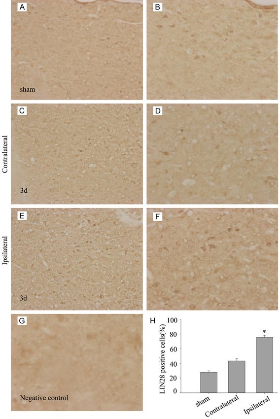
Figure 4. Immunofluorescence staining of LIN28 with different phenotype-specific markers. Sections were labeled
with LIN28 (green, A, E, I), neuronal marker NeuN (red, B), astrocyte marker GFAP (red, F) and microglia marker Iba-
1 (red, J). The yellow color visualized in the merged images represented the colocalization of LIN28 with different
phenotype-specific markers (C, G, K) as well as the sham group (D, H, L). Scale bars 50 μm (A-L).
The correlation of LIN28 with the proliferation were transfected with LIN28 specific, non-
of astrocytes induced by LPS specific siRNA and vehicle. Western blot was
performed to examine LIN28 expression
As reported, LIN28 was involved in astrocytes after transfected for 48 h, and LIN28 specific-
inflammation through NF-kB signaling pathway siRNA obviously down-regulated LIN28 ex-
during spinal cord injury [21], so we hypothe- pression (Figure 6E). After being transfected
sized whether LIN28 is involved in astrocytes for 30 h, primary astrocytes were then sub-
activation during ICH. Therefore, we used LPS jected to LPS treatment for another 18 h and
stimulate primary astrocytes which was a typi- western blot was performed to test the ex-
cal model of astrocytes activation. Different pression of LIN28, PCNA, and GFAP. The result
concentration of LPS was used to stimulating showed that the expression of LIN28, PCNA,
primary astrocytes and western blot was per- and GFAP were reduced after LIN28 knocked
formed to detect the expression of LIN28. As down and LPS stimulation (Figure 6G). Based
shown in Figure 6A, the expression of LIN28 on the above experiments, we have sufficient
changed along with the dose of LPS and reasons to draw the conclusion that LIN28
maximum at the concentration of 1 µg/ml. was involved on astrocyte proliferation.
Next, we used 1 µg/ml LPS to stimulate primary
astrocytes for different time points. The result Discussion
indicated that the expression of LIN28 was
increased at 12 h and peaked at 18 h. The The injury mechanisms of ICH include physical
expression of PCNA and astrocyte-specific trauma and mass effect, cerebral blood flow
glial fibrillary acidic protein (GFAP) were also reduction, thrombin, erythrocytes, haemoglo-
increased at 12 h and peaked at 18 h and 24 h bin, iron, inflammation, and complement [10,
(Figure 6C). The parallel expression of LIN28 22]. The inflammatory response surrounding
with PCNA and GFAP implied LIN28 was as- the haematoma involves enzyme activation,
sociated with astrocytes activation. To further mediator release, inflammatory cell migration,
confirm the role of LIN28, primary astrocytes glial activation, brain tissue breakdown and
1141 Int J Clin Exp Pathol 2020;13(5):1136-1145Ding et al: Lin 28 is associated with astrocyte proliferation Figure 5. Correlations of LIN28 with astrocyte proliferation following ICH. (A) Western blot analysis showed the ex- pression of GFAP and PCNA increased and peaked at day 3. (B) The bar graph indicated the density of GFAP and PCNA versus GAPDH at each time point. Data are presented as mean ± SEM (*,#P
Ding et al: Lin 28 is associated with astrocyte proliferation

                                                                                The animal ICH model used
                                                                                in our experiment deserves
                                                                                to be mentioned. The most
                                                                                commonly used ICH model
                                                                                was intracerebral injection of
                                                                                collagenase to cause vessel
                                                                                rupture or direct injection of
                                                                                blood into the brain [10, 32].
                                                                                The two models have advan-
                                                                                tages and self deficits. The
                                                                                collagenase-induced haemor-
                                                                                rhage is due to disrupt vascu-
                                                                                lature, but widespread dis-
                                                                                solution of the endothelial
                                                                                basement membrane might
                                                                                cause disruption of several
                                                                                blood vessels, which is differ-
                                                                                ent from the spontaneous
                                                                                ICH. In addition, the collage-
                                                                                nase has toxic impacts on
                                                                                brain parenchymal cells. How-
                                                                                ever, the intraparenchymal
                                                                                blood injection mimics the
                                                                                effects of an intracerebral
                                                                                haematoma but it lacks the
                                                                                ruptured blood vessels which
                                                                                is the primary cause of ICH
                                                                                [9, 10, 32]. Therefore, rele-
                                                                                vant models of spontaneous
                                                                                ICH in animals are greatly
Figure 6. HERPUD1 modulated cell proliferation in vitro. Primary cultured       needed.
astrocytes stimulated with different concentration of LPS, and LIN28 expres-
sion maxed at the concentration of 1 µg/ml (A, B). We used 1 µg/ml LPS         In summary, our current study
to stimulate primary astrocytes at different time points. The expression of
LIN28 was increased at 12 h and peaked at 18 h. The expression of PCNA
                                                                               might provide an innovative
and GFAP were also increased at 12 h and peaked at 18 h (C, D). Western        way to illustrate the underly-
blot analysis showed siRNA silenced LIN28 in primary cultured astrocytes       ing molecular and cellular
(E, F). The knockdown of LIN28 induced down-regulated levels of PCNA and       mechanisms following ICH-
GFAP expression (G). The bar graph indicated the relative density of LIN28,    induced astrocytes activation
PCNA and GFAP versus GAPDH (H). Data are mean ± SD, n=3, P
Ding et al: Lin 28 is associated with astrocyte proliferation
People’s Hospital, 2666 Ludang Road, Suzhou [13] Gong C, Boulis N, Qian J, Turner DE, Hoff JT
215200, Jiangsu Province, China. Tel: +86-0512- and Keep RF. Intracerebral hemorrhage-in-
82881198; E-mail: qzg66666666@163.com; Dr. duced neuronal death. Neurosurgery 2001;
Xingjuan Fan, Department of Neurology, Affiliated 48: 875-82.
Hospital of Nantong University, 20 Xisi Road, [14] Moss EG, Lee RC and Ambros V. The cold
shock domain protein LIN-28 controls develop-
Nantong 226001, Jiangsu Province, China. Tel: +86-
mental timing in C. elegans and is regulated by
0513-85052222; E-mail: ntfanxj@163.com
the lin-4 RNA. Cell 1997; 88: 637-46.
[15] Yang DH and Moss EG. Temporally regulated
References
expression of Lin-28 in diverse tissues of the
developing mouse. Gene Expr Patterns 2003;
[1] Feigin VL, Lawes CM, Bennett DA, Barker-Collo
3: 719-26.
SL and Parag V. Worldwide stroke incidence
[16] Li N, Zhong X, Lin X, Guo J, Zou L, Tanyi JL,
and early case fatality reported in 56 popula-
Shao Z, Liang S, Wang LP, Hwang WT, Katsaros
tion-based studies: a systematic review. Lan-
D, Montone K, Zhao X and Zhang L. Lin-28 ho-
cet Neurol 2009; 8: 355-69.
mologue A (LIN28A) promotes cell cycle pro-
[2] van Asch CJ, Luitse MJ, Rinkel GJ, van der
gression via regulation of cyclin-dependent ki-
Tweel I, Algra A and Klijn CJ. Incidence, case
fatality, and functional outcome of intracere- nase 2 (CDK2), cyclin D1 (CCND1), and cell
bral haemorrhage over time, according to age, division cycle 25 homolog A (CDC25A) expres-
sex, and ethnic origin: a systematic review and sion in cancer. J Biol Chem 2012; 287: 17386-
meta-analysis. Lancet Neurol 2010; 9: 167-76. 97.
[3] LoPresti MA, Bruce SS, Camacho E, Kunchala [17] Xu B, Zhang K and Huang Y. Lin28 modulates
S, Dubois BG, Bruce E, Appelboom G and Con- cell growth and associates with a subset of cell
nolly ES. Hematoma volume as the major de- cycle regulator mRNAs in mouse embryonic
terminant of outcomes after intracerebral stem cells. RNA 2009; 15: 357-61.
hemorrhage. J Neurol Sci 2014; 345: 3-7. [18] Balzer E and Moss EG. Localization of the de-
[4] Kuramatsu JB, Huttner HB and Schwab S. Ad- velopmental timing regulator Lin28 to mRNP
vances in the management of intracerebral complexes, p-bodies and stress granules. RNA
hemorrhage. J Neural Transm (Vienna) 2013; Biol 2007; 4: 16-25.
120 Suppl 1: S35-41. [19] Viswanathan SR and Daley GQ. Lin28: a mi-
[5] Ariesen MJ, Claus SP, Rinkel GJ and Algra A. croRNA regulator with a macro role. Cell 2010;
Risk factors for intracerebral hemorrhage in 140: 445-9.
the general population: a systematic review. [20] Okauchi M, Hua Y, Keep RF, Morgenstern LB
Stroke 2003; 34: 2060-5. and Xi G. Effects of deferoxamine on intracere-
[6] Jackson CA and Sudlow CL. Is hypertension a bral hemorrhage-induced brain injury in aged
more frequent risk factor for deep than for lo- rats. Stroke 2009; 40: 1858-63.
bar supratentorial intracerebral haemorrhage? [21] Yue Y, Zhang D, Jiang S, Li A, Guo A, Wu X, Xia
J Neurol Neurosurg Psychiatry 2006; 77: 1244- X, Cheng H, Tao T and Gu X. LIN28 expression
52. in rat spinal cord after injury. Neurochem Res
[7] Söderholm M, Inghammar M, Hedblad B, 2014; 39: 862-74.
Egesten A and Engström G. Incidence of stroke [22] Xue M and Del Bigio MR. Intracerebral injec-
and stroke subtypes in chronic obstructive pul- tion of autologous whole blood in rats: time
monary disease. Eur J Epidemiol 2016; 31: course of inflammation and cell death. Neuro-
159-68. sci Lett 2000; 283: 230-2.
[8] Wilson D, Charidimou A and Werring DJ. Ad- [23] Wang X and Lo EH. Triggers and mediators of
vances in understanding spontaneous intrace- hemorrhagic transformation in cerebral isch-
rebral hemorrhage: insights from neuroimag- emia. Mol Neurobiol 2003; 28: 229-44.
ing. Expert Rev Neurother 2014; 14: 661-78. [24] Wang J and Doré S. Inflammation after intrace-
[9] Keep RF, Hua Y and Xi G. Intracerebral haem- rebral hemorrhage. J Cereb Blood Flow Metab
orrhage: mechanisms of injury and therapeu- 2007; 27: 894-908.
tic targets. Lancet Neurol 2012; 11: 720-31. [25] Panickar KS and Norenberg MD. Astrocytes in
[10] Xi G, Keep RF and Hoff JT. Mechanisms of cerebral ischemic injury: morphological and
brain injury after intracerebral haemorrhage. general considerations. Glia 2005; 50: 287-
Lancet Neurol 2006; 5: 53-63. 98.
[11] Qureshi AI, Mendelow AD and Hanley DF. Intra- [26] Eddleston M and Mucke L. Molecular profile of
cerebral haemorrhage. Lancet 2009; 373: reactive astrocytes--implications for their role
1632-44. in neurologic disease. Neuroscience 1993; 54:
[12] Aronowski J and Zhao X. Molecular pathophysi- 15-36.
ology of cerebral hemorrhage: secondary brain [27] Dong Y and Benveniste EN. Immune function
injury. Stroke 2011; 42: 1781-6. of astrocytes. Glia 2001; 36: 180-90.
1144 Int J Clin Exp Pathol 2020;13(5):1136-1145Ding et al: Lin 28 is associated with astrocyte proliferation
[28] Swanson RA, Ying W and Kauppinen TM. Astro- [31] Liu L, Rudin M and Kozlova EN. Glial cell prolif-
cyte influences on ischemic neuronal death. eration in the spinal cord after dorsal rhizoto-
Curr Mol Med 2004; 4: 193-205. my or sciatic nerve transection in the adult rat.
[29] Pyo H, Yang MS, Jou I and Joe EH. Wortmannin Exp Brain Res 2000; 131: 64-73.
enhances lipopolysaccharide-induced induc- [32] Rosenberg GA, Mun-Bryce S, Wesley M and Ko-
ible nitric oxide synthase expression in microg- rnfeld M. Collagenase-induced intracerebral
lia in the presence of astrocytes in rats. Neuro- hemorrhage in rats. Stroke 1990; 21: 801-7.
sci Lett 2003; 346: 141-4.
[30] Ridet JL, Malhotra SK, Privat A and Gage FH.
Reactive astrocytes: cellular and molecular
cues to biological function. Trends Neurosci
1997; 20: 570-7.
1145 Int J Clin Exp Pathol 2020;13(5):1136-1145You can also read