LRRC17 regulates the bone metabolism of human bone marrow mesenchymal stem cells from patients with idiopathic necrosis of femoral head through ...
←
→
Page content transcription
If your browser does not render page correctly, please read the page content below
EXPERIMENTAL AND THERAPEUTIC MEDICINE 22: 666, 2021

                LRRC17 regulates the bone metabolism of human
               bone marrow mesenchymal stem cells from patients
                with idiopathic necrosis of femoral head through
                 Wnt signaling pathways: A preliminary report
                         DA SONG1,2, ZHEN‑SONG WU3, QI XU2, KAI WANG2, MING‑TAO XU2,
                               CHENG‑ZHI HA2, CHAO ZHANG2 and DA‑WEI WANG2

        1
         Department of Orthopedics, Liaocheng People's Hospital, Cheeloo College of Medicine, Shandong University;
                  2
                   Department of Orthopedics, Liaocheng People's Hospital, Liaocheng, Shandong 252000;
           3
            Department of Joint Surgery, Zaozhuang Municipal Hospital, Zaozhuang, Shandong 277100, P.R. China

                                      Received October 29, 2020; Accepted March 19, 2021

                                                   DOI: 10.3892/etm.2021.10098

Abstract. Idiopathic necrosis of the femoral head (INFH) is         the expression of LRRC17 was lower in the bone tissue and
a common disease with unknown cause. Its successful treat‑          hBMSCs from patients with INFH compared with patients
ment relies on the repair of the necrotic bone. The application     with FNF (P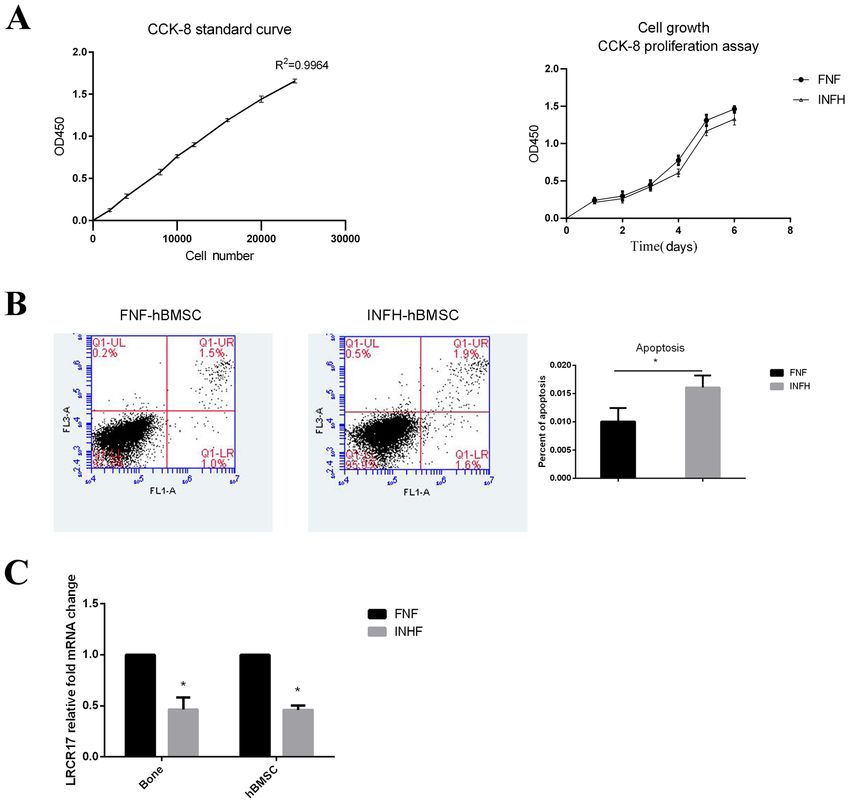
2                SONG et al: LRRC17 REGULATES THE BONE METABOLISM OF hBMSCs FROM PATIENTS WITH INFH

are important for making and repairing skeletal tissues. It has   gradient ethanol (100, 95, 85 and 75%), and stained with hema‑
strong self‑renewal abilities and can undergo chondrogenic,       toxylin solution at 25˚C for 10 min. After that, the sections
osteogenic and adipogenic differentiation. It also main‑          were dipped in 1% hydrochloric acid for 2‑5 sec, 1% ammonia
tains the balance of bone metabolism in the human skeletal        for 1 min, and then 0.5% eosin for 5‑10 sec at 25˚C. Finally,
system (7). Recently, the decrease in hBMSC and the change        sections were dehydrated with 95 and 100% ethanol and
in cell behavior have been linked to the occurrence and devel‑    mounted with xylene‑based glue. Blocking was not performed.
opment of osteonecrosis of the femoral head (8). This suggests    Sections were observed under a light microscope at a magnifi‑
that BMSC maybe a great target for INFH treatment. Wnt            cation of x200 and images were captured.
signaling has been implicated in controlling BMSC fate, but it        Bone marrow were harvested from five patients with INFH
has not been well investigated in hBMSC from INFH patients.       (three males and two females; average age, 61.33±7.2 years old)
    Recently, Wang et al conducted a microRNA expression          and five patients with femoral neck fracture (FNF; two males
profiling of BMSCs in patients with femoral head osteone‑         and three females; average age, 67.33±8.6 years old) between
crosis compared to femoral neck fracture. They found a group      February 2017 and February 2018, who received femoral head
of differentially expressed microRNAs and predicted their         replacement in the hospital. According to the Ficat staging
target genes. Among these, the expression of Leucine‑rich         system (12), all patients with INFH were at stages III or IV.
repeat‑containing 17 (LRRC17) was significantly lower in          The exclusion criteria were: i) Medical history of cancer;
hBMSCs obtained from patients with femoral head osteo‑            ii) other metabolic bone disease; iii) previous hip surgery or
necrosis compared to femoral neck fracture (9). LRRC17            infectious disease; and iv) long‑term use of hormones and
is a secretory protein containing five leucine‑rich repeat        alcohol. hBMSCs harvested from five patients with INFH or
domains. It was first characterized in bone metabolism as         FNF (INFH‑hBMSC or FNF‑hBMSC) were pooled together
an inhibitor of receptor activator of nuclear factor‑κ B ligand   for following experiments.
(RANKL)‑induced osteoclast differentiation (10). RANKL                All experimental procedures were conducted in strict
is the ligand of the NF‑κ B receptor activator, which is an       conformity with the work described, and carried out in accor‑
indispensable biomolecule in osteoclast activation and prolif‑    dance with the Code of Ethics of the World Medical Association
eration (10). LRRC17 negatively regulates RANKL to inhibit        (Declaration of Helsinki) for experiments involving humans.
bone degradation. Later, Kim et al demonstrated that post‑
menopausal women with lower LRRC17 level had a 3.32‑fold          Isolation and culture of hBMSCs. The hBMSCs were
higher odds ratio for osteoporotic fracture and associated with   isolated by subjecting to Ficoll (1.077 g/ml; Tianjin Haoyang
a 46% higher risk of osteoporotic fracture than the group         Biological Products Technology Co., Ltd.) density gradient
with higher LRRC17 levels, suggesting LRRC17's potential as       separation. The mononuclear fraction was collected and plated
a marker for osteoporotic fracture (11). However, the role of     in T‑25 flasks (Corning, Inc.) using complete DMEM/F‑12
LRRC17 in the INFH has never been fully investigated.             containing 10% fetal bovine serum and 1% penicillin and strep‑
    In this study, we aimed to investigate the role of LRRC17     tomycin (Gibco; Thermo Fisher Scientific, Inc.) medium at 37˚C
in hBMSCs derived from INFH patients. By manipulating the         with 5% CO2. After three days, the medium was changed to
expression of LRRC17, we explored the potential mechanisms        remove non‑adherent cells. Then, the media was changed every
with a focus on the Wnt signaling pathways. This study may        three days. When the primary cells reached 70‑80% confluence,
help us understand the effect of LRRC17 on the pathogenesis       the cells were passaged by trypsinization (Hyclone; Cyvita), and
of INFH and identify a potential treatment target for INFH        cultured based on the aforementioned method.
patients.
                                                                  Flow cytometry. The cell‑specific surface markers were
Materials and methods                                             examined by flow cytometry. The hBMSCs were dissociated
                                                                  and fixed in 70% ethanol at 4˚C for 24 h. Anti‑CD34‑FITC
Clinical samples. The present study was approved by the           (cat. no. 560942), anti‑CD44‑FITC (cat. no. 560977),
Ethics Committee of Liaocheng People's Hospital, and all          anti‑CD45‑FITC (cat. no. 560976) and anti‑CD90‑FITC
patients who participated in this study provided a signed         (cat. no. 561969) (all ready to use; BD Biosciences PharMingen)
written informed consent.                                         antibodies were used and incubated at 25˚C for 20 min. The
                                                                  data analysis was conducted using the BD FACSDiva software
Hematoxylin‑eosin staining. The surgically resected femoral       (version. 8.0.1; BD Biosciences).
head was semi‑dissected from the coronal face, and the appear‑
ance of the specimen and the pathology of the section were        Osteogenic induction and evaluation. To induce osteogenic
observed. A bone grain of ~10x10x5 mm was taken from the          differentiation, hBMSCs were incubated in DMEM supple‑
necrotic area (INFH‑derived) and normal area (FNF‑derived)        mented with dexamethasone (100 nM), β ‑glycerophosphate
under naked eye observation. All the bone blocks were fixed       (10 mM) and ascorbic acid 2‑phosphate (200 µM) (all from
in 10% formalin solution at 4˚C for 3 days, decalcified with      Sigma‑Aldrich; Merck KGaA), and 10% fetal bovine serum
10% EDTA Tris‑HCl (pH 7.4) buffer and changed every day           (Hyclone; Cyvita). The medium was changed every three
at 4˚C. When acupuncturing specimen had no resistance, this       days. After osteogenic induction for 14 days, cells were
indicated that the bone tissue had been decalcified completely.   stained with 2% alizarin red S (Sigma‑Aldrich; Merck KGaA)
The specimen was embedded in paraffin and cut into sections       at 25˚C for 15 min to evaluate the calcium deposition. Matrix
with a thickness of 4‑5 µm. Sections were dewaxed using           mineral‑bound staining was detected as a bright orange‑red
xylene for three times with 5 min each, rehydrated using          color under a light microscope (magnification, x200).
EXPERIMENTAL AND THERAPEUTIC MEDICINE 22: 666, 2021 3
Adipogenic induction and evaluation. To induce adipogenic FNF‑hBMSCs. Then, 1x10⁶ cells from each group were resus‑
differentiation, lipid induction A solution was prepared pended with 1X binding buffer, and 5 µl of Annexin V‑FITC
by supplementing DMEM with dexamethasone (1 µM), was added and incubated for 15 min at room temperature in
3‑isobutyl‑1‑methylxanthine (0.5 mM), insulin (10 µg/ml), the dark. Propidium iodide (PI) staining with a volume of 5 µl
rosiglitazone (0.5 µM) and indomethacin (100 µM) (all from was added at 5 min before detection. Then, cells were analyzed
Sigma‑Aldrich; Merck KGaA) and 10% fetal bovine serum by flow cytometry (Becton, Dickinson and Company).
(Hyclone; Cyvita). Then, B solution was prepared by adding
insulin (20 µg/ml) to the DMEM and 10% fetal bovine Reverse transcription‑quantitative polymerase chain reaction
serum. Subsequently, 2x10⁵/ml hBMSCs were incubated with (RT‑qPCR). TRIzol RNA isolation reagents (Thermo Fisher
the A solution for three days, and this was switched to the Scientific, Inc.) was used to extract the total RNA. A NanoDrop
B solution for one day. This was repeated for four times. In 2000 Spectrophotometer (Thermo Fisher Scientific, Inc.)
order to evaluate the adipogenesis, oil red O (Sigma‑Aldrich; was used to measure the concentration and purity of the
Merck KGaA) staining was performed at 25˚C for 15 min and total RNA. Then, 1 µg RNA was converted to cDNA using a
observed under a light microscope at a magnification of x200. PrimeScript™ RT reagent kit (Takara Bio, Inc.) with a gDNA
Eraser at 37˚C for 15 min, then 85˚C for 5 sec and 4˚C continu‑
Cell transfection. The INFH‑hBMSCs were randomly divided ously, and amplified in the ABI 7500 Sequence Detection
into four groups: LRRC17 overexpression group and its System (Applied Biosystems; Thermo Fisher Scientific, Inc.)
vector control group, and LRRC17 knockout group and its using a SYBR® Premix Ex Taq™ II kit (all obtained from
negative control group. For the overexpression or knockout, Takara Biotechnology Co., Ltd.). The thermal profile of the
INFH‑hBMSCs were transfected with lentiviral particles of RT‑qPCR was: Pre‑incubation at 95˚C for 30 sec for one cycle,
GV492‑LRRC17 or GV493‑LRRC17 short hairpin (sh)RNA, followed by 40 cycles incubated at 95˚C for 5 sec, and 60˚C
and the respective control vectors. The quantity of lentiviral for 34 sec. The polymerase chain reaction (PCR) primers were
plasmid used for transfection was 9 µg. The ratio of the lenti‑ synthesized by Sangon Biotech Co., Ltd. The primer sequences
viral plasmid: Packaging vector: Envelope was 9:13:4. The are summarized in Table I. The housekeeping gene GAPDH
duration of transfection into cells was 3 days and the multi‑ was used as an internal control. The relative expression levels
plicity of infection (MOI) was 70%. of each gene were calculated using 2‑ΔΔCq (13). Each experi‑
The lentiviral supernatants that contained lentiviral vectors ment was evaluated by the same PCR reactions for three times.
for LRRC17 overexpression or knockout, and the respective
controls were purchased from Shanghai GeneChem Co., Ltd. Western blotting. The protein expression was measured after
Positive clones were selected with 200 ng/ml of puromycin transfecting hBMSCs with lentivirus that contained the
for three days. After five days, the infection efficiency was LRRC17 gene. The expression levels of osteoprotegerin (OPG),
observed under a fluorescence microscope (cat. no. CKX71; bone morphogenetic protein 2 (BMP2), peroxisome prolifer‑
Olympus Corporation). When the transduction was over, the ator‑activated receptor γ (PPARγ), CCAAT/enhancer‑binding
subsequent experiments were performed immediately. protein α (CEBPα), Wnt3a, β ‑catenin, Wnt5a and Rankl
proteins were measured for all groups of hBMSCs.
Dickkopf‑related protein 1 (DKK1) and Wnt inhibition PMSF was used to lyse the cell precipitation. The cell lysates
treatment. INFH‑hBMSCs were seeded at 1x10³ in a 6‑well were centrifuged at 10,000 x g for 4 min at 4˚C, and the BCA
plate, and randomly divided into four groups: LRRC17 protein assay kit was used to measure the protein concentrations
overexpression treatment with or without 100 ng/ml DKK1 (all were obtained from Beyotime Institute of Biotechnology).
(MedChemExpress) treatment, and LRRC17‑knockout cells The same amount of protein (10 µg) and Prestained Protein
with or without 10 µM SP600125 (Beyotime Institute of Ladder (4 µl) were injected into the 10% sodium dodecyl
Biotechnology). Then, cells were transfected for the LRRC17 sulfate‑polyacrylamide gel electrophoresis lanes, and electro‑
overexpression or knockout cells at 24 h after plating, followed phoresis was performed for 1 h at 100 V. Then, the proteins
by DKK1 or SP600125 treatment after 48 h. Cells were were transferred onto polyvinylidene fluoride membranes
harvested after 24 h for downstream analysis. (EMD Millipore) for 1.5 h at 200 mA. Next, 5% skimmed milk
(dissolved in TBST) was used to block the non‑specific binding
In vitro cell proliferation assay. In order to generate the stan‑ membranes at room temperature for 1 h. Then, the membranes
dard curve, hBMSCs were trypsinized and seeded at 2x10³, were incubated with the appropriate primary antibodies
4x10³, 8x10³, 1.0x10⁴, 1.2x10⁴, 1.6x10⁴, 2.0x10⁴ and 2.4x10⁴ cells overnight at 4˚C: Anti‑LRRC17 (1:600), anti‑CEBPα (1:750),
per well in a 96‑well plate. Each well was repeated for three anti‑ β ‑catenin (1:30,000) (all obtained from Proteintech
times. After 2 h, cells were incubated with Cell Counting Kit‑8 Group, Inc.); anti‑OPG (1:1,000; ABclonal Biotech Co.,
(CCK‑8) (Dojindo Molecular Technologies, Inc.) for another Ltd.); anti‑Wnt5a (1:1,000), anti‑BMP2 (1:1,000), anti‑PPARγ
2 h at 25˚C and detected using an enzyme‑labeling instrument. (1:1,000), anti‑RANKL (1:700), anti‑Wnt3a (1:1,000), mouse
The INFH‑hBMSCs or FNF‑hBMSCs were trypsinized, monoclonal anti‑β‑actin antibody (1:10,000) (all obtained from
and 5x10³ cells per well were plated in a 96‑well plate. For each Beyotime Institute of Biotechnology). TBST was used to wash
of the following six days, cells were incubated with CCK‑8 for the membranes three times, for 10 min each. The membranes
2 h at 37˚C and detected using an enzyme labeling instrument. were incubated with species‑specific horseradish peroxidase
coupled with the secondary antibodies (goat anti‑mouse or
Cell apoptosis assay. Annexin V‑FITC (BD Biosciences) anti‑rabbit IgG; cat. nos. SA00001‑1 and SA00001‑2, respec‑
was used to evaluate the apoptosis in INFH‑hBMSCs or tively) at 1:5,000 for 1 h at room temperature. After washing4 SONG et al: LRRC17 REGULATES THE BONE METABOLISM OF hBMSCs FROM PATIENTS WITH INFH
Table I. Sequences of primers.
Gene Refseq accession number Direction Sequence, 5' to 3'
LRRC17 NM_001031692 Forward AAAGTGCCAAACAACATCCCT
Reverse TGGGTCGAAGTTGGTTGATTTT
OPG NM_002546.4 Forward GCCCCTTGCCCTGACCACTAC
Reverse TGCGATTGCACTCCTGCTTGAC
BMP2 NM_001200.4 Forward GTCCTGAGCGAGTTCGAGTTGC
Reverse GTGGTCTGGGGCGGGTGAG
CEBPα NM_004364.4 Forward TCGGTGGACAAGAACAGCAACG
Reverse GGCGGTCATTGTCACTGGTCAG
PPARγ NM_015869.4 Forward GCTGAATCCAGAGTCCGCTGAC
Reverse ATCGCCCTCGCCTTTGCTTTG
Wnt3a NM_033131.4 Forward TCGGAGATGGTGGTGGAGAAGC
Reverse GGGTTGGGCTCGCAGAAGTTG
Wnt5a NM_003392.5 Forward GACTTCCGCAAGGTGGGTGATG
Reverse GTCTTGTGTGGTGGGCGAGTTG
β‑catenin NM_001904.4 Forward TACTGTCCTTCGGGCTGGTGAC
Reverse GCTTCTTGGTGTCGGCTGGTC
Rankl NM_003701.4 Forward CAGCATCGCTCTGTTCCTGTA
Reverse CTGCGTTTTCATGGAGTCTCA
GAPDH NM_002046.7 Forward CGGAGTCAACGGATTTGGTCGTAT
Reverse AGCCTTCTCCATGGTGGTGAAGAC
for 30 min, an ECL western blotting kit (Beyotime Institute of Spindle‑shaped, had a long fusiform, uniform in size and
Biotechnology) was used to develop the blots. The quantifica‑ easily adherent to the plate, arranged in a certain direction,
tion of the protein bands was visualized using the AlphaView and rich in cytoplasm with a strong refraction (Fig. 1B).
analysis system (ProteinSimple). The β ‑actin antibody was
used as the protein loading control. The LRRC17, CEBPα, Characterization of hBMSCs by surface marker expression
β‑catenin, OPG, Wnt5a, PPARγ, BMP2, RANKL and Wnt3a and multi‑lineage differentiation. The expression levels of
protein expression was normalized against β‑actin. MSC surface markers (CD44 and CD90) and hematopoietic
markers (CD45 and CD34) were analyzed in hBMSCs from
Statistical analysis. The data are presented as mean ± stan‑ both groups by flow cytometry (14). The results showed that
dard deviation (SD), and at least three independent hBMSCs from both INFH and FNF groups consistently
experiments were performed. The quantitative data was expressed CD44 and CD90 and were negative for CD45 and
collected and statistically analyzed using the SPSS 20.0 soft‑ CD34 (Fig. 1C).
ware (IBM Corp.) by one‑way ANOVA between two groups In order to evaluate the adipogenic differentiation,
followed by SNK‑q and LSD test. The level of statistical the lipid droplet accumulation was assessed by oil red O
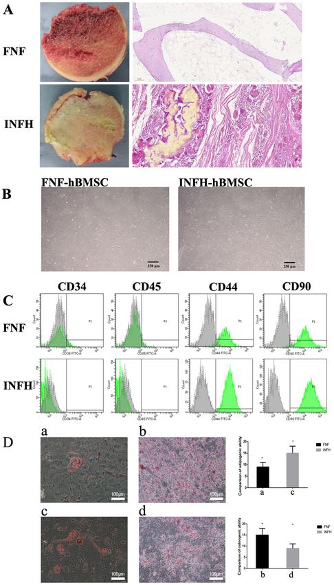
significance was set at PEXPERIMENTAL AND THERAPEUTIC MEDICINE 22: 666, 2021 5 Figure 1. Characterization of hBMSCs from patients with INFH or FNF. (A) The longitudinal section and hematoxylin and eosin staining of femoral head tissues in patients with FNF or INFH. (B) The primary hBMSCs for INFH and FNF, respectively. (C) The characterization of hBMSC surface markers in patients with FNF or INFH. (D) The two images on the left represent the comparison of hBMSC adipogenic induction, and the differentiation between patients with FNF (a) and patients with INFH (c). The two images on the right represent the comparison of the osteogenic differentiation of hBMSCs in patients with FNF (b) and patients with INFH (d). Scale bar, 100 µm. hBMSCs, human bone marrow mesenchymal stem cells; INFH, idiopathic necrosis of the femoral head; FNF, femoral neck fracture. were seeded at the same density on day 0 and cultured for the culture. The cell number of FNF‑hBMSCs was comparable 6 days. After generating a standard curve with R2=0.9964, the to INFH‑hBMSCs for the first 3 days, then became higher CCK‑8 levels of each sample was measured. The data showed from day 4 with a statistically significant difference observed that hBMSCs from both groups continued to proliferate during on day 4 (P
6                    SONG et al: LRRC17 REGULATES THE BONE METABOLISM OF hBMSCs FROM PATIENTS WITH INFH

Figure 2. Cell proliferation and apoptosis in hBMSCs from patients with INFH or FNF. (A) The left presents the standard proliferation curve of patients with
FNF; the right presents the comparison of hBMSC proliferation curves between patients with FNF and INFH. (B) The comparison of hBMSC apoptosis in
patients with FNF (1%) or INFH (1.6%) (FNF, 0.01±0.002386; INFH, 0.016±0.002111; P=0.0310). (C) Comparison of the mRNA expression of LRRC17 in
femoral head tissues and hBMSCs of patients with FNF or INFH. *P
EXPERIMENTAL AND THERAPEUTIC MEDICINE 22: 666, 2021                                                        7

Figure 3. Effect of LRRC17 expression on osteogenic and adipogenic differentiation of INFH‑hBMSCs. (A and B) Changes in LRRC17, BMP2, OPG, CEBPα,
PPARγ mRNA and protein expression after the overexpression and interference with LRRC17. *P
8                   SONG et al: LRRC17 REGULATES THE BONE METABOLISM OF hBMSCs FROM PATIENTS WITH INFH

Figure 4. Effect of LRRC17 on Wnt signaling in the INFH‑hBMSCs. (A and B) Changes in β‑catenin, Wnt3a, Rankl and Wnt5a mRNA and protein expres‑
sion after the overexpression and interference with LRRC17. (C) Changes in β ‑catenin and Wnt3a protein expression after the overexpression of LRRC17
and the addition of DKK1. (D) Changes in Rankl and Wnt5a protein expression after the interference of LRRC17 and the addition of Sp600125. *P
EXPERIMENTAL AND THERAPEUTIC MEDICINE 22: 666, 2021                                            9

also expressed lower levels of LRRC17 and were highly apop‑         precursor differentiation and JNK phosphorylation in
totic compared with FNF‑hBMSCs. The overexpression of               osteoclasts. Furthermore, JNK appears to be involved in the
LRRC17 promoted osteogenesis, and inhibited adipogenesis,           crosstalk between RANK and Ror2‑mediated signals (30,31).
while the knockout of LRRC17 reversed these changes in the          These results suggest that Wnt5a is produced by osteoblasts,
INFH‑hBMSCs; this may be mediated through both canonical            and that this promotes osteoclast differentiation through
and non‑canonical Wnt signaling pathways. The results               the non‑canonical Wnt pathway in osteoclast precursors.
suggested that LRRC17 may be a potential target for the stem        Together with the present findings, LRRC17 overexpression
cell therapeutic treatment of INFH.                                 may inhibit RANKL‑induced osteoclast differentiation by
    The present study data demonstrated that LRRC17 has an          inhibiting Wnt5a, while LRRC17 interference may enhance
important role in the pathogenesis of INFH. Abnormal bone           RANKL‑induced osteoclast differentiation by promoting
metabolism is part of the pathogenesis of femoral head osteo‑       Wnt5a.
necrosis (15). During INFH, the proliferative and osteogenic            The potential mechanism on how LRRC17 regulates Wnt
differentiation abilities of hBMSCs decrease. This diminishes       signaling was investigated. Proteins that contain LRR domains
the bone‑repair ability of the body, further leading to subchon‑    control a variety of physiological processes, including bone
dral bone weakening, stress fracture, and even articular surface    metabolism. For example, glycan and decorin are highly
collapse. Consistently, it was also found that the osteogenic       expressed in the extracellular bone matrix, affecting the
differentiation of INFH‑hBMSCs was significantly decreased          differentiation and proliferation of osteocytes. Mice with
and adipogenic differentiation ability was increased compared       leucine proteoglycan deficiency develop osteoporosis, which
with FNF‑hBMSCs. LRRC17 expression was significantly                is characterized by failure to reach the peak bone mass due to
lower in patients with INFH. Overexpression of LRRC17               decreased bone formation (8).
promoted osteogenesis, and inhibited the adipogenesis of                One of the limitations of the present study was the small
INFH‑hBMSCs. Therefore, targeting LRRC17 provides a                 number of patient samples. In future studies, the present find‑
promising therapeutic target for stem cell treatment of patients    ings require further validation in hBMSCs derived from a
with INFH.                                                          larger number of patients with INFH or FNF and specimens
    Previous studies have confirmed that Wnt signaling              with different causes of avascular necrosis.
pathways participate in regulating MSC self‑renewal and                 Another limitations of the present study is the lack of
osteogenic differentiation, and maintaining the balance             experiments in the FNF‑hBMSCs; the hBMSCs from patients
between osteoblast and osteoclast differentiation (16,17). This     with FNF should be included in the LRRC17 overexpression or
has important clinical significance for bone injury repair,         knockout experiments as a control. Based on the present find‑
tissue renewal, and intra‑articular homeostasis (18). Wnt signal    ings that LRRC17 was decreased in INFH‑hBMSCs, it can be
pathways are evolutionarily conserved, and can be mainly            anticipated that a similar trend of changes could be observed
classified as follows: Canonical‑ or Wnt/β‑catenin‑dependent        in the FNF‑hBMSCs, with the manipulation of LRRC17 as in
pathway, and the non‑canonical or β ‑catenin‑independent            the INFH‑hBMSCs.
pathway. These two pathways intersect with each other, and              In conclusion, the present study is the first to expound the
their association is very complicated, and has not been fully       pathogenesis of INFH from the perspective of bone metabo‑
elaborated currently (19). The members of the Wnt family            lism. The modulation of the LRRC17 gene may delay or even
are involved in bone regeneration, including the proliferation      change the process of INFH, providing a new target for the
and differentiation of osteoblasts, and their progenitors (20).     treatment of osteonecrosis of the femoral head.
The Wnt/β ‑catenin signaling stimulates the generation of
osteoblasts by promoting the osteogenic differentiation of          Acknowledgements
MSCs, while suppressing the other two lineages differen‑
tiation (21). It has been shown that MSC osteogenesis can be        Not applicable.
induced by increasing the transcription of β‑catenin (22). The
overexpression of the Wnt/β‑catenin signal in periosteal cells      Funding
can increase intramembranous ossification and endochondral
ossification (23,24).                                               No funding was received.
    Wnt3a can regulate the proliferation of differentiated osteo‑
blasts and their progenitors, and promote the differentiation       Availability of data and materials
of MSCs into osteoblasts under appropriate conditions (25).
In addition, Wnt3a binds to the Frizzled and LRP5 or LRP6           The datasets used and/or analyzed during the present study are
receptor complex, inhibits GSK‑3β, and promotes the accu‑           available from the corresponding author on reasonable request.
mulation of β‑catenin in osteoblasts (26,27). The accumulated
β ‑catenin translocates into the nucleus, and together with         Authors' contributions
TCF/LEF, induces the expression of OPG (28). Furthermore,
Wnt3a exhibits an inhibitory effect on osteoclast formation,        DWW and DS designed and performed most of the investiga‑
which is mediated through the canonical pathway (29). The           tion, data analysis, confirm the authenticity of all the raw data
present results also revealed that LRRC17 may promote osteo‑        and wrote the manuscript; ZSW and QX provided pathological
genesis by increasing the Wnt3a levels in patients with INFH.       assistance; KW, MTX, CZH and CZ contributed to the inter‑
    Wnt5a stimulates non‑canonical Wnt signals in osteo‑            pretation of the data and analyses. All of the authors have read
clast precursors, and promotes RANKL‑induced osteoclast             and approved the final manuscript.
10 SONG et al: LRRC17 REGULATES THE BONE METABOLISM OF hBMSCs FROM PATIENTS WITH INFH
Ethics approval and consent to participate 14. Tan SL, Ahmad TS, Selvaratnam L and Kamarul T: Isolation,
characterization and the multi‑lineage differentiation potential of
rabbit bone marrow‑derived mesenchymal stem cells. J Anat 222:
The present study was approved by the Ethics Committee 437‑450, 2013.
of Liaocheng People's Hospital (approval no. 2018010). All 15. Ko JY, Wang FS, Wang CJ, Wong T, Chou WY and Tseng SL:
Increased dickkopf‑1 expression accelerates bone cell apoptosis
procedures performed in studies involving human participants in femoral head osteonecrosis. Bone 46: 584‑591, 2010.
were in accordance with the ethical standards of the institu‑ 16. Phetfong J, Sanvoranart T, Nartprayut K, Nimsanor N,
tional and/or national research committee and with the 1964 Seenprachawong K, Prachayasittikul V and Supokawej A:
Osteoporosis: The current status of mesenchymal stem cell‑based
Helsinki declaration and its later amendments or comparable therapy. Cell Mol Biol Lett 21: 12, 2016.
ethical standards. All patients who participated in this study 17. Albers J, Keller J, Baranowsky A, Beil FT, Catala‑Lehnen P,
provided a signed written informed consent. Schulze J, Amling M and Schinke T: Canonical Wnt signaling
inhibits osteoclastogenesis independent of osteoprotegerin.
J Cell Biol 200: 537‑549, 2013.
Patient consent for publication 18. To r n e r o ‑E st eba n P, Pe r a lt a ‑ Sa st r e A, He r r a n z E,
Rodriguez‑Rodriguez L, Mucientes A, Abasolo L, Marco F,
Fernandez‑Gutierrez B and Lamas JR: Altered expression of wnt
Not applicable. signaling pathway components in osteogenesis of mesenchymal
stem cells in osteoarthritis patients. PLoS One 10: e0137170,
Competing interests 2015.
19. Duchartre Y, Kim YM and Kahn M: The Wnt signaling pathway
in cancer. Crit Rev Oncol Hematol 99: 141‑149, 2016.
All authors declare that they have no competing interests. 20. Chen J, Tu X, Esen E, Joeng KS, Lin C, Arbeit JM, Rüegg MA,
Hall MN, Ma L and Long F: WNT7B promotes bone formation
in part through mTORC1. PLoS Genet 10: e1004145, 2014.
References 21. Rodda SJ and McMahon AP: Distinct roles for Hedgehog and
canonical Wnt signaling in specification, differentiation and
1. Mankin HJ: Nontraumatic necrosis of bone (osteonecrosis). maintenance of osteoblast progenitors. Development 133:
N Engl J Med 326: 1473‑1479, 1992. 3231‑3244, 2006.
2. Maillefert JF, Tavernier C, Toubeau M and Brunotte F: 22. Krause U, Harris S, Green A, Ylostalo J, Zeitouni S, Lee N
Non‑traumatic avascular necrosis of the femoral head. J Bone and Gregory CA: Pharmaceutical modulation of canonical Wnt
Joint Surg Am 78: 473‑474, 1995. signaling in multipotent stromal cells for improved osteoinduc‑
3. Yamaguchi R, Yamamoto T, Motomura G, Ikemura S and tive therapy. Proc Natl Acad Sci USA 107: 4147‑4152, 2010.
Iwamoto Y: Incidence of nontraumatic osteonecrosis of the 23. Nagayama M, Iwamoto M, Hargett A, Kamiya N, Tamamura Y,
femoral head in the Japanese population. Arthritis Rheum 63: Young B, Morrison T, Takeuchi H, Pacifici M, Enomoto‑Iwamoto M
3169‑3173, 2011. and Koyama E: Wnt/beta‑catenin signaling regulates cranial base
4. Wang W, Sun QM, Zhang FQ, Zhang QL, Wang LG and development and growth. J Dent Res 87: 244‑249, 2008.
Wang WJ: Core decompression combined with autologous bone 24. Tamamura Y, Otani T, Kanatani N, Koyama E, Kitagaki J,
marrow stem cells versus core decompression alone for patients Komori T, Yamada Y, Costantini F, Wakisaka S, Pacifici M, et al:
with osteonecrosis of the femoral head: A meta‑analysis. Int Developmental regulation of Wnt/beta‑catenin signals is required
J Surg 69: 23‑31, 2019. for growth plate assembly, cartilage integrity, and endochondral
5. Andriolo L, Merli G, Tobar C, Altamura SA, Kon E and ossification. J Biol Chem 280: 19185‑19195, 2005.
Filardo G: Regenerative therapies increase survivorship of 25. Cho HH, Kim YJ, Kim SJ, Kim JH, Bae YC, Ba B and Jung JS:
avascular necrosis of the femoral head: A systematic review and Endogenous Wnt signaling promotes proliferation and suppresses
meta‑analysis. Int Orthop 42: 1689‑1704, 2018. osteogenic differentiation in human adipose derived stromal
6. Mutijima E, Maertelaer VD, Deprez M, Malaise M and cells. Tissue Eng 12: 111‑121, 2006.
Hauzeur JP: The apoptosis of osteoblasts and osteocytes in 26. Ring L, Neth P, Weber C, Steffens S and Faussner A:
femoral head osteonecrosis: Its specificity and its distribution. β ‑catenin‑dependent pathway activation by both promiscuous
Clin Rheumatol 33: 1791‑1795, 2014. ‘canonical’ WNT3a‑, and specific ‘noncanonical’ WNT4‑ and
7. Boregowda SV, Krishnappa V, Strivelli J, Haga CL, Booker CN WNT5a‑FZD receptor combinations with strong differences in
and Phinney DG: Basal p53 expression is indispensable for LRP5 and LRP6 dependency. Cell Signal 26: 260‑267, 2014.
mesenchymal stem cell integrity. Cell Death Differ 25: 679‑692, 27. Zhang S, Chen X, Hu Y, Wu J, Cao Q, Chen S and Gao Y:
2018. All‑trans retinoic acid modulates Wnt3A‑induced osteogenic
8. Kim T, Kim K, Lee SH, So HS, Lee J, Kim N and Choi Y: differentiation of mesenchymal stem cells via activating the
Identification of LRRc17 as a negative regulator of receptor PI3K/AKT/GSK3β signalling pathway. Mol Cell Endocrinol 422:
activator of NF‑kappaB ligand (RANKL)‑induced osteoclast 243‑253, 2016.
differentiation. J Biol Chem 284: 15308‑15316, 2009. 28. Yamane T, Kunisada T, Tsukamoto H, Yamazaki H, Niwa H,
9. Wang A, Ren M, Song Y, Wang X, Wang Q, Yang Q, Liu H, Takada S and Hayashi SI: Wnt signaling regulates hemopoiesis
Du Z, Zhang G and Wang J: MicroRNA expression profiling of through stromal cells. J Immunol 167: 765‑772, 2001.
bone marrow mesenchymal stem cells in steroid‑induced osteo‑ 29. Takahashi N, Maeda K, Ishihara A, Uehara S and Kobayashi Y:
necrosis of the femoral head associated with osteogenesis. Med Regulatory mechanism of osteoclastogenesis by RANKL and
Sci Monit 24: 1813‑1825, 2018. Wnt signals. Front Biosci (Landmark Ed) 16: 21‑30, 2011.
10. Hong N, Kim BJ, Kim CH, Baek KH, Min YK, Kim DY, Lee SH, 30. Oishi I, Suzuki H, Onishi N, Takada R, Kani S, Ohkawara B,
Koh JM, Kang MI and Rhee Y: Low plasma level of leucine‑rich Koshida I, Suzuki K, Yamada G, Schwabe GC, et al: The receptor
repeat‑containing 17 (LRRc17) is an independent and additive tyrosine kinase Ror2 is involved in non‑canonical Wnt5a/JNK
risk factor for osteoporotic fractures in postmenopausal women. signalling pathway. Genes Cells 8: 645‑654, 2003.
J Bone Miner Res 31: 2106‑2114, 2016. 31. Sato A, Yamamoto H, Sakane H, Koyama H and Kikuchi A:
11. Kim BJ, Lee SH and Koh JM: Potential biomarkers to improve Wnt5a regulates distinct signalling pathways by binding to
the prediction of osteoporotic fractures. Endocrinol Metab Frizzled2. EMBO J 29: 41‑54, 2010.
(Seoul) 35: 55‑63, 2020.
12. Ficat RP: Idiopathic bone necrosis of the femoral head. Early This work is licensed under a Creative Commons
diagnosis and treatment. J Bone Joint Surg Br 67: 3‑9, 1985. Attribution-NonCommercial-NoDerivatives 4.0
13. Schmittgen TD and Livak KJ: Analyzing real‑time PCR data by International (CC BY-NC-ND 4.0) License.
the comparative C(T) method. Nat Protoc 3: 1101‑1108, 2008.You can also read