Pink-color sign in esophageal squamous neoplasia, and speculation regarding the underlying mechanism
←
→
Page content transcription
If your browser does not render page correctly, please read the page content below
Online Submissions: http://www.wjgnet.com/esps/                                     World J Gastroenterol 2013 July 21; 19(27): 4300-4308
   wjg@wjgnet.com                                                                         ISSN 1007-9327 (print) ISSN 2219-2840 (online)
   doi:10.3748/wjg.v19.i27.4300                                                                       © 2013 Baishideng. All rights reserved.

                                                                                                                  ORIGINAL ARTICLE

Pink-color sign in esophageal squamous neoplasia, and
speculation regarding the underlying mechanism

Ryu Ishihara, Hiromitsu Kanzaki, Hiroyasu Iishi, Kengo Nagai, Fumi Matsui, Takeshi Yamashina,
Noriko Matsuura, Takashi Ito, Mototsugu Fujii, Sachiko Yamamoto, Noboru Hanaoka, Yoji Takeuchi,
Koji Higashino, Noriya Uedo, Masaharu Tatsuta, Yasuhiko Tomita, Shingo Ishiguro

Ryu Ishihara, Hiroyasu Iishi, Kengo Nagai, Fumi Matsui,                       were evaluated histologically. The following histologic
Takeshi Yamashina, Noriko Matsuura, Takashi Ito, Mo-                          features that were possibly associated with the pink-
totsugu Fujii, Sachiko Yamamoto, Noboru Hanaoka, Yoji                         color sign were evaluated. The keratinous layer and
Takeuchi, Koji Higashino, Noriya Uedo, Masaharu Tatsuta,                      basal cell layer were classified as present or absent.
Department of Gastrointestinal Oncology, Osaka Medical Center
                                                                              Cellular atypia was classified as high grade, moder-
for Cancer and Cardiovascular Diseases, Osaka 537-8511, Japan
                                                                              ate grade or low grade, based on nuclear irregularity,
Hiromitsu Kanzaki, Department of Gastroenterology and Hepa-
tology, Okayama University Graduate School of Medicine,                       mitotic figures, loss of polarity, chromatin pattern and
Okayama 700-0082, Japan                                                       nuclear/cytoplasmic ratio. Vascular change was as-
Yasuhiko Tomita, Department of Pathology, Osaka Medical                       sessed based on dilatation, tortuosity, caliber change
Center for Cancer and Cardiovascular Diseases, Osaka 537-8511,                and variability in shape. Vessels with these four findings
Japan                                                                         were classified as positive for vascular change. Endo-
Shingo Ishiguro, PCL Osaka Inc., Osaka 537-8511, Japan                        scopic images of the lesions were captured immediately
Author contributions: Ishihara R, Kanzaki H, Ishiguro S, Nagai                after iodine staining, 2-3 min after iodine staining and
K, Matsui F, Yamashina T, Matsuura N, Ito T, Fujii M, Yamamoto                after complete fading of iodine staining. Quantitative
S, Hanaoka N, Takeuchi Y, Higashino K, Uedo N, Iishi H, Tatsuta               analysis of color changes after iodine staining was also
M and Tomita Y contributed to Conception and design, and final
                                                                              performed.
approval of the article; Ishihara R, Kanzaki H and Ishiguro S con-
tributed to data analysis and interpretation; Ishihara R contributed
to drafting of the article.                                                   RESULTS: A total of 61 superficial esophageal neo-
Correspondence to: Ryu Ishihara, MD, Department of Gas-                       plasms in 54 patients were included in the study. The
trointestinal Oncology, Osaka Medical Center for Cancer and                   lesions were located in the cervical esophagus in one
Cardiovascular Diseases, 3-3, Nakamichi 1-chome, Higashinari-                 case, the upper thoracic esophagus in 10 cases, the
ku, Osaka 537-8511, Japan. isihara-ry@mc.pref.osaka.jp                        mid-thoracic esophagus in 33 cases, and the lower tho-
Telephone: +81-6-69721181 Fax: +81-6-69814067                                 racic esophagus in 17 cases. The median diameter of
Received: March 7, 2013          Revised: May 16, 2013                        the lesions was 20 mm (range: 2-74 mm). Of the 61 le-
Accepted: May 18, 2013                                                        sions, 28 were classified as pink-color sign positive and
Published online: July 21, 2013                                               33 as pink-color sign negative. The histologic diagno-
                                                                              sis was high-grade intraepithelial neoplasia (HGIN) or
                                                                              cancer invading into the lamina propria in 26 of the 28
                                                                              pink-color sign positive lesions. There was a significant
Abstract                                                                      association between pink-color sign positive epithelium
AIM: To investigate the reasons for the occurrence of                         and HGIN or invasive cancer (P = 0.0001). Univariate
the pink-color sign of iodine-unstained lesions.                              analyses found that absence of the keratinous layer
                                                                              and cellular atypia were significantly associated with
METHODS: In chromoendoscopy, the pink-color sign                              the pink-color sign. After Bonferroni correction, there
of iodine-unstained lesions is recognized as useful for                       were no significant associations between the pink-color
the diagnosis of esophageal squamous cell carcinoma.                          sign and presence of the basal membrane or vascular
Patients with superficial esophageal neoplasms treated                        change. Multivariate analyses found that only absence
by endoscopic resection were included in the study.                           of the keratinous layer was independently associated
Areas of mucosa with and without the pink-color sign                          with the pink-color sign (OR = 58.8, 95%CI: 5.5-632).

            WJG|www.wjgnet.com                                         4300                             July 21, 2013|Volume 19|Issue 27|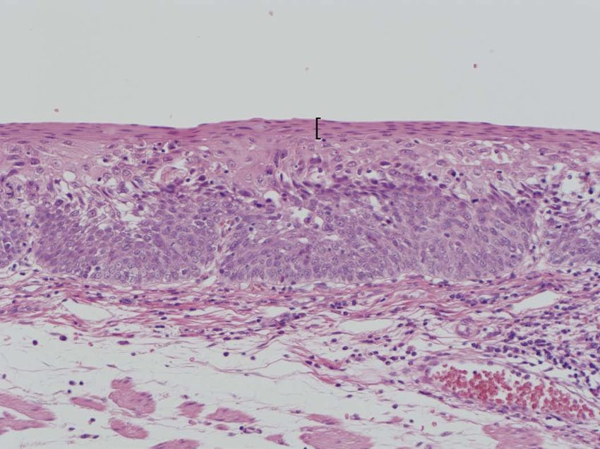
Ishihara R et al . Pink-color sign in esophageal cancer

Quantitative analysis was performed on 10 superficial                    can be expected after treatment with esophagectomy[2,3],
esophageal neoplasms with both pink-color sign posi-                     chemoradiotherapy[4,5] or endoscopic resection[6-9] if the
tive and negative areas in 10 patients. Pink-color sign                  cancer is detected at an early stage.
positive mucosa had a lower mean color value in the                          Conventional endoscopy has limited usefulness for
late phase (pinkish color) than in the early phase (yel-                 the treatment of esophageal cancer, because it is not easy
lowish color), and had similar mean color values in the                  to identify early neoplastic changes[10,11]. Chromoendos-
late and final phases. These findings suggest that pink-                 copy with iodine staining is reported to be more sensitive
color positive mucosa underwent color fading from the                    for the early diagnosis of esophageal squamous cell car-
color of the iodine (yellow) to the color of the mucosa
                                                                         cinoma[12,13], but has low specificity and requires multiple
(pink) within 2-3 min after iodine staining. Pink-color
                                                                         biopsy specimens[10,14]. A dramatic color change after
sign negative mucosa had similar mean color values in
                                                                         iodine staining, from the initial yellow color to a pink
the late and early phases (yellowish color), and had a
lower mean color value in the final phase (pinkish col-
                                                                         color 2-3 min later, is known as the pink-color sign, and
or) than in the late phase. These findings suggest that                  is useful for identifying cancerous lesions[15,16]. This sign
pink-color sign negative mucosa did not undergo color                    has been reported to dramatically improve specificity for
fading during the 2-3 min after iodine staining, and                     esophageal high-grade intraepithelial neoplasia (HGIN)
underwent color fading only after spraying of sodium                     and invasive cancer[15,16]. Choosing adequate biopsy sites is
thiosulfate.                                                             sometimes difficult, especially in patients with scattered-
                                                                         type staining of the esophagus, which is characterized by
CONCLUSION: The pink-color sign was associated                           multiple Lugol-voiding lesions[17]. Some of these iodine-
with absence of the keratinous layer. This sign may be                   unstained lesions may indicate inflammation or low-grade
caused by early fading of iodine staining.                               intraepithelial neoplasia (LGIN), and in such cases a lack
                                                                         of iodine staining is not a good indication for taking
© 2013 Baishideng. All rights reserved.                                  a biopsy specimen. Because of its high specificity, the
                                                                         pink-color sign is a good indicator for choosing adequate
Key words: Chromoendoscopy; Esophageal cancer;                           biopsy sites in patients with scattered-type staining. How-
Esophageal squamous neoplasia; Iodine staining; Pink-                    ever, the mechanism underlying the occurrence of the
color sign                                                               pink-color sign has not been fully investigated. Improved
                                                                         understanding of this mechanism may improve our un-
Core tip: The pink-color sign of iodine-unstained lesions
                                                                         derstanding of the characteristics of the relevant lesions,
is useful for the diagnosis of esophageal squamous
                                                                         and increase the likelihood of accurate diagnosis. This
cell carcinoma. We investigated histologic findings of
                                                                         study therefore aimed to clarify the histologic changes
esophageal neoplasms, and found that absence of the
                                                                         responsible for the occurrence of the pink-color sign in
keratinous layer because of neoplastic cell proliferation
may be responsible for the pink-color sign. Quantitative
                                                                         esophageal squamous neoplasia.
analysis of color showed that pink-color sign positive
mucosa underwent early color fading from the color of                    MATERIALS AND METHODS
the iodine (yellow) to the color of the mucosa (pink)
within 2-3 min. Based on these results, we speculated                    Endoscopic examination and resection
on the mechanism underlying the pink-color sign.                         The current clinical investigation was conducted during
These findings may improve our understanding of the                      routine endoscopic procedures for resection of esopha-
characteristics of esophageal neoplasms.                                 geal squamous lesions. Patients with superficial esopha-
                                                                         geal neoplasia confirmed by histologic examination were
                                                                         included in the study. Superficial esophageal neoplasia
Ishihara R, Kanzaki H, Iishi H, Nagai K, Matsui F, Yamashina T,          was defined as a lesion limited to the submucosa. Typi-
Matsuura N, Ito T, Fujii M, Yamamoto S, Hanaoka N, Takeuchi              cal endoscopic findings were superficial protruding type,
Y, Higashino K, Uedo N, Tatsuta M, Tomita Y, Ishiguro S. Pink-           superficial flat type and superficial excavated type. If a le-
color sign in esophageal squamous neoplasia, and speculation             sion had a large broad-based protrusion, crater and stiff-
regarding the underlying mechanism. World J Gastroenterol                ened wall, it was diagnosed as advanced cancer. Patients
2013; 19(27): 4300-4308 Available from: URL: http://www.wjg-
                                                                         were excluded if they had previously undergone surgery,
net.com/1007-9327/full/v19/i27/4300.htm DOI: http://dx.doi.
                                                                         chemotherapy or radiotherapy for esophageal cancer. The
org/10.3748/wjg.v19.i27.4300
                                                                         endoscopic procedures were performed using a high-
                                                                         resolution magnifying upper-gastrointestinal endoscope
                                                                         (GIF-Q240Z or GIF-H260Z; Olympus, Tokyo, Japan).
                                                                         The structure enhancement function of the video pro-
INTRODUCTION                                                             cessor was set at level B8 for narrow-band imaging (NBI).
Esophageal cancer is the sixth most common cause of                      A black soft hood (MB-162 for GIF-Q240Z or MB-46
cancer-related mortality worldwide[1]. The overall survival              for GIF-H260Z; Olympus) was mounted on the tip of
of patients with esophageal cancer remains poor, regard-                 the endoscope to maintain an adequate distance between
less of histologic type. However, a favorable prognosis                  the tip of the endoscopic zoom lens and the mucosal

           WJG|www.wjgnet.com                                     4301                            July 21, 2013|Volume 19|Issue 27|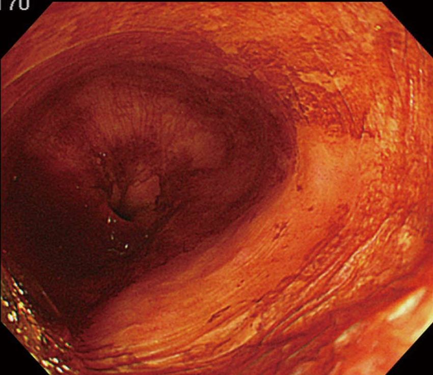
Ishihara R et al . Pink-color sign in esophageal cancer
(Ishihara R or Kanzaki H). Procedures that were per-
A
formed without these two endoscopists in attendance
were excluded from the study. Written informed consent
was obtained from all patients before endoscopic exami-
nation and resection of lesions. Institutional review board
approval was granted for a retrospective chart review and
analysis of the data.
Histologic evaluation
All specimens were cut into 2-mm slices and embedded
50 μm
in paraffin. Sections were cut from the paraffin blocks
and stained with hematoxylin and eosin. The pink-color
sign positive and negative lesions marked by the marker
B
dots were examined histologically. The depth of cancer
involvement was classified according to the Japanese
Classification of Esophageal Carcinoma[18], and intraepi-
thelial neoplasms were classified as LGIN or HGIN,
according to the World Health Organization classifica-
tion[19]. Intraepithelial cancer was included in HGIN.
The following histologic features that were possibly
associated with the pink-color sign were evaluated. The
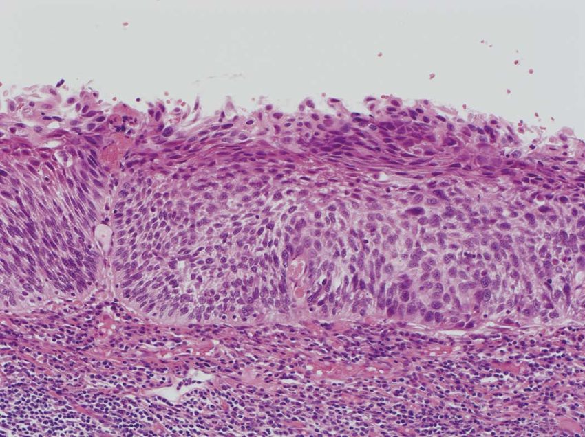
keratinous layer (Figure 1) and basal cell layer were classi-
50 μm
fied as present or absent. Cellular atypia was classified as
high grade, moderate grade or low grade, based on nuclear
irregularity, mitotic figures, loss of polarity, chromatin pat-
Figure 1 Histologic findings of esophageal lesions. A: Esophageal lesion tern and nuclear/cytoplasmic ratio. Vascular change was
with keratinous layers, shown by black parentheses; B: Esophageal lesion with-
assessed based on dilatation, tortuosity, caliber change and
out keratinous layers.
variability in shape. Vessels with these four findings were
classified as positive for vascular change[20]. All histologic
surface during observations. The endoscopic procedure assessments were performed by the same pathologist (SI),
was performed under intravenous sedation with midazol- who was blinded to the endoscopic and clinical findings.
am (Dormicam; Yamanouchi Pharma, Tokyo, Japan) and
pentazocine (Pentazin; Sankyo Pharmaceuticals, Tokyo, Quantitative analysis of color
Japan). This study included esophageal cancers those have both
The esophageal mucosa was initially examined us- pink-color sign positive and pink-color sign negative
ing white-light imaging or NBI. A catheter was then areas. The esophageal mucosa was initially examined
used to spray 20-40 mL of 0.6%-1.2% iodine solution with white-light imaging or NBI. A catheter was used to
until the esophageal mucosa was evenly stained, and the spray 20-40 mL of 1.2% iodine solution until the normal
subsequent color changes were observed. Immediately esophageal mucosa was evenly stained, and the subse-
after spraying of the iodine solution, the normal mucosa quent color changes were examined. Sodium thiosulfate
was dark brown, whereas abnormal mucosa suspicious solution was then sprayed to relieve symptoms caused
of dysplasia or cancer was yellow. The iodine-unstained by the iodine, which also accelerated the color fading by
(yellow) areas were classified as pink-color sign positive reduction of the iodine.
or pink-color sign negative. In pink-color sign positive The digital images were used to perform quantitative
mucosa, the yellow areas changed to pink within 2-3 min analysis of the color changes after iodine staining. Endo-
of spraying, and in pink-color sign negative mucosa, scopic images of the lesions were captured immediately
these areas remained yellow after 3 min. Representative after iodine staining, 2-3 min after iodine staining and after
areas (2-5 mm diameter) were marked with marker dots, complete fading of iodine staining, taking care to ensure
and the lesions were resected by endoscopic submucosal that all images were obtained from a similar direction and
dissection or endoscopic mucosal resection. Iodine stain- a similar distance, to enable accurate analysis (Figure 2). All
ing and marking of pink-color sign positive and nega- the images were captured under the instruction of one
tive areas were performed immediately before resection. endoscopist (Ishihara R). The images were stored in bit-
Considering the potential disadvantages of intraoperative map format (.bmp) with a resolution of 640 × 480 pixels.
endoscopy and iodine staining, patients requiring surgical A small region of interest was chosen in both the pink-
resection were excluded. To ensure that the study pro- color sign positive and pink-color sign negative areas.
tocol was strictly followed, marking before endoscopic These regions of interest were carefully chosen to be of
resection and confirmation of marking after endoscopic similar size in each area (Figure 3). The red-green-blue
resection were performed by one of two endoscopists components of each region of interest were calculated
WJG|www.wjgnet.com 4302 July 21, 2013|Volume 19|Issue 27|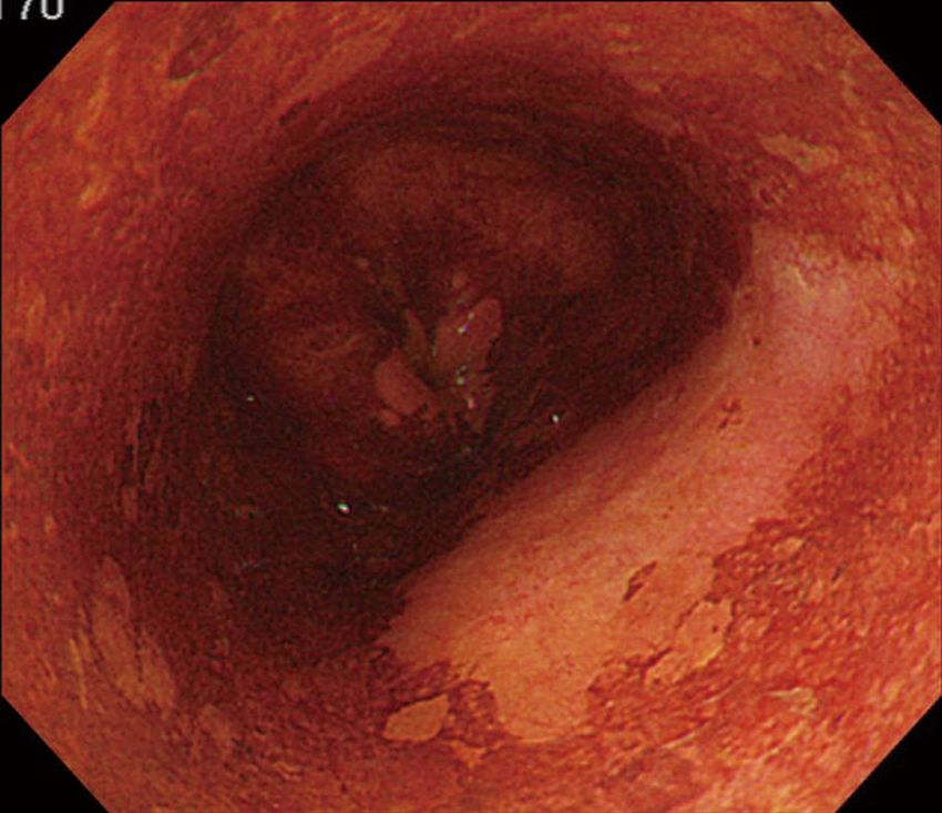
Ishihara R et al . Pink-color sign in esophageal cancer

 A                                                        B                                                       C

Figure 2 Endoscopic image of type IIc esophageal cancer. A: Endoscopic image of type IIc esophageal cancer immediately after iodine staining; B: Endoscopic
image of type IIc esophageal cancer 2 min after iodine staining. The pink-color sign is observed on the right side of the lesion; C: Endoscopic image of type IIc esoph-
ageal cancer 3 min after sodium thiosulfate spraying.

                                                                                        was resected from a patient, each lesion was considered
                                                                                        separately for the purposes of statistical analysis. Univari-
                                                                                        ate analyses of the relationships between the pink-color
        R: 229.9                                                                        sign and the histologic findings were performed using
        G: 130.2                                                                        the χ 2 test with Yates’ correction. Factors independently
        B: 75.5
                                                                                        associated with the pink-color sign were identified using
                   R: 217.3                                                             multivariate logistic regression analysis. The model fit was
                   G: 98.3                                                              assessed using the Hosmer-Lemeshow test. A two-sided P
                   B: 79.6                                                              value of < 0.05 was considered statistically significant. A
                                                                                        Bonferroni-adjusted P-value was used for multiple com-
                                                                                        parisons to control for experimental errors due to multiple
                                                                                        testing. All analyses were performed using SPSS software
                                                                                        version 11.0 (SPSS Inc, Chicago, IL, United States).
Figure 3 A small region of interest was chosen in both the pink-color sign
positive and pink-color sign negative areas. These regions of interest were
carefully chosen to be of similar size in each area. The red-green-blue compo-          RESULTS
nents of each region of interest were calculated using Image J software.
                                                                                        Histologic evaluation of pink-color sign positive areas
                                                                                        A total of 97 patients with superficial esophageal cancer
using Image J software (National Institutes of Health,                                  were treated by endoscopic resection at the Osaka Medi-
Bethesda, MD, United States), and color diagrams were                                   cal Center for Cancer and Cardiovascular Diseases from
created using the graphing function of Microsoft Excel                                  May 31, 2011 to March 1, 2012. Of these, 10 patients were
(Microsoft Corp., Redmond, WA, United States).                                          excluded because of previous radiation, 29 were excluded
     The LU’V’ color system is a uniform color space that                               because endoscopic resection was performed without the
was adopted by the Commission Internationale de l’E’                                    attendance of the two endoscopists (Ishihara R or Kanzaki
clairage in 1976 and is used to systematically represent                                H) and 4 were excluded because no images were captured
the different colors[21,22]. A color system is a model for                              at 2-3 min after iodine staining. A total of 61 superficial
representing colors in terms of intensity values. Gener-                                esophageal neoplasms in 54 patients were included in the
ally, colors are described using color systems with three                               study. The lesions were located in the cervical esophagus
or four dimensions (red-green-blue or cyan-magenta-                                     in one case, the upper thoracic esophagus in 10 cases, the
yellow-black). The diagram of the LU’V’ system has U’                                   mid-thoracic esophagus in 33 cases, and the lower thoracic
and V’ coordinates that represent the chromaticity values                               esophagus in 17 cases. The median diameter of the le-
of each color. This color space is designed to be per-                                  sions was 20 mm (range: 2-74 mm) (Table 1). Of the 61
ceptually uniform, meaning that a given change in value                                 lesions, 28 were classified as pink-color sign positive and
roughly corresponds to the same perceptual difference                                   33 as pink-color sign negative. The histologic diagnosis
over any part of the space. Using this system, color can                                was HGIN or cancer invading into the lamina propria in
be quantified and evaluated on a two-dimensional plane.                                 26 of the 28 pink-color sign positive lesions. Two lesions
However, determining the range of color is challenging                                  that were classified as pink-color sign positive were diag-
because there is no clear border between colors.                                        nosed as LGIN. One of these lesions had a thin kerati-
                                                                                        nous layer and mild cellular atypia. This lesion showed an
Statistical analysis                                                                    obscured pink-color sign, which was classified as positive
The relationships between the pink-color sign and each                                  in this study. In retrospect, it is possible that this lesion
histologic finding were analyzed. If more than one lesion                               should have been classified as pink-color sign negative.

               WJG|www.wjgnet.com                                                4303                                    July 21, 2013|Volume 19|Issue 27|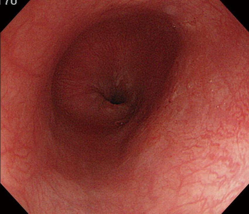
Ishihara R et al . Pink-color sign in esophageal cancer

 Table 1 Characteristics of patients and lesions                                   Table 3 Associations between pink-color sign and histologic
                                                                                   findings (univariate analysis)
 Characteristics                                    n
 Gender                                                                                                     Pink-color-sign     Pink-color-sign     P value
  Male                                              50                                                         positive            negative
  Female                                             4                             Keratinous layer                                                 < 0.0001
 Age (yr)                                                                           Present                        7                     32
  Median (range)                                 67 (45-82)                         Absent                        21                      1
 Lesion location                                                                   Cellular atypia1                                                   0.004
  Cervical esophagus                                 1                              Mild                           1                     13
  Upper thoracic esophagus                          10                              Moderate                      20                     19
  Middle thoracic esophagus                         33                              Severe                         7                      1
  Lower thoracic esophagus                          17                             Presence of basal membrane                                         0.018
 Lesion size (mm)                                                                   Yes                           14                     27
  Median (range)                                 20 (2-74)                          No                            14                      6
 Histological diagnosis of the marked area                                         Vascular change                                                    0.070
  LGIN                                              21                              Severe                        20                     15
  HGIN                                              37                              Mild                           8                     18
  LPM                                                3
                                                                               1
                                                                               Cellular atypia was classified based on nuclear irregularity, mitotic fig-
LGIN: Low-grade intraepithelial neoplasia; HGIN: High-grade intraepi-          ures, loss of polarity, chromatin pattern and nuclear/cytoplasmic ratio.
thelial neoplasia; LPM: Cancer invading into the lamina propria.

                                                                                   Table 4 Associations between pink-color sign and histologic
 Table 2 Associations between histologic diagnosis, keratinous                     findings (multivariate analysis)
 layer and pink-color sign
                                                                                                                       OR (95%CI)             P value
                      HGIN or invasive cancer     LGIN        P value              Keratinous layer                                               0.001
 Pink-color-sign                                              0.0001                Present                                   1
   Positive                      26                 2                               Absent                             58.8 (5.5-632)
   Negative                      14                19                              Cellular atypia1                                               0.580
 Keratinous layer                                             0.0007                Mild                                      1
   Present                       19                20                               Moderate                            3.5 (0.3-35.5)
   Absent                        21                 1                               Severe                              3.4 (0.07-165)
                                                                                   Presence of basal membrane
HGIN: High-grade intraepithelial neoplasia; LGIN: Low-grade intraepi-               Yes                                       1                   0.610
thelial neoplasia.                                                                  No                                  1.6 (0.3-9.3)
                                                                                   Vascular change
                                                                                    Mild                                      1                   0.770
The other lesion did not have a keratinous layer and had                            Severe                              1.3 (0.3-6.3)
moderate cellular atypia. However, this lesion showed
surface differentiation and was diagnosed as LGIN be-
                                                                               1
                                                                               Cellular atypia was classified based on nuclear irregularity, mitotic fig-
                                                                               ures, loss of polarity, chromatin pattern and nuclear/cytoplasmic ratio.
cause obvious cytological abnormalities were confined to
the lower half of the squamous epithelium. There was a
significant association between pink-color sign positive                       to December 5, 2012. Of these, 29 were diagnosed with
epithelium and HGIN or invasive cancer (P = 0.0001)                            esophageal squamous cell carcinoma. Fourteen of these
(Table 2). There was also a significant association be-                        29 patients were excluded for the following reasons: his-
tween the presence of a keratinous layer and HGIN or                           tory of chemoradiotherapy (8 patients), the entire lesion
invasive cancer (P = 0.0007).                                                  was pink-color sign positive (4 patients), or the pink-color
    Univariate analyses (Table 3) found significant as-                        sign was negative (2 patients). Ten of the 15 patients
sociations between the pink-color sign and absence of                          that had lesions with both pink-color sign positive and
the keratinous layer or cellular atypia. After Bonferroni                      pink-color sign negative areas were examined under the
correction, there were no significant associations between                     instruction of the endoscopist (Ishihara R) and were in-
the pink-color sign and presence of the basal membrane                         cluded in the analysis. Endoscopic images of the lesions
or vascular change. Multivariate analyses (Table 4) showed                     immediately after iodine staining (early phase), 2-3 min
that absence of the keratinous layer was independently                         after iodine staining (late phase) and after complete fading
associated with the pink-color sign (OR = 58.8, 95%CI:                         of iodine staining (final phase) were analyzed (Figure 4).
5.5-632). Hosmer-Lemeshow testing indicated that the                           The mean U’ and V’ values of the pink-color sign posi-
model achieved a sufficient goodness-of-fit (P = 0.678).                       tive and negative areas are shown in Figure 5. Pink-color
                                                                               sign positive mucosa had a lower mean V’ value in the
Quantitative analysis of the pink-color sign                                   late phase (pinkish color) than in the early phase (yellowish
A total of 1373 patients underwent esophagogastro-                             color), and had similar mean U’ and V’ values in the late
duodenoscopy at the Osaka Medical Center for Cancer                            and final phases (Figure 5A). These findings suggest that
and Cardiovascular Diseases from September 21, 2012                            pink-color positive mucosa underwent color fading from

             WJG|www.wjgnet.com                                         4304                                    July 21, 2013|Volume 19|Issue 27|
Ishihara R et al . Pink-color sign in esophageal cancer

 A                        Immediately after iodine staining                                A                     Immediately after iodine staining
                                                                                                                 2-3 min after iodine staining
                          2-3 min after iodine staining
                                                                                                                 3-5 min after sodium thiosulfate spraying
                          3-5 min after sodium thiosulfate spraying
     0.54                                                                                      0.54
                                  Yellow                                                                                   Yellow

     0.52                                                         Red                          0.52                                                          Red

     0.50                                                                                      0.50
V'

                                                                                           V'
     0.48                                      Pink                                            0.48                                      Pink

     0.46                                                                                      0.46
        0.15               0.20                  0.25                0.30                         0.15              0.20                 0.25                0.30
                                         U'                                                                                     U'

 B                    Immediately after iodine staining                                    B                     Immediately after iodine staining
                      2-3 min after iodine staining                                                              2-3 min after iodine staining
                      3-5 min after sodium thiosulfate spraying                                                  3-5 min after sodium thiosulfate spraying

  0.54                                                                                         0.54
                                Yellow                                                                                     Yellow

  0.52                                                              Red                        0.52                                                          Red

  0.50                                                                                         0.50
                                                                                           V'
V'

  0.48                                        Pink                                             0.48                                      Pink

  0.46                                                                                         0.46
     0.15                0.20                  0.25                 0.30                          0.15              0.20                 0.25                0.30
                                      U'                                                                                        U'

 Figure 4 The U’ and V’ values of the pink-color sign positive mucosa and                  Figure 5 Mean U’ and V’ values of the mucosa in the early, late and final
 negative mucosa in the early, late and final phases were plotted on a color               phases were plotted on a color diagram. A: Pink-color sign positive mucosa
 diagram. A: Pink-color sign positive mucosa; B: Pink-color sign negative mucosa.          had a lower mean V’ value in the late phase (pinkish color) than in the early
                                                                                           phase (yellowish color), suggesting that the pink-color sign positive mucosa
                                                                                           underwent a color change from yellow to pink. The mucosa had similar mean
 the color of the iodine (yellow) to the color of the muco-                                U’ and V’ values in the late and final phases, suggesting that the color of pink-
 sa (pink) within 2-3 min after iodine staining. Pink-color                                color sign positive mucosa in the late stage was similar to the color of mucosa
                                                                                           after complete fading of iodine staining; B: Pink-color sign negative mucosa had
 sign negative mucosa had similar mean U’ and V’ values
                                                                                           similar mean U’ and V’ values in the early and late phases (yellowish color),
 in the late and early phases (yellowish color), and had a                                 suggesting that pink-color sign negative mucosa did not change in color during
 lower mean V’ value in the final phase (pinkish color)                                    this time period.
 than in the late phase (Figure 5B). These findings suggest
 that pink-color sign negative mucosa did not undergo
 color fading during the 2-3 min after iodine staining, and                                procedures were of high quality to obtain accurate results.
 underwent color fading only after spraying of sodium                                      Marking before endoscopic resection and confirmation
 thiosulfate.                                                                              of marking after endoscopic resection were performed
                                                                                           by one of two endoscopists (Ishihara R or Kanzaki H) to
                                                                                           accurately identify the region of interest. All endoscopic
 DISCUSSION                                                                                images were captured under the instruction of one en-
 Analysis of the endoscopic and histologic findings of                                     doscopist (Ishihara R) to ensure that they were captured
 this study found that absence of the keratinous layer was                                 under similar conditions. This may have caused some se-
 independently associated with the pink-color sign. Quan-                                  lection bias, but it was felt necessary to limit the number
 titative analysis of color changes found that pink-color                                  of endoscopists involved for this detailed analysis.
 sign positive mucosa changed from yellowish to pinkish                                        Figure 6 shows our speculated mechanism for the
 within 2-3 min after iodine staining, suggesting that the                                 occurrence of the pink-color sign. Locally administered
 mucosa underwent early color fading from the color of                                     iodine is usually absorbed into the epithelium by passive
 the iodine (yellow) to the color of the mucosa (pink).                                    diffusion[23]. In normal esophageal epithelium, absorbed
     This study did not include all patients who met our                                   iodine combines with glycogen in the micro-granules of
 inclusion criteria, because we wanted to ensure that all                                  the prickle cells[24]. The resulting glycogen-iodine complex

                WJG|www.wjgnet.com                                                  4305                                    July 21, 2013|Volume 19|Issue 27|
Ishihara R et al . Pink-color sign in esophageal cancer

                      Histologic image                                                 Endoscopic image

                                                                   Before iodine
                                                                     staining

                            Cancer
                                                                                            Cancer
 Iodine penetrates into the epithelium by passive diffusion

                                                                   Iodine staining

                     Original color of iodine     Iodine glycogen
                                                      reaction

                Iodine exudes from the epithelium

                                                                    2-3 min after
                                                                   iodine staining

 Color fade by reductive reaction of sodium thiosulfate solution

                                                                    After sodium
                                                                     thiosulfate
                                                                      spraying

Figure 6 Speculated mechanism of the pink-color sign.

gives the epithelium a brown color. In the epithelium of                             epithelium. The keratin layer of squamous epithelium has
neoplastic lesions, the prickle-cell layer is usually replaced                       a barrier function[29,30], and may play an important role in
by neoplastic cells, and no glycogen-iodine complex is                               preventing leakage of iodine from the epithelium. Dis-
formed. The yellow iodine solution therefore gives the                               ruption of the normal epithelial structure, especially of
epithelium a yellow color.                                                           the keratin layer, may increase early leakage of iodine and
    In the epithelium of neoplastic lesions, the yellow                              early color fading. Considering the association between
color may fade because of reduction of the iodine, ab-                               the absence of the keratinous layer and the pink-color
sorption of iodine into the bloodstream, or leakage of                               sign, this may be the mechanism underlying the occur-
iodine into the esophageal lumen. Application of sodium                              rence of the sign.
thiosulfate solution reduces the adverse effects of iodine                               Neoplastic lesions show less staining when exposed
staining and accelerates color fading by reduction of the                            to iodine solution than normal mucosa. However, both
iodine[25]. However, iodine is a strong oxidizing agent[26],                         cancer and LGIN result in iodine-unstained areas. As-
and reduction of iodine requires a strong reducing agent.                            sessment of iodine staining without assessment of the
Absorption of iodine by the blood stream may occur in                                pink-color sign is therefore not very accurate for the
the esophagus[27]. However, iodine is mainly absorbed in                             diagnosis of cancer[10,14]. The keratinous layer was found
the small intestine[28] and the amount of iodine absorbed                            to be absent in areas where most of the epithelium was
by the esophagus is not large. Leakage of iodine into the                            replaced by neoplastic cells. Most areas that were pink-
esophageal lumen may therefore be the main cause of                                  color sign positive had a proliferation of neoplastic cells
color fading, rather than reduction of iodine or absorp-                             in the upper half of the epithelium. The pink-color sign
tion of iodine into the bloodstream.                                                 was associated with HGIN or cancer, those are both
    Epithelium plays an important role in regulating the                             characterized by abnormal cells in the upper half of the
permeability of mucosa, and serves as a barrier between                              epithelium. Accurate endoscopic diagnosis of esophageal
the outside world and the internal milieu of the organ-                              lesions is therefore possible by assessment of the pink-
ism[29,30]. The barrier function of epithelium is determined                         color sign after iodine staining.
by its microstructure, and varies among different types of                               Differentiating HGIN or cancer from LGIN is im-

             WJG|www.wjgnet.com                                               4306                            July 21, 2013|Volume 19|Issue 27|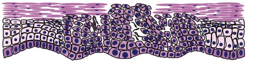
Ishihara R et al . Pink-color sign in esophageal cancer

portant when deciding on a treatment strategy, because                                       of diagnosis of esophageal neoplasms.
resection of the lesion is required for HGIN and can-
cer[19]. Histologic diagnosis of biopsy specimens may re-                                    REFERENCES
sult in misdiagnosis if the correct area was not biopsied.
                                                                                             1      Parkin DM, Bray F, Ferlay J, Pisani P. Global cancer statis-
Areas that are pink-color sign positive should always be
                                                                                                    tics, 2002. CA Cancer J Clin 2005; 55: 74-108 [PMID: 15761078
biopsied, because this finding is closely associated with                                           DOI: 10.3322/canjclin.55.2.74]
HGIN and cancer. Moreover, the keratinous layer is usu-                                      2      Kodama M, Kakegawa T. Treatment of superficial cancer of
ally absent in areas that are pink-color sign positive, and it                                      the esophagus: a summary of responses to a questionnaire
may therefore be relatively easy to biopsy neoplastic cells                                         on superficial cancer of the esophagus in Japan. Surgery
                                                                                                    1998; 123: 432-439 [PMID: 9551070]
from these areas. However, if further studies confirm                                        3      Igaki H, Kato H, Tachimori Y, Daiko H, Fukaya M, Yajima S,
that the accuracy of endoscopic diagnosis is similar to                                             Nakanishi Y. Clinicopathologic characteristics and survival
that of biopsy diagnosis, cancer could eventually be di-                                            of patients with clinical Stage I squamous cell carcinomas of
agnosed based on the pink-color sign without a need for                                             the thoracic esophagus treated with three-field lymph node
                                                                                                    dissection. Eur J Cardiothorac Surg 2001; 20: 1089-1094 [PMID:
biopsy.                                                                                             11717009]
    In conclusion, the pink-color sign was closely associ-                                   4      Yamamoto S, Ishihara R, Motoori M, Kawaguchi Y, Uedo
ated with absence of the keratinous layer. The pink-color                                           N, Takeuchi Y, Higashino K, Yano M, Nakamura S, Iishi
sign may be caused by early leakage of iodine into the                                              H. Comparison between definitive chemoradiotherapy and
                                                                                                    esophagectomy in patients with clinical stage I esophageal
esophageal lumen because of impaired barrier function                                               squamous cell carcinoma. Am J Gastroenterol 2011; 106:
of the epithelium.                                                                                  1048-1054 [PMID: 21343920 DOI: 10.1038/ajg.2011.42]
                                                                                             5      Kato H, Sato A, Fukuda H, Kagami Y, Udagawa H, Togo
                                                                                                    A, Ando N, Tanaka O, Shinoda M, Yamana H, Ishikura S. A
COMMENTS
COMMENTS                                                                                            phase II trial of chemoradiotherapy for stage I esophageal
                                                                                                    squamous cell carcinoma: Japan Clinical Oncology Group
Background                                                                                          Study (JCOG9708). Jpn J Clin Oncol 2009; 39: 638-643 [PMID:
A dramatic color change after iodine staining, from the initial yellow color to                     19549720 DOI: 10.1093/jjco/hyp069]
a pink color 2-3 min later, is known as the pink-color sign, and is useful for               6      Ishihara R, Tanaka H, Iishi H, Takeuchi Y, Higashino K,
identifying cancerous lesions of the esophagus. This sign has been reported to                      Uedo N, Tatsuta M, Yano M, Ishiguro S. Long-term outcome
dramatically improve specificity for esophageal squamous high-grade intraepi-                       of esophageal mucosal squamous cell carcinoma without
thelial neoplasia and invasive cancer.                                                              lymphovascular involvement after endoscopic resection.
Research frontiers                                                                                  Cancer 2008; 112: 2166-2172 [PMID: 18348303 DOI: 10.1002/
The mechanism underlying the occurrence of the pink-color sign has not been                         cncr.23418]
fully investigated. Improved understanding of this mechanism may improve our                 7      Fujishiro M, Yahagi N, Kakushima N, Kodashima S, Mu-
understanding of the characteristics of the relevant lesions, and increase the                      raki Y, Ono S, Yamamichi N, Tateishi A, Shimizu Y, Oka M,
likelihood of accurate diagnosis.                                                                   Ogura K, Kawabe T, Ichinose M, Omata M. Endoscopic sub-
Innovations and breakthroughs                                                                       mucosal dissection of esophageal squamous cell neoplasms.
This study clarified the histologic changes responsible for the occurrence of the                   Clin Gastroenterol Hepatol 2006; 4: 688-694 [PMID: 16713746
pink-color sign in esophageal squamous neoplasia as follows. Analysis of the                        DOI: 10.1016/j.cgh.2006.03.024]
endoscopic and histologic findings of this study found that absence of the kera-             8      Takahashi H, Arimura Y, Masao H, Okahara S, Tanuma T,
tinous layer was independently associated with the pink-color sign. Quantitative                    Kodaira J, Kagaya H, Shimizu Y, Hokari K, Tsukagoshi H,
analysis of color changes found that pink-color sign positive mucosa changed                        Shinomura Y, Fujita M. Endoscopic submucosal dissection is
from yellowish to pinkish within 2-3 min after iodine staining, suggesting that the                 superior to conventional endoscopic resection as a curative
mucosa underwent early color fading from the color of the iodine (yellow) to the                    treatment for early squamous cell carcinoma of the esopha-
color of the mucosa (pink). The pink-color sign may be caused by early leakage                      gus (with video). Gastrointest Endosc 2010; 72: 255-264, 264.
of iodine into the esophageal lumen because of impaired barrier function of the                     e1-2 [PMID: 20541198 DOI: 10.1016/j.gie.2010.02.040]
epithelium.                                                                                  9      Yamashina T, Ishihara R, Nagai K, Matsuura N, Matsui F,
                                                                                                    Ito T, Fujii M, Yamamoto S, Hanaoka N, Takeuchi Y, Hi-
Applications
                                                                                                    gashino K, Uedo N, Iishi H. Long-term outcome and meta-
The pink-color sign is a good indicator for choosing adequate biopsy sites,
                                                                                                    static risk after endoscopic resection of superficial esopha-
because of its high specificity. Improved understanding of this mechanism may
                                                                                                    geal squamous cell carcinoma. Am J Gastroenterol 2013; 108:
improve their understanding of the characteristics of the relevant lesions, and
                                                                                                    544-551 [PMID: 23399555 DOI: 10.1038/ajg.2013.8]
increase the likelihood of accurate diagnosis.
                                                                                             10     Hashimoto CL, Iriya K, Baba ER, Navarro-Rodriguez T,
Terminology                                                                                         Zerbini MC, Eisig JN, Barbuti R, Chinzon D, Moraes-Filho
Pink-color sign: A dramatic color change after iodine staining, from the initial                    JP. Lugol’s dye spray chromoendoscopy establishes early
yellow color to a pink color 2-3 min later. Keratinous layer: The outer layer of                    diagnosis of esophageal cancer in patients with primary
the squamous epithelium, which contain a tough, fibrous protein. This layer acts                    head and neck cancer. Am J Gastroenterol 2005; 100: 275-282
as a protective barrier against outside elements.                                                   [PMID: 15667482]
Peer review                                                                                  11     Dawsey SM, Fleischer DE, Wang GQ, Zhou B, Kidwell JA,
This study investigated the detailed histologic findings of esophageal neo-                         Lu N, Lewin KJ, Roth MJ, Tio TL, Taylor PR. Mucosal iodine
plasms, and found that absence of the keratinous layer because of neoplastic                        staining improves endoscopic visualization of squamous
cell proliferation may be responsible for the pink-color sign. Quantitative analy-                  dysplasia and squamous cell carcinoma of the esophagus in
sis of color changes showed that pink-color sign positive mucosa changed                            Linxian, China. Cancer 1998; 83: 220-231 [PMID: 9669803]
from yellowish to pinkish within 2-3 min after iodine staining, suggesting that              12     Shiozaki H, Tahara H, Kobayashi K, Yano H, Tamura S,
the mucosa underwent early color fading from the color of the iodine (yellow) to                    Imamoto H, Yano T, Oku K, Miyata M, Nishiyama K. Endo-
the color of the mucosa (pink). Based on these results, the authors speculated                      scopic screening of early esophageal cancer with the Lugol
on the mechanism underlying the pink-color sign. These findings may improve                         dye method in patients with head and neck cancers. Cancer
understanding of the characteristics of these lesions, and increase the accuracy                    1990; 66: 2068-2071 [PMID: 1699649]

               WJG|www.wjgnet.com                                                     4307                                  July 21, 2013|Volume 19|Issue 27|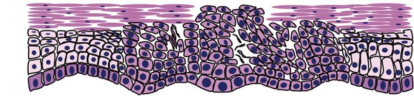
Ishihara R et al . Pink-color sign in esophageal cancer

13    Freitag CP, Barros SG, Kruel CD, Putten AC, Dietz J, Gru-                20   Yoshida T, Inoue H, Usui S, Satodate H, Fukami N, Kudo
      ber AC, Diehl AS, Meurer L, Breyer HP, Wolff F, Vidal R,                      SE. Narrow-band imaging system with magnifying endos-
      Arruda CA, Luz LP, Fagundes RB, Prolla JC. Esophageal                         copy for superficial esophageal lesions. Gastrointest Endosc
      dysplasias are detected by endoscopy with Lugol in patients                   2004; 59: 288-295 [PMID: 14745410]
      at risk for squamous cell carcinoma in southern Brazil. Dis              21   Chung KL, Yang WJ, Yan WM. Efficient edge preserving
      Esophagus 1999; 12: 191-195 [PMID: 10631911 DOI: 10.1046/                     algorithm for color contrast enhancement with application
      j.1442-2050.1999.00046.x]                                                     to color image segmentation. J Vis Commun Image Represent
14    Yokoyama A, Ohmori T, Makuuchi H, Maruyama K,                                 2008; 19: 299-310 [DOI: 10.1016/j.jvcir.2008.02.002]
      Okuyama K, Takahashi H, Yokoyama T, Yoshino K, Hayas-                    22   Schanda J. Colorimetry: Understanding the CIE System.
      hida M, Ishii H. Successful screening for early esophageal                    New York: John Wiley & Sons Inc, 2007: 61-64
      cancer in alcoholics using endoscopy and mucosa iodine                   23   Dela Cruz F, Brown DH, Leikin JB, Franklin C, Hryhorczuk
      staining. Cancer 1995; 76: 928-934 [PMID: 8625217]                            DO. Iodine absorption after topical administration. West J
15    Shimizu Y, Omori T, Yokoyama A, Yoshida T, Hirota J, Ono                      Med 1987; 146: 43-45 [PMID: 3825108]
      Y, Yamamoto J, Kato M, Asaka M. Endoscopic diagnosis                     24   Silverman S, Barbosa J, Kearns G. Ultrastructural and histo-
      of early squamous neoplasia of the esophagus with iodine                      chemical localization of glycogen in human normal and hy-
      staining: high-grade intra-epithelial neoplasia turns pink                    perkeratotic oral epithelium. Arch Oral Biol 1971; 16: 423-434
      within a few minutes. J Gastroenterol Hepatol 2008; 23: 546-550               [PMID: 5281213]
      [PMID: 17573830 DOI: 10.1111/j.1440-1746.2007.04990.x]                   25   Kondo H, Fukuda H, Ono H, Gotoda T, Saito D, Takahiro
16    Ishihara R, Yamada T, Iishi H, Kato M, Yamamoto S, Yama-                      K, Shirao K, Yamaguchi H, Yoshida S. Sodium thiosulfate
      moto S, Masuda E, Tatsumi K, Takeuchi Y, Higashino K,                         solution spray for relief of irritation caused by Lugol’s stain
      Uedo N, Tatsuta M, Ishiguro S. Quantitative analysis of the                   in chromoendoscopy. Gastrointest Endosc 2001; 53: 199-202
      color change after iodine staining for diagnosing esophageal                  [PMID: 11174292 DOI: 10.1067/mge.2001.110730]
      high-grade intraepithelial neoplasia and invasive cancer.                26   Knight CA, Stanley WM. THE EFFECT OF SOME CHEMI-
      Gastrointest Endosc 2009; 69: 213-218 [PMID: 18718584 DOI:                    CALS ON PURIFIED INFLUENZA VIRUS. J Exp Med 1944;
      10.1016/j.gie.2008.04.052]                                                    79: 291-300 [PMID: 19871371 DOI: 10.1084/jem.79.3.291]
17    Shimizu Y, Tukagoshi H, Fujita M, Hosokawa M, Kato                       27   Vorherr H, Vorherr UF, Mehta P, Ulrich JA, Messer RH.
      M, Asaka M. Metachronous squamous cell carcinoma of                           Vaginal absorption of povidone-iodine. JAMA 1980; 244:
      the esophagus arising after endoscopic mucosal resection.                     2628-2629 [PMID: 7431610 DOI: 10.1001/jama.1980.0331023
      Gastrointest Endosc 2001; 54: 190-194 [PMID: 11474389 DOI:                    0030018]
      10.1067/mge.2001.116877]                                                 28   Small MD, Bezman A, Longarini AE, Fennell A, Zamcheck
18    Japan Esophageal Society. Japanese Classification of                          N. Absorption of Potassium Iodide from Gastro-Intestinal
      Esophageal Cancer, tenth edition: part I. Esophagus 2009; 6:                  Tract. Proc Soc Exp Biol Med 1961; 106: 450-452
      1-25 [DOI: 10.1007/s10388-009-0169-0]                                    29   Powell DW. Barrier function of epithelia. Am J Physiol 1981;
19    Gabbert HE, Shimoda T, Hainaut P, Nakamura Y, Field JK,                       241: G275-G288 [PMID: 7032321]
      Inoue H. Squamous cell carcinoma of the esophagus. In:                   30   Kalinin AE, Kajava AV, Steinert PM. Epithelial barrier func-
      Hamilton SR, Aaltonen LA, editors. Pathology and Genetics                     tion: assembly and structural features of the cornified cell
      of the Digestive System: World Health Organization Clas-                      envelope. Bioessays 2002; 24: 789-800 [PMID: 12210515 DOI:
      sification. Lyon: IARC press, 2000: 11–19                                     10.1002/bies.10144]

                                                                                         P- Reviewers Kawakami K, Oka S S- Editor Wen LL
                                                                                                              L- Editor A E- Editor Ma S

             WJG|www.wjgnet.com                                         4308                               July 21, 2013|Volume 19|Issue 27|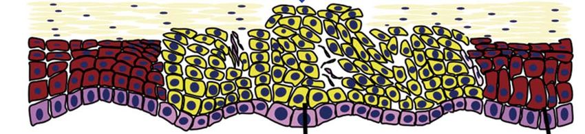
Published by Baishideng Publishing Group Co., Limited
                                  Flat C, 23/F., Lucky Plaza,
                    315-321 Lockhart Road, Wan Chai, Hong Kong, China
                                     Fax: +852-65557188
                                  Telephone: +852-31779906
                                E-mail: bpgoffice@wjgnet.com
                                    http://www.wjgnet.com

                                                                          I S S N 1 0  0 7  -   9  3 2  7
                                                                                                     27

                                                                      9   7 7 10  0 7   9 3 2 0 45

Baishideng Publishing Group Co., Limited                  © 2013 Baishideng. All rights reserved.
You can also read