Long non-coding RNA OTUD6B-AS1 overexpression inhibits the proliferation, invasion and migration of colorectal cancer cells via downregulation of ...
←
→
Page content transcription
If your browser does not render page correctly, please read the page content below
ONCOLOGY LETTERS 21: 193, 2021

        Long non-coding RNA OTUD6B-AS1 overexpression inhibits
           the proliferation, invasion and migration of colorectal
            cancer cells via downregulation of microRNA-3171
                                      WEI WANG1*, XIA CHENG2,3* and JIANHUA ZHU1

                   1
                    Department of Emergency Traumatic Surgery, Shanghai Pudong New District Zhoupu Hospital
                (Shanghai University of Medicine and Health Sciences Affiliated Zhoupu Hospital), Shanghai 201318;
                               2
                                 Graduate School, Dalian Medical University; 3Department of Oncology,
                      Affiliated Zhongshan Hospital of Dalian University, Dalian, Liaoning 116000, P.R. China

                                        Received June 24, 2020; Accepted October 13, 2020

                                                       DOI: 10.3892/ol.2021.12454

Abstract. Colorectal cancer (CRC) is a common digestive              strated that OTUD6B‑AS1 expression was low in CRC cells
system malignancy and a major cause of cancer‑associated             and tissues. Overexpression of OTUD6B‑AS1 inhibited
mortality worldwide. Aberrant expression of long non‑coding          the proliferation, invasion and migration of HCT116 cells.
RNAs has been reported in several types of cancer. The aim of        Furthermore, miR‑3171 was demonstrated to be a direct target
the present study was to investigate the role of ovarian tumor       of OTUD6B‑AS1 using a luciferase reporter assay. The rescue
domain containing 6B antisense RNA1 (OTUD6B‑AS1)                     assays revealed that miR‑3171 mimics markedly reversed
in CRC and its underlying mechanisms. OTUD6B‑AS1                     the inhibitory effects of OTUD6B‑AS1 overexpression on
expression in CRC cell lines was examined using reverse              proliferation, invasion and migration of CRC cells. Overall,
transcription‑quantitative PCR. Furthermore, The Cancer              these findings demonstrated that OTUD6B‑AS1 overexpres‑
Genome Atlas database was utilized to examine the expres‑            sion inhibited the proliferation, invasion and migration of
sion levels of OTUD6B‑AS1 in CRC tissues. Following                  HCT116 cells via downregulation of miR‑3171, suggesting
OTUD6B‑AS1 overexpression, Cell Counting Kit‑8 and colony            that OTUD6B‑AS1 may serve as a novel biomarker for CRC
formation assays were used to detect the proliferation ability       treatment.
of HCT116 cells. The expression levels of proliferation‑related
protein Ki67 were determined using immunofluorescence                Introduction
staining. Subsequently, Transwell and wound healing assays
were used to evaluate the invasion and migration of HCT116           Colorectal cancer (CRC) was the third most common cause of
cells, respectively. The expression levels of migration‑related      cancer‑associated mortality worldwide, and caused approxi‑
proteins (MMP2 and MMP9) were measured using western                 mately 900,000 deaths in 2013 (1). Due to technological
blotting. Additionally, a luciferase reporter assay was used to      advances in colonoscopy and other screening measures, the
verify the potential interaction between OTUD6B‑AS1 and              health condition of patients with CRC has improved in recent
microRNA‑3171 (miR‑3171). Subsequently, rescue assays were           years. However, it has been estimated that the overall inci‑
performed to clarify the regulatory effects of OTUD6B‑AS1            dence of CRC worldwide will increase by 60% to >2.2 million
and miR‑3171 on CRC development. The results demon‑                  cases and 1.1 million deaths by 2030 (2). Notably, CRC inci‑
                                                                     dence and mortality rates are increasing rapidly in developing
                                                                     countries (3). Furthermore, the incidence of CRC in the young
                                                                     generation is rising, as epidemiological studies have noted the
                                                                     rising numbers of adolescents and adults 200 nucleotides, and do not exhibit
                                                                     any capacity to encode proteins (6). lncRNAs are indispens‑
Key words: colorectal cancer, ovarian tumor domain containing 6B     able regulators in the process of gene expression, and emerging
antisense RNA1, proliferation, invasion, migration                   evidence has revealed the involvement of lncRNAs in cancer
                                                                     development and progression, since several studies have
                                                                     identified that the aberrant expression of lncRNAs is closely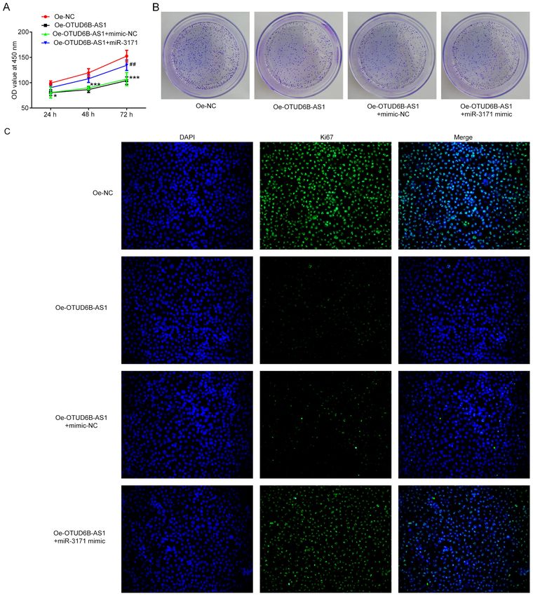
2                                 WANG et al: ROLES OF OTUD6B-AS1 IN COLORECTAL CANCER

associated with biological behaviors of malignant carcinoma       transfected with the aforementioned oligonucleotides using
cells, such as proliferation, invasion and metastasis (7,8).      Lipofectamine® 3000 (Invitrogen; Thermo Fisher Scientific,
lncRNA ovarian tumor domain containing 6B antisense RNA1          Inc.) according to the manufacturer's protocols at 37˚C for
(OTUD6B‑AS1) is oriented in an antisense direction relative       48 h. At 48 h after transfection, HCT116 cells were harvested
to the protein‑coding gene OTUD6B on the opposite DNA             for further experiments, and successful transfection was veri‑
strand (9). OTUD6BAS1 is located on chromosome 8q21.3             fied using reverse transcription‑quantitative PCR (RT‑qPCR).
and has 2,179 bp (NR_110439, ENST00000524003.1) (10). A
previous study has demonstrated that OTUD6B‑AS1 expres‑           RT‑qPCR. Total RNA was extracted from HCT116 cells using
sion is downregulated in clear cell renal cell carcinoma tissue   TRIzol® reagent (Invitrogen; Thermo Fisher Scientific, Inc.).
samples, while overexpression of OTUD6B‑AS1 inhibits cell         Total RNA was then reverse transcribed into cDNA at 42˚C for
proliferation, migration and invasion of clear cell renal cell    30 min using a reverse transcription kit (PrimeScript™ RT
carcinoma (11). However, to the best of our knowledge, the role   Reagent Kit; Takara Bio, Inc.). qPCR was performed using
of OTUD6B‑AS1 in CRC has not yet been determined.                 iTaq™ Universal SYBR® ‑Green Supermix (Bio‑Rad
    MicroRNAs (miRNAs/miRs) are highly conserved endog‑           Laboratories, Inc.) on an ABI 7500 instrument (Applied
enous non‑coding RNAs that are ~22 nucleotides long. They         Biosystems; Thermo Fisher Scientific, Inc.). The following ther‑
have been demonstrated to serve important roles in a variety      mocycling conditions were used: Pre‑denaturation at 95˚C for
of biological processes, including development, differentiation   10 min, denaturation at 95˚C for 15 sec and annealing at 60˚C
and signaling (12‑15). Dysregulated miRNAs may function           for 1 min (40 cycles). The sequences of the gene‑specific primers
as either tumor suppressors or oncogenes in carcinoma by          used in the present study were as follows: lncRNA OTUD6B‑AS1
targeting each one of these features (16). Using Starbase, it     forward, 5'‑AGCACACCCAGTCAGAAACCAG‑3' and reverse,
was predicted that OTUD6B‑AS1 can bind to miR‑3171.               5'‑TCTACAAACGGGAATGTCG‑3'; miR‑3171 forward,
Studies have demonstrated that miR‑3171 expression is             5'‑AGATGTATGGAATCTGTATATA‑3' and reverse, 5'‑GAA
abnormally increased in bladder cancer and hepatocellular         CATGTCTGCGTATCTC‑3'; GAPDH forward, 5'‑TGTGGG
carcinoma tissues (17,18). Therefore, it was hypothesized that    CATCAATGGATTTGG‑3' and reverse, 5'‑ACACCATGTATT
OTUD6B‑AS1 could exert certain effects on the proliferation,      CCGGGTCAAT‑3'; and U6 forward, 5'‑TCTGCTCCTATCC
invasion and migration of CRC cells by regulating miR‑3171.       CAATTACCTG‑3' and reverse, 5'‑ACTCCCGGATCTCTTCT
    To the best of our knowledge, the present study was the       AAGTTG‑3'. GAPDH and U6 were used as internal controls for
first to investigate the role of OTUD6B‑AS1 in CRC, and to        OTUD6B‑AS1 and miR‑3171, respectively, and relative expres‑
examine whether OTUD6B‑AS1 could affect the proliferation,        sion was calculated based on the 2‑ΔΔCq method (20).
invasion and migration of CRC cells by regulating miR‑3171.
                                                                  Cell Counting Kit‑8 (CCK‑8) assay. HCT116 cells were seeded
Materials and methods                                             into a 96‑well plate at a density of 2x10⁴ cells/well and cultured
                                                                  at 37˚C with 5% CO2. Following transfection for 24, 48 and
The Cancer Genome Atlas (TCGA) database analysis. Human           72 h, 10 µl CCK‑8 solution [OBiO Technology (Shanghai)
RNA‑sequencing data from colorectal cancer projects, which        Corp., Ltd.] was added to each well. Following incubation at
included 482 patients with CRC and 155 normal tissues (project    37˚C for 4 h, the absorbance at 450 nm was detected using a
no. TCGA‑COAD) were obtained by TCGA (https://portal.gdc.         spectrophotometer (Thermo Fisher Scientific, Inc.).
cancer.gov/) analysis in the UALCAN database (ualcan.path.
uab.edu/) (19). A Mann‑Whitney test was used to determine         Colony formation assay. The HCT116 cells (0.5x10³ cells/well)
the statistical significance of the difference in OTUD6B‑AS1      were seeded in a six‑well plate and cultured for 10 days after
expression between normal and tumor samples.                      treatment. Subsequently, colonies were fixed with 10% form‑
                                                                  aldehyde for 10 min at room temperature and stained with
Cell culture. CRC cell lines (Caco2, HCT116, LoVo, SW480 and      0.5% crystal violet for 5 min at room temperature. The number
SNU‑C1) and the normal intestinal epithelial cell line (HIEC)     of colonies was counted using ImageJ software (version 1.52r;
were purchased from American Type Culture Collection. All         National Institutes of Health) and images were captured under
cell lines were routinely maintained in RPMI‑1640 medium          a fluorescence inversion microscope (Olympus Corporation).
(HyClone; Cytiva) supplemented with 10% FBS (Gibco;
Thermo Fisher Scientific, Inc.), 100 U/ml penicillin and          Immunofluorescence staining. Treated HCT116 cells were
100 µg/ml streptomycin (Invitrogen; Thermo Fisher Scientific,     fixed with 4% paraformaldehyde for 30 min at 37˚C, and then
Inc.). Cells were cultured in a humidified incubator at 37˚C      0.5% Triton X‑100 was used to permeabilize the cells at room
with 5% CO2. The culture medium was replaced every 3 days.        temperature for 20 min. After blocking with 5% BSA (Beyotime
Cells were passaged when 80% confluence was reached.              Institute of Biotechnology) for 1 h at room temperature, slides
                                                                  were incubated overnight at 4˚C with a primary antibody against
Cell transfection. The OTUD6B‑AS1 overexpression plasmid          Ki67 (cat. no. ab15580; dilution, 1:1,000; Abcam). Subsequently,
(Oe‑OTUD6B‑AS1; 1 µg) and empty vector (Oe‑NC;                    the slides were incubated with a fluorescent secondary anti‑
1 µg) were obtained from Shanghai GenePharma Co., Ltd.            body (cat. no. BA1105; dilution, 1:10,000; Boster Biological
miR‑3171 mimics (40 nM; cat. no. miR10015046‑1‑5) and             Technology) for 1 h in a wet box at room temperature in the
corresponding scrambled mimic negative control (mimic‑NC;         dark. Finally, DAPI was used to counterstain the nuclei at room
40 nM; cat. no. miR1N0000001‑1‑5) were purchased from             temperature for 15 min. Images were captured under a fluores‑
Guangzhou RiboBio Co., Ltd. Cells (1x10⁶ cells/well) were         cence inversion Olympus microscope (Olympus Corporation).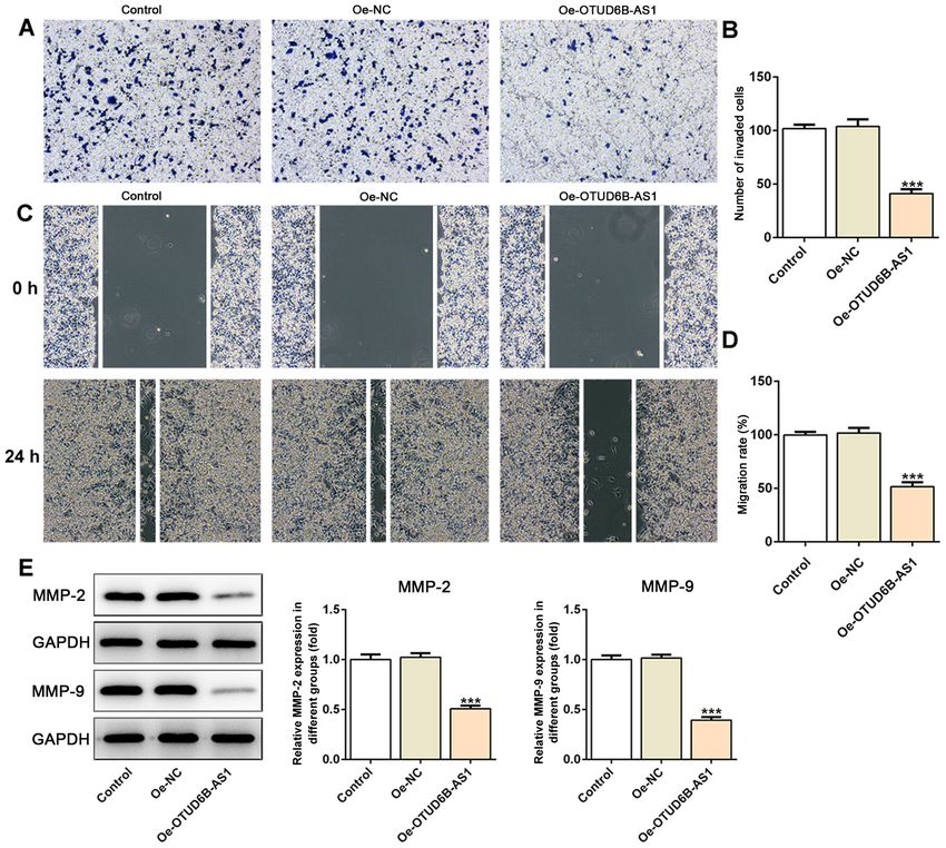
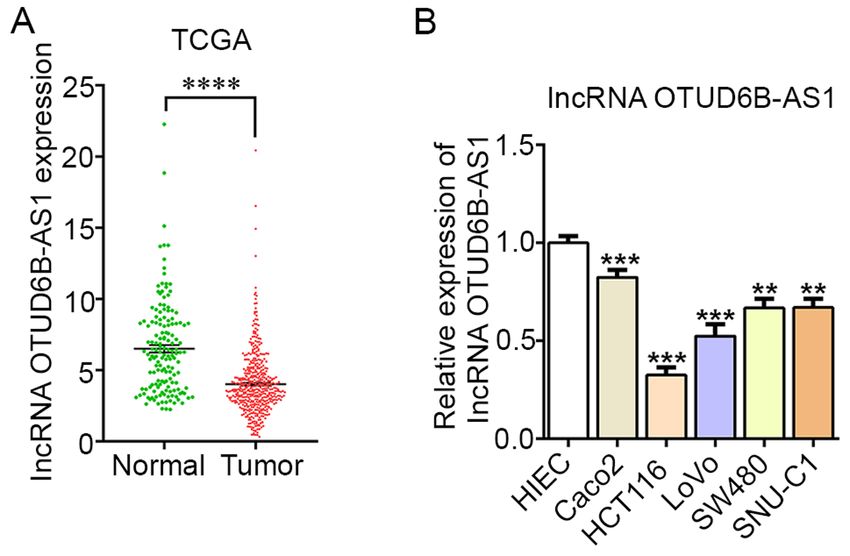
ONCOLOGY LETTERS 21: 193, 2021                                                                  3

Luciferase reporter assay. Potential target genes of lncRNA
OTUD6B‑AS1 were predicted using an online bioinformatics
software Starbase 2.0 (http://starbase.sysu.edu.cn/) (21).
HCT116 cells were reseeded into 24‑well plates and cultured
for 24 h. The fragments of OTUD6B‑AS1 containing predicted
wild‑type (WT) and mutant (MUT) miR‑3171 binding
sequences were amplified by Shanghai GenePharma Co., Ltd.,
and inserted into the luciferase reporter gene of the pmirGLO
vector (Promega Corporation) to produce the reporter
plasmids OTUD6B‑AS1‑WT and OTUD6B‑AS1‑MUT,
respectively. Subsequently, the cells (1x10 4 cells/well) were
co‑transfected with plasmids and miR‑3171 mimic (40 nM;           Figure 1. OTUD6B‑AS1 is expressed at low levels in CRC tissues and several
cat. no. miR10015046‑1‑5; Guangzhou RiboBio Co., Ltd.)            CRC cell lines (Caco2, HCT116, LoVo, SW480 and SNU‑C1). (A) TCGA was
or mimic‑NC vector (40 nM; cat. no. miR1N0000001‑1‑5;             used to determine the mRNA expression levels of OTUD6B‑AS1 in CRC tumor
Guangzhou RiboBio Co., Ltd.) using Lipofectamine® 2000            tissues and adjacent non‑tumor tissues. ****P
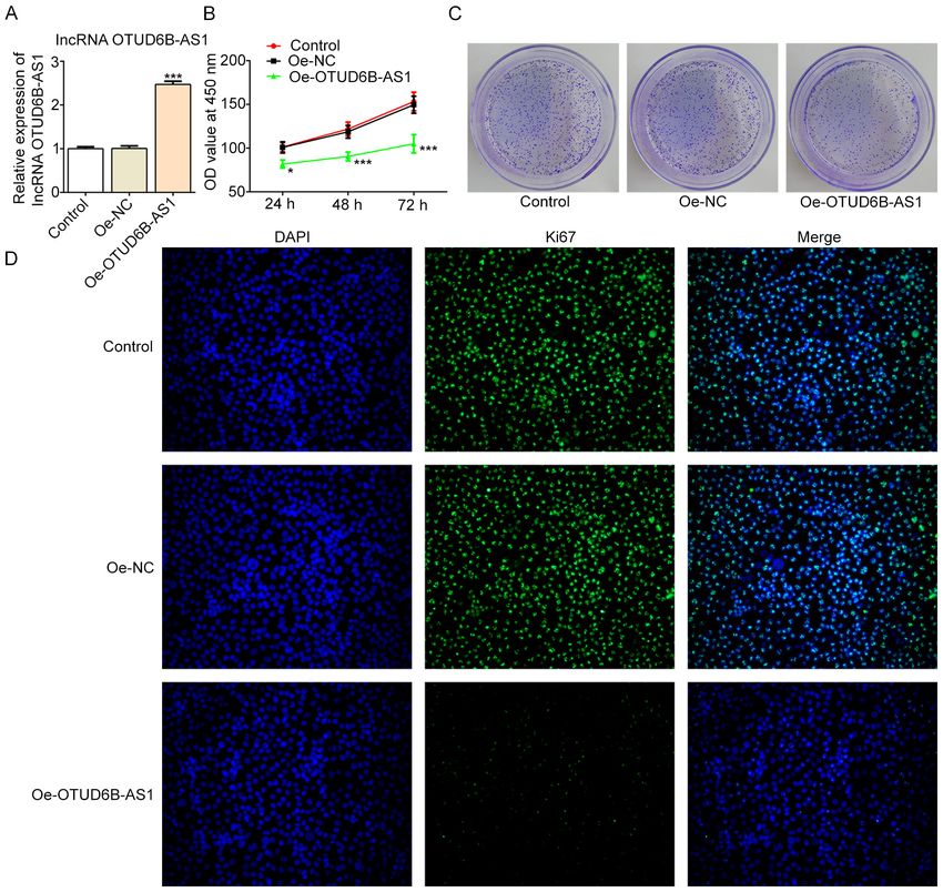
4                                        WANG et al: ROLES OF OTUD6B-AS1 IN COLORECTAL CANCER

Figure 2. Overexpression of OTUD6B‑AS1 inhibits the proliferation of HCT116 cells. (A) mRNA expression levels of OTUD6B‑AS1 were determined using
reverse transcription‑quantitative PCR following transfection with OTUD6B‑AS overexpression plasmid. The proliferation of HCT116 cells was measured
using (B) Cell Counting Kit‑8 and (C) colony formation assays. (D) Expression levels of proliferation‑related protein Ki67 were detected by immunofluores‑
cence staining. Magnification, x200. *P
ONCOLOGY LETTERS 21: 193, 2021                                                                   5

Figure 3. Overexpression of OTUD6B‑AS1 suppresses the invasion and migration of HCT116 cells. (A) The invasive ability of HCT116 cells was detected
using a Transwell assay. Magnification, x100. (B) Relative number of invaded cells. (C) The migratory activity of cells was evaluated using a scratch wound
healing assay. Magnification, x200. (D) Relative migration rate. (E) Expression levels of migration‑related proteins were measured by western blot analysis.
All band images presented were obtained from the same representative gel. ***P
6                                       WANG et al: ROLES OF OTUD6B-AS1 IN COLORECTAL CANCER

Figure 5. miR‑3171 mimic reverses the inhibitory effect of OTUD6B‑AS1 overexpression on the proliferation of HCT116 cells. The proliferation of HCT116
cells was examined using (A) Cell Counting Kit‑8 and (B) colony formation assays following co‑transfection of OTUD6B‑AS1 overexpression plasmid and
miR‑3171 mimic. (C) Expression of proliferation‑related protein Ki67 was detected by immunofluorescence staining. Magnification, x200. *P
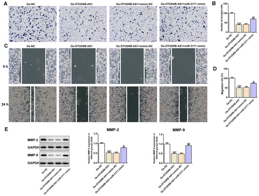
ONCOLOGY LETTERS 21: 193, 2021                                                                     7

Figure 6. miR‑3171 overexpression alleviates the inhibitory effects of OTUD6B‑AS1 overexpression on the invasion and migration of HCT116 cells. (A) Invasive
ability of HCT116 cells detected using a Transwell assay. Magnification, x100. (B) Relative number of invaded cells. (C) Representative images and (D) relative
quantification of cell migration, as examined using a scratch assay. Magnification, x100. (E) Expression levels of migration‑related proteins were examined by
western blotting. All band images presented were obtained from the same representative gel. ***P
8                                 WANG et al: ROLES OF OTUD6B-AS1 IN COLORECTAL CANCER

carcinoma cells (11). When analyzing data from TCGA, it           comprehensive analysis is required in the future. Additionally,
was revealed that the levels of OTUD6B‑AS1 in CRC tissues         the use of only one TCGA dataset to investigate OTUD6B‑AS1
were notably decreased compared with those in the adjacent        expression is another limitation of the present study, and future
non‑tumor tissues, suggesting a potential antitumor effect        studies should improve upon this and the other limitations.
of OTUD6B‑AS1 in CRC progression. It is well‑known that
abnormal and uncontrolled proliferation is a characteristic       Acknowledgements
of cancer cells (32). Furthermore, invasion and migration are
considered to be two dominant processes for tumor metastasis,     Not applicable.
which leads to cancer‑associated mortality (33). Thus, inter‑
ruption of the aforementioned processes is an effective method    Funding
for the inhibition of cancer metastasis. Therefore, the present
study aimed to investigate whether OTUD6B‑AS1 can have            No funding was received.
an effect on the proliferation, invasion and migration of CRC
cells. The present study demonstrated that OTUD6B‑AS1             Availability of data and materials
expression was decreased in CRC cells compared with in a
normal intestinal epithelial cell line, which was in accordance   The datasets used and/or analyzed during the current study are
with the results of TCGA database analysis. Furthermore,          available from the corresponding author on reasonable request.
overexpression of OTUD6B‑AS1 inhibited the proliferation,
migration and invasion of CRC cells.                              Authors' contributions
    Evolutionarily conserved miRNAs can suppress gene
expression at the posttranscriptional level (34). Furthermore,    WW and XC searched the literature, designed the experiments
they are considered to mediate the expression levels of ≥30%      and performed the experiments. WW and JZ analyzed the
of all protein‑coding genes, and are involved in most cellular    data and wrote the manuscript. JZ revised the manuscript. All
processes (35). Over the past few decades, the aberrant           authors read and approval the final manuscript.
expression profile of miRNAs has been frequently recog‑
nized in the peripheral blood and tumor tissue specimens          Ethics approval and consent to participate
from patients with CRC, hinting at the oncogenic effects of
miRNAs in various types of cancer (36,37). The upregula‑          Not applicable.
tion of miR‑135b has been commonly detected in patients
with CRC by functional screening, and is associated with the      Patient consent for publication
clinical stage and cancer‑specific survival of patients (38).
Additionally, increased expression levels of miR‑31 in patients   Not applicable.
with CRC are associated with poor prognosis (39). It has been
hypothesized that the inhibition of dysregulated miRNAs           Competing interests
could decrease the activity of CRC cells (40). In the present
study, a binding site between OTUD6B‑AS1 and miR‑3171             The authors declare that they have no competing interests.
was predicted using the Starbase database, and miR‑3171
expression was upregulated in HCT116 cells. Therefore, it was     References
hypothesized that OTUD6B‑AS1 could inhibit CRC progres‑
sion via inhibition of miR‑3171 expression. To further verify      1. Kuipers EJ, Grady WM, Lieberman D, Seufferlein T, Sung JJ,
                                                                      Boelens PG, van de Velde CJ and Watanabe T: Colorectal cancer.
this hypothesis, another RT‑qPCR assay was performed and              Nat Rev Dis Primers 1: 15065‑15065, 2015.
revealed that miR‑3171 expression was markedly reduced             2. Arnold M, Sierra MS, Laversanne M, Soerjomataram I, Jemal A
following OTUD6B‑AS1 overexpression. In a series of assays,           and Bray F: Global patterns and trends in colorectal cancer
                                                                      incidence and mortality. Gut 66: 683‑691, 2017.
it was observed that the additional treatment with miR‑3171        3. Zhou LL, Zou MD and Li WM: Recent advances in colorectal
mimics following OTUD6B‑AS1 overexpression in HCT116                  cancer‑specific nucleic acid aptamers for diagnostic and thera‑
cells promoted the proliferation, invasion and migration of           peutic applications. Sci Adv Mater 12: 38‑43, 2020.
                                                                   4. The Lancet Oncology: Colorectal cancer: A disease of the
HCT116 cells, suggesting that OTUD6B‑AS1 overexpression               young? Lancet Oncol 18: 413, 2017.
inhibited the proliferation, invasion and migration of HCT116      5. Kasi PM, Shahjehan F, Cochuyt JJ, Li Z, Colibaseanu DT and
cells via downregulation of miR‑3171 expression.                      Merchea A: Rising proportion of young individuals with rectal
                                                                      and colon cancer. Clin Colorectal Cancer 18: e87‑e95, 2019.
    In conclusion, to the best of our knowledge, the present       6. Xu M, Xu X, Pan B, Chen X, Lin K, Zeng K, Liu X, Xu T, Sun L,
study was the first to investigate the role of OTUD6B‑AS1 in          Qin J, et al: LncRNA SATB2‑AS1 inhibits tumor metastasis and
CRC cells, and to reveal that lncRNA OTUD6B‑AS1 over‑                 affects the tumor immune cell microenvironment in colorectal
                                                                      cancer by regulating SATB2. Mol Cancer 18: 135, 2019.
expression inhibited the proliferation, invasion and migration     7. Quinn JJ and Chang HY: Unique features of long non‑coding
of HCT116 cells, at least partially, by targeting miR‑3171.           RNA biogenesis and function. Nat Rev Genet 17: 47‑62, 2016.
Therefore, OTUD6B‑AS1 may serve as a potential novel               8. Rathinasamy B and Velmurugan BK: Role of lncRNAs in the
                                                                      cancer development and progression and their regulation by
biomarker and target for the diagnosis and treatment of CRC.          various phytochemicals. Biomed Pharmacother 102: 242‑248,
However, the use of only one CRC cell line in the cell function       2018.
experiments and mechanism experiments, and lack of data            9. Takata M, Pachera E, Frank‑Bertoncelj M, Kozlova A, Jüngel A,
                                                                      Whitfield ML, Assassi S, Calcagni M, de Vries‑Bouwstra J,
obtained from clinical samples and investigations in animal           Huizinga TW, et al: OTUD6B‑AS1 might be a novel regulator
models were limitations of the present study. Therefore, a            of apoptosis in systemic sclerosis. Front Immunol 10: 1100, 2019.
ONCOLOGY LETTERS 21: 193, 2021 9
10. Wang Z, Xia F, Feng T, Jiang B, Wang W and Li X: OTUD6B‑AS1 27. Heo JB, Lee YS and Sung S: Epigenetic regulation by long
inhibits viability, migration, and invasion of thyroid carcinoma by noncoding RNAs in plants. Chromosome Res 21: 685‑693, 2013.
targeting miR‑183‑5p and miR‑21. Front Endocrinol (Lausanne) 11: 28. Zhang H, Chen Z, Wang X, Huang Z, He Z and Chen Y: Long
136, 2020. non‑coding RNA: A new player in cancer. J Hematol Oncol 6:
11. Wang G, Zhang ZJ, Jian WG, Liu PH, Xue W, Wang TD, 37, 2013.
Meng YY, Yuan C, Li HM, Yu YP, et al: Novel long noncoding 29. Ma Y, Yang Y, Wang F, Moyer MP, Wei Q, Zhang P, Yang Z,
RNA OTUD6B‑AS1 indicates poor prognosis and inhibits clear Liu W, Zhang H, Chen N, et al: Long non‑coding RNA
cell renal cell carcinoma proliferation via the Wnt/β ‑catenin CCAL regulates colorectal cancer progression by activating
signaling pathway. Mol Cancer 18: 15, 2019. Wnt/β ‑catenin signalling pathway via suppression of activator
12. Mahesh G and Biswas R: MicroRNA‑155: A master regulator of protein 2α. Gut 65: 1494‑1504, 2016.
inflammation. J Interferon Cytokine Res 39: 321‑330, 2019. 30. Cai B, Song XQ, Cai JP and Zhang S: HOTAIR: A cancer‑related
13. Ambros V: The functions of animal microRNAs. Nature 431: long non‑coding RNA. Neoplasma 61: 379‑391, 2014.
350‑355, 2004. 31. Galamb O, Barták BK, Kalmár A, Nagy ZB, Szigeti KA,
14. Wu Y, Yang L, Yu M and Wang J: Identification and expression Tulassay Z, Igaz P and Molnár B: Diagnostic and prognostic
analysis of microRNAs during ovule development in rice (Oryza potential of tissue and circulating long non‑coding RNAs in
sativa) by deep sequencing. Plant Cell Rep 36: 1815‑1827, 2017. colorectal tumors. World J Gastroenterol 25: 5026‑5048, 2019.
15. Rane S, Sayed D and Abdellatif M: MicroRNA with a 32. Wang AH, Fan WJ, Fu L and Wang XT: LncRNA PCAT‑1
MacroFunction. Cell Cycle 6: 1850‑1855, 2007. regulated cell proliferation, invasion, migration and apoptosis in
16. Iorio MV and Croce CM: MicroRNA dysregulation in cancer: colorectal cancer through targeting miR‑149‑5p. Eur Rev Med
Diagnostics, monitoring and therapeutics. A comprehensive Pharmacol Sci 23: 8310‑8320, 2019.
review. EMBO Mol Med 9: 852, 2017. 33. Zhang H, Song Y, Yang C and Wu X: Overexpression of lncRNA
17. Wei Y, He R, Wu Y, Gan B, Wu P, Qiu X, Lan A, Chen G, TUSC7 reduces cell migration and invasion in colorectal cancer.
Wang Q, Lin X, et al: Comprehensive investigation of aberrant Oncol Rep 41: 3386‑3392, 2019.
microRNA profiling in bladder cancer tissues. Tumour Biol 37: 34. Lin J, Chuang CC and Zuo L: Potential roles of microRNAs and
12555‑12569, 2016. ROS in colorectal cancer: Diagnostic biomarkers and therapeutic
18. Wang Z, Zhao Y, Wang Y and Jin C: Circular RNA circHIAT1 targets. Oncotarget 8: 17328‑17346, 2017.
inhibits cell growth in hepatocellular carcinoma by regulating 35. Liu Z, Wang Y, Borlak J and Tong W: Mechanistically linked
miR‑3171/PTEN axis. Biomed Pharmacother 116: 108932, 2019. serum miRNAs distinguish between drug induced and fatty liver
19. Guo S, Zhu KX, Yu WH, Wang T, Li S, Wang YX, Zhang CC, disease of different grades. Sci Rep 6: 23709, 2016.
Guo JQ, et al: SH3PXD2A‑AS1/miR‑330‑5p/UBA2 ceRNAnetwork 36. Gmerek L, Martyniak K, Horbacka K, Krokowicz P, Scierski W,
mediates the progression of colorectal cancer through regulating Golusinski P, Golusinski W, Schneider A and Masternak MM:
the activity of the Wnt/beta‑catenin signaling pathway. Environ MicroRNA regulation in colorectal cancer tissue and serum.
Toxicol: Oct 19, 2020 (Epub ahead of print). doi: 10.1002/tox.23038. PLoS One 14: e0222013, 2019.
20. Livak KJ and Schmittgen TD: Analysis of relative gene expression 37. Nagy ZB, Barták BK, Kalmár A, Galamb O, Wichmann B,
data using real‑time quantitative PCR and the 2(‑Delta Delta Dank M, Igaz P, Tulassay Z and Molnár B: Comparison of
C(T)) method. Methods 25: 402‑408, 2001. circulating miRNAs expression alterations in matched tissue
21. Li JH, Liu S, Zhou H, Qu LH and Yang JH: starBase v2.0: and plasma samples during colorectal cancer progression. Pathol
Decoding miRNA‑ceRNA, miRNA‑ncRNA and protein‑RNA Oncol Res 25: 97‑105, 2019.
interaction networks from large‑scale CLIP‑Seq data. Nucleic 38. Valeri N, Braconi C, Gasparini P, Murgia C, Lampis A,
Acids Res 42: D92‑D97, 2014. Paulus‑Hock V, Hart JR, Ueno L, Grivennikov SI, Lovat F, et al:
22. Torre LA, Bray F, Siegel RL, Ferlay J, Lortet‑Tieulent J and MicroRNA‑135b promotes cancer progression by acting as a
Jemal A: Global cancer statistics, 2012. CA Cancer J Clin 65: downstream effector of oncogenic pathways in colon cancer.
87‑108, 2015. Cancer Cell 25: 469‑483, 2014.
23. Hua RX, Zhuo ZJ, Zhu J, Zhang SD, Xue WQ, Zhang JB, 39. Sun D, Yu F, Ma Y, Zhao R, Chen X, Zhu J, Zhang CY, Chen J
Xu HM, Li XZ, Zhang PF, He J, et al: XPG Gene Polymorphisms and Zhang J: MicroRNA‑31 activates the RAS pathway and
contribute to colorectal cancer susceptibility: A two‑stage functions as an oncogenic MicroRNA in human colorectal cancer
case‑control study. J Cancer 7: 1731‑1739, 2016. by repressing RAS p21 GTPase activating protein 1 (RASA1).
24. He D, Ma L, Feng R, Zhang L, Jiang Y, Zhang Y and Liu G: J Biol Chem 288: 9508‑9518, 2013.
Analyzing large‑scale samples highlights significant association 40. Yin Y, Yan ZP, Lu NN, Xu Q, He J, Qian X, Yu J, Guan X,
between rs10411210 polymorphism and colorectal cancer. Jiang BH and Liu LZ: Downregulation of miR-145 associated
Biomed Pharmacother 74: 164‑168, 2015. with cancer progression and VEGF transcriptional activation
25. Global Burden of Disease Cancer Collaboration; Fitzmaurice C, by targeting N-RAS and IRS1. Biochim Biophys Acta 1829:
Allen C, Barregard L, Bhutta ZA, Brenner H, Dicker DJ, Chimed- 239-247, 2013.
Orchir O, Dandona R, Dandona L, Fleming T, et al: Global,
regional, and national cancer incidence, mortality, years of life This work is licensed under a Creative Commons
lost, years lived with disability, and disability‑adjusted life‑years Attribution-NonCommercial-NoDerivatives 4.0
for 32 cancer groups, 1990 to 2015: A Systematic Analysis for the International (CC BY-NC-ND 4.0) License.
Global Burden of Disease Study. JAMA Oncol 3: 524‑548, 2017.
26. Tang J, Yan T, Bao Y, Shen C, Yu C, Zhu X, Tian X, Guo F,
Liang Q, Liu Q, et al: LncRNA GLCC1 promotes colorectal
carcinogenesis and glucose metabolism by stabilizing c‑Myc.
Nat Commun 10: 3499, 2019.You can also read