Voluntary exercise and cardiac remodeling in a myocardial infarction model
←
→
Page content transcription
If your browser does not render page correctly, please read the page content below
Open Medicine 2020; 15: 545–555
Research Article
Hamad Al Shahi#, Tomoyasu Kadoguchi#, Kazunori Shimada*, Kosuke Fukao,
Satoshi Matsushita, Tatsuro Aikawa, Shohei Ouchi, Tomoyuki Shiozawa, Shuhei Takahashi,
Yayoi Sato-Okabayashi, Koji Akita, Kikuo Isoda, Tetsuro Miyazaki, Hiroyuki Daida
Voluntary exercise and cardiac remodeling in a
myocardial infarction model
https://doi.org/10.1515/med-2020-0109 expression levels of IL-1β, IL-6, follistatin-like 1, fibro-
received November 8, 2019; accepted March 10, 2020 blast growth factor 21, and mitochondrial function-
Abstract: We investigated the effects of voluntary related genes were significantly elevated in skeletal
exercise after myocardial infarction (MI) on cardiac muscle compared with the Sed mice. The plasma levels
function, remodeling, and inflammation. Male C57BL/6J of IL-6 were also significantly elevated in the Ex-MI
mice were divided into the following four groups: group compared with the Sed-MI groups. These findings
sedentary + sham (Sed-Sh), sedentary + MI (Sed-MI), suggest that voluntary exercise after MI may improve in
exercise + sham (Ex-Sh), and exercise + MI (Ex-MI). MI cardiac remodeling associated with anti-inflammatory
induction was performed by ligation of the left coronary effects in the myocardium and myokine production in
artery. Exercise consisting of voluntary wheel running the skeletal muscles.
started after the operation and continued for 4 weeks. Keywords: voluntary exercise, myocardial infarction,
The Ex-MI mice had significantly increased cardiac cardiac remodeling, inflammation, myokine
function compared with the Sed-MI mice. The Ex-MI
mice showed significantly reduced expression levels of
tumor necrosis factor-α, interleukin (IL)-1β, IL-6, and IL-
10 in the infarcted area of the left ventricle compared 1 Introduction
with the Sed-MI mice. In the Ex-MI mice, the expression
levels of fibrosis-related genes including collagen I and Left ventricular (LV) remodeling leads to chronic heart
III were decreased compared to the Sed-MI mice, and the failure and remains a major source of morbidity and
mortality after myocardial infarction (MI). An MI results
in instant tissue damage due to myocardial ischemia,
# The first two authors have equal contribution to this paper. followed by biochemical changes that are triggered by
reperfusion and pathological remodeling [1]. The loss of
myocardial tissue and the consequently increased
* Corresponding author: Kazunori Shimada, Department of hemodynamic load on the remaining myocardium
Cardiovascular Medicine, Juntendo University Graduate School of
induce hypertrophy of the myocytes, myocardial extra-
Medicine, Hongo 2-1-1, Bunkyo-ku, Tokyo, Japan; Sportology
Center, Juntendo University Graduate School of Medicine, Tokyo,
cellular matrix remodeling, and rearrangement of the
Japan, tel: +81-(0)3-3813-3111, fax: +81-(0)3-5689-06, myocytes [2]. However, despite the progress in our
e-mail: shimakaz@juntendo.ac.jp understanding of the pathophysiological processes of MI
Hamad Al Shahi, Tomoyasu Kadoguchi, Kosuke Fukao, Tatsuro and the use of pharmacological interventions in recent
Aikawa, Shohei Ouchi, Tomoyuki Shiozawa, Shuhei Takahashi, decades, post-MI mortality remains high [3].
Yayoi Sato-Okabayashi, Koji Akita, Kikuo Isoda, Tetsuro Miyazaki,
Inflammatory responses are known to play a major
Hiroyuki Daida: Department of Cardiovascular Medicine, Juntendo
University Graduate School of Medicine, Tokyo, Japan role in the initiation, progression, and destabilization of
Tomoyasu Kadoguchi, Hiroyuki Daida: Sportology Center, Juntendo atherosclerosis [4]. An acute MI triggers an acute
University Graduate School of Medicine, Tokyo, Japan inflammatory response – which helps to repair the heart –
Kosuke Fukao: Graduate School of Health and Sports Science, but excessive inflammatory responses lead to adverse LV
Juntendo University, Chiba, Japan
remodeling and heart failure [5]. In addition to local
Satoshi Matsushita: Department of Cardiovascular Surgery,
Juntendo University Graduate School of Medicine, Tokyo, Japan
inflammation in the myocardium, patients who have
Hiroyuki Daida: Faculty of Health Science, Juntendo University, experienced an acute MI have been shown to exhibit an
Tokyo, Japan amplified systemic inflammatory response, that includes
Open Access. © 2020 Hamad Al Shahi et al., published by De Gruyter. This work is licensed under the Creative Commons Attribution 4.0
Public License.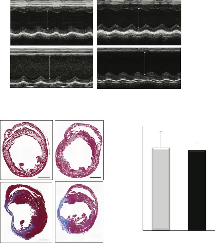
546 Hamad Al Shahi et al.
elevations of circulating inflammatory levels of cytokines, anesthetized with an intraperitoneal injection of pento-
chemokines, and cell adhesion molecules, and the barbital sodium (50 mg/kg) and then intubated and
activation of various types of immune cells [6–8]. connected to a rodent respirator. The chest cavity was
Exercise training is a non-pharmacological intervention opened via a left thoracotomy to expose the heart, and
for the improvement of cardiac function and skeletal muscle LAD was visualized by microscopy and permanently
function [9], and it is also associated with reductions of the ligated with a 7-0 silk suture at the site of its emergence
risk of coronary artery disease and heart failure through from the left atrium. Complete occlusion of the vessel
direct and indirect mechanisms by which exercise con- was confirmed by the presence of myocardial blanching
tributes an anti-inflammatory effect [10]. We and another in the perfusion bed. After operation, the sedentary
research group demonstrated that in atherosclerotic mice, groups were maintained under usual care, whereas the
voluntary exercise ameliorated the progression of athero- mice in the exercise groups were allowed to exercise
sclerotic lesion formation by exerting anti-inflammatory immediately by engaging in voluntary wheel running for
effects [11,12]. In addition, the benefits of exercise training 28 days as described [11].
on skeletal muscle induce mitochondrial adaptations that
are characterized mainly by increased mitochondrial bio-
genesis and the regulation of energy metabolism [13]. These 2.3 Echocardiography
results suggested that skeletal muscle might mediate the
anti-inflammatory effects of exercise via secretion of proteins Echocardiographic evaluations were performed at base-
that could counteract the harmful effects of excessive line and on day 28 after the operation. The anterior and
inflammation. We thus conducted this study to assess the posterior LV wall thickness, LV end-diastolic diameter
effects of voluntary post-MI exercise on cardiac function, (LVEDD), and LV end-systolic diameter (LVESD), the
remodeling, and inflammation, including myokine produc- ejection fraction (EF), and the fractional shortening (FS)
tion, in a mouse model. percentages were measured in mice anesthetized with
1.5% isoflurane. M-mode tracings were obtained with the
use of a transthoracic 2D M-mode echocardiographic
system (Vevo 770, VisualSonics, Toronto, Canada) [15].
2 Methods
2.4 Histology and immunohistochemistry
2.1 Experimental groups analyses
Eight-week-old male C57BL/6J mice were randomly assigned Heart tissue was fixed in 10% buffered formalin,
to the following four groups: sham-operated mice (Sh) and embedded in paraffin, and cut into 5-µm-thick sections.
mice with MI that were sedentary (Sed-Sh and Sed-MI) and The sections were stained with Masson’s trichrome to
sham-operated mice and mice with MI that were subjected determine the percentage of the infarct size, and the
to voluntary exercise training (Ex-Sh and Ex-MI) for 28 days. morphology of the infarcted size was identified using
After the 28-day training period, echocardiography was ImageJ software (ver. 1.47v; U.S. National Institutes of
performed for all mice. The mice were then sacrificed, and Health, Bethesda, MD). The percentage of the infarct size
the heart and skeletal muscle tissues were excised. Total was assessed as the total infarct circumference divided
RNA and protein were isolated from these tissues. All animal by the total LV circumference × 100, as described [16].
experiments were reviewed and approved by the Immunohistochemistry staining was performed by the
Institutional Animal Care and Use Committee at Juntendo immune-peroxidase method using paraffin-embedded
University, Graduate School of Medicine. tissue sections. After inhibition of endogenous perox-
idase activity, the sections were incubated with primary
rat anti-mouse Mac-3 antibody (#550292, BD Bioscience,
Franklin Lakes, NJ), or rabbit anti-human CD3 antibody
2.2 Experimental procedures and exercise (#20006172F, Dako Autostainer/Autostainer Plus, Dako,
protocol Glostrup, Denmark) and then incubated at 4°C overnight
with the respective secondary antibodies. Following
Mice were subjected to MI by ligation of the left anterior visualization with 3,3′-diaminobenzidine (#10107863,
descending coronary artery (LAD) or to a sham operation Sigma–Aldrich, St. Louis, MO) according to the manu-
without ligation [14]. In brief, the mouse was facturer’s instructions, the sections were finally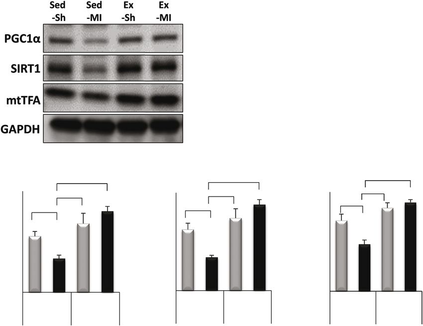
Myokine and cardiac remodeling 547
counterstained with Mayer’s hematoxylin (Wako, Tokyo). the chemiluminescence detection reagent in the Amersham
All images were digitized using a microscope (Olympus ECL Western Blotting Analysis System (GE Healthcare,
AX80; Olympus Optical, Tokyo) equipped with a high- Chalfont St Giles, UK) for enhanced chemiluminescence.
resolution camera (Nikon D2X; Nikon, Tokyo). Equal loading of protein was verified by immunoblotting
with GAPDH (Cell Signaling). The proteins were quantified
(band × volume) using a LAS-3000 Imaging System
2.5 RNA extraction and quantitative RT-PCR (FujiFilm, Kanagawa, Japan).
Total RNA from the heart and skeletal muscle tissue were
isolated using the RNeasy Mini Kit (Qiagen, Valencia, CA). 2.7 Measurement of plasma levels of cytokines
We prepared complementary DNA from the total RNA by
using reverse transcriptase (Applied Biosystems, Foster The plasma levels of tumor necrosis factor-alpha (TNF-α),
City, CA) [17]. Specific mRNAs were amplified using SYBR interleukin (IL)-1β, IL-6, and IL-10 were determined using
Premix Ex Taq II (Takara Biotechnology, Shiga, Japan) in commercial multiplexed fluorescent bead-based immu-
an ABI PRISM 7500 thermal cycler (Applied Biosystems). noassays (Milliplex Map Kit; EMD Millipore, Billerica, MA)
Quantitative real-time polymerase chain reaction (PCR) and measured by a Luminex 200 xPONENT 3.1 System
was performed using a 7500 Real-Time PCR system (EMD Millipore). In the experimental design, the base kit
(Applied Biosystems). The relative amounts of mRNA can be used with any combination of the four analyte-
were calculated by the comparative computed tomo- specific bead sets for greater flexibility. All the samples
graphy method with glyceraldehyde-3-phosphate dehy- were measured in duplicate. The assays were performed
drogenase (GAPDH) mRNA as the invariant control. according to the manufacturer’s instructions.
2.6 Immunoblotting 2.8 Statistical analysis
Immunoblotting was performed as described [18]. In brief, Data are expressed as the mean and standard error of the
skeletal muscle tissue samples were homogenized in 1× cell mean (SEM) and tested for significance using Student’s
lysis buffer (Cell Signaling, Danvers, MA), supplemented t-test or a one-way analysis of variance with Tukey’s test
with 1× complete protease inhibitor cocktail (Roche, Basel, for post-hoc analysis. All statistics were carried out using
Switzerland), and 1 mmol/L phenylmethylsulfonic fluoride. GraphPad Prism® ver. 6.0e (GraphPad Software, San
After sonification and centrifugation at 15 000g for 10 min at Diego, CA). Results were considered significant when the
4°C, the supernatants were collected. Protein aliquots were p-value was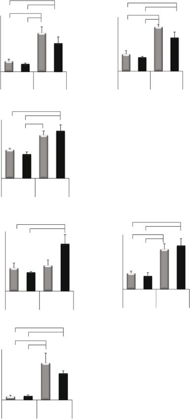
548 Hamad Al Shahi et al.
Table 1: Characteristics of WT mice after sham or MI operation groups (Table 2). The values of LVEDD and LVESD were
significantly lower in the Ex-MI group compared with the
Sedentary Exercise Sed-MI group (p < 0.05) (Figure 1a and Table 2). However,
Sham MI Sham MI there were no significant differences in the EF or FS values
between the Ex-MI and Sed-MI groups. The infarct size
n 11 10 10 9
showed no significant difference between the Sed-MI and
Initial 23.5 ± 0.3 23.1 ± 0.3 23.8 ± 0.3 23.1 ± 0.3
BW, g Ex-MI groups (Figure 1b and c).
Final BW, g 27.6 27.8 25.2 25.8
± 0.5* ± 0.5* ± 0.5*† ± 0.4*†
HW, mg 112 ± 3 160 ± 7# 123 ± 4 151 ± 4#
LW, mg 129 ± 3 160 ± 9# 134 ± 3 139 ± 4†
3.3 Exercise suppressed inflammation in
HW/BW 4.0 ± 0.1 5.7 ± 0.2# 4.9 ± 0.1 5.9 ± 0.1#
LW/BW 4.7 ± 0.1 5.8 ± 0.3# 5.3 ± 0.1 5.4 ± 0.2 the myocardium at 28 days post-MI
Results are mean ± SEM. *p < 0.05 vs initial BW, #p < 0.05 vs
#,†
Immunohistochemistry staining of Mac-3-positive cells
corresponding sham, †p < 0.05 vs Sed-MI. BW, body weight; HW, on cardiac tissue sections was carried out 28 days after
heart weight; LW, lung weight; MI, myocardial infarction.
MI. The infiltration of Mac-3 cells in the border zone of
the LV of the Ex-MI mice was significantly decreased
the sham group. Voluntary exercise had no significant effect compared with the Sed-MI group (66.7 ± 9.8% vs 147.4 ±
on the survival rate (Sed-MI: 76%, Ex-MI 68%, p = 0.42). The 15.0%, respectively, p < 0.001) (Figure 2a and b).
exercise distances per day at 28 days after MI were not However, the staining of CD3-positive cells in the border
significantly different between the Ex-Sh and Ex-MI groups zone of the LV at 28 days post-MI showed no significant
(13 ± 0.3 vs 10 ± 0.2 km/day, respectively; p = 0.33). difference between the Ex-MI and Sed-MI groups (11 ±
Moreover, the total exercise distance over the 28 days after MI 1.7% vs 14.8 ± 1.7%, respectively, p = 0.17) (Figure 2c and
was significantly different between the Ex-Sh and Ex-MI d). The mRNA expression levels of TNF-α, IL-1β, IL-6, IL-
groups (117 ± 5 vs 98 ± 4 km, respectively; p < 0.05). 10, and IL-27 of the infarcted area of the LV and the
border zone of the LV in the Ex-MI group were
significantly decreased compared with those of Sed-MI
3.2 Exercise improved cardiac remodeling group (p < 0.001, Figure 3a).
after MI
3.4 Exercise reduced cardiac fibrosis
Heart rates were not significantly different among the four after MI
groups (Table 2). The values of LVEDD and LVESD were
significantly higher and the EF and FS values were In the infarcted area, the mRNA expression levels of
significantly lower at 28 days post-MI in the Sed-MI and various fibrosis-related genes including transforming
Ex-MI groups compared with the corresponding sham growth factor (TGF) β1, TGFβ2, collagen (COL) I,
Table 2: Echocardiographic measurements
Baseline 4 weeks
Sedentary Exercise Sedentary Exercise
Sham MI Sham MI Sham MI Sham MI
n 18 18 17 15 18 18 17 15
HR, bpm 391 ± 9 392 ± 13 373 ± 8 382 ± 8 419 ± 9 412 ± 16 399 ± 6 388 ± 8
LVEDD, mm Hg 3.96 ± 0.04 3.98 ± 0.04 4.12 ± 0.08 3.95 ± 0.04 4.15 ± 0.08 6.32 ± 0.09*# 4.27 ± 0.07 5.66 ± 0.05*#†
LVEDS, mm Hg 2.90 ± 0.04 2.92 ± 0.04 3.07 ± 0.08 2.93 ± 0.04 2.92 ± 0.10 5.72 ± 0.10*# 3.15 ± 0.07 5.00 ± 0.06*#†
EF, % 52.5 ± 1.0 52.2 ± 1.5 50.6 ± 1.5 51.4 ± 0.9 57.0 ± 2.0 20.3 ± 1.0*# 52.4 ± 1.5 24.8 ± 1.0*#
FS, % 26.6 ± 0.6 26.5 ± 0.9 25.5 ± 0.9 25.9 ± 0.6 29.9 ± 1.5 9.4 ± 0.5*# 26.8 ± 1.0 11.6 ± 0.5*#
#,†
Results are mean ± SEM. *p < 0.05 vs baseline, #p < 0.05 vs corresponding sham, †p < 0.05 vs Sed-MI at 28 days post-MI. bpm: beats
per minute, EF: ejection fraction, FS: fraction shortening, HR: heart rate, LVEDD: LV end-diastolic diameter, LVESD: LV end-systolic
diameter, MI: myocardial infarction.Myokine and cardiac remodeling 549
(a) Sed
d Exx
Sh
MI
(b) (c)
Seed Ex
40
35
Sh 30
25
Infarct size, %
20
15
10
MI
5
0
Sed-MI Ex-- MI
Figure 1: Exercise improved cardiac remodeling after MI. M-mode echocardiographic images obtained from Sed-Sh, Sed-MI, Ex-Sh, and Ex-
MI mice at 28 days post-operation (a). Trichrome-stained heart sections (28 days post-MI) (b). Percentage of infarct size in Sed-MI (n = 4)
and Ex-MI (n = 4) (c). Scale bar, 1 mm. Results are given as mean ± SEM. Arrows indicate LV chamber diameter. Ex: exercise, MI: myocardial
infarction, Sed: sedentary, Sh: sham.
COLIII, and connective tissue growth factor (CTGF) were exercise-related genes, such as PGC1α, SIRT1, and
significantly increased in the Sed-MI and Ex-MI groups mtTFA, we performed immunoblotting and RT-PCR
compared with the Sham groups (p < 0.001). Compared analysis (Figures 4a, b and 5a). The voluntary exercise
with the Sed-MI group, the Ex-MI group showed produced a shift toward a greater expression of PGC1α,
significant reductions of the expression of TGFβ2, COLI, SIRT1, and mtTFA in the Ex-Sh and Ex-MI groups
COLIII, and CTGF in the infarcted area (p < 0.05) and compared with the Sed-MI group as revealed by
significant reductions in the expression of TGFβ2, COLI, immunoblotting (p < 0.001). We also observed a
and COLIII in the border zone (p < 0.05). TGFβ1 and significant increase in the mRNA expressions of PGC1α,
CTGF showed no significant differences between the Sed- SIRT1, and mtTFA in the Ex-Sh and Ex-MI groups
MI and Ex-MI groups in the border zone of the LV compared with the Sed-Sh and Sed-MI groups (p <
(Figure 3b). 0.05) (Figure 5a).
We also investigated the mRNA expressions in
3.5 Exercise promoted mitochondrial skeletal muscle of myokines, i.e., IL-6, follistatin-like
function and myokine expression in (FSTL) 1, and fibroblast growth factor (FGF) 21 and their
skeletal muscle potential post-MI roles (Figure 5b). Interestingly, dra-
matic increases of FSTL1 and FGF21 mRNA expression
To determine whether voluntary exercise provided a were observed in the Ex-Sh and Ex-MI mice compared to
stimulus to the expression of various cytokines and the Sed-Sh and Sed-MI mice (p < 0.05). The IL-6 mRNA550 Hamad Al Shahi et al.
(a)
Sed Ex
100 μm
m
50 μm
m
(b) **
** *
Mac 3 posive cell (Cells/mm2)
18
80
16
60
14
40
12
20
10
00
8
80
6
60
4
40
2
20
0
Sh MI Sh MI
SSed Ex
(c)
Sed Ex
100 μm
50 μm
m
(d) ***
*
18
8
CD3 posive cell (Cells/mm2)
16
6
14
4
12
2
10
0
8
6
4
2
0
Sh MI Sh MI
M
Seed Ex
Figure 2: Exercise suppresses inflammation in the myocardium at 28 days post-MI. Immunohistochemistry staining of inflammatory Mac3
cells (macrophage cells) (a). Results of the quantitative analysis of Mac3-positive cells at 28 days post-MI (n = 4–5 each) (b).
Immunohistochemistry staining of inflammatory CD3 cells (lymphocytes) in the border zone at 28 days post-MI (c). The results of the
quantitative analysis of CD3-positive cells at 28 days post-MI (n = 4–5 each) (d). Results are given as mean ± SEM. *p < 0.05, **p < 0.001.
Ex: exercise, MI: myocardial infarction, Sed: sedentary, Sh: sham.Myokine and cardiac remodeling 551
(a) TNFα IL1β expression demonstrated a significant increase only in
**
**
the Ex-MI mice compared with the Sed-Sh and Sed-MI
Relave mRNA expression
Relave mRNA Expression
25 ** ** ** ** 45 ** *
**
40
**
mice (p < 0.05).
20 35
30
15 25
20
10 15
5 10
5
0 0 3.6 Exercise-induced higher IL-6 plasma
Sh IA BZ Sh IA B
BZ SSh IA BZ Sh IA B
BZ
Sed Ex Sed Ex
levels after MI
IL6 ILL10
** The plasma level of IL-6 in the Ex-MI group was
**
significantly increased compared with those of the Sed-
Relave mRNA Expression
** **
Relave mRNA Expression
100 ** ** 25
** **
80
*
20 ** Sh and Sed-MI groups (p < 0.05, Figure 6). The plasma
60 15 level of TNF-α showed no significant differences between
40 10 the groups.
20 5
0
0
Sh IA BZ Sh IA BZ Sh IA BZZ Sh IA BZ
SSed Ex Sed Ex
4 Discussion
(b) TGFβ1 TGFFβ2
**
** ** ** ** This study results in a mouse model demonstrated that
Relave mRNA Expression
7
Relave mRNA Expression
25 ** ** ** ***
6 **
voluntary exercise attenuated cardiac remodeling,
20
5 modulated inflammatory responses, and induced an
p
4 15
3
improvement in mitochondrial function and an increase
10
2 in myokine expression in the skeletal muscle after MI.
1 5
Other animal studies showed that exercise can
0 0
Sh A
IA BZ Sh IA BZ Sh IA BZZ Sh IA BZ promote a protective reaction against irreversible tissue
Sed Ex Sed Ex damage produced by ischemic injury in the myocardium
[19,20]. Moreover, exercise training may attenuate post-
COLII C
COLIII
* ** MI remodeling independent of the preconditioning
60
Relave mRNA Expression
Relave mRNA Expression
** ** ** ** ** ** ** **
50 *
40 effect. Other investigations revealed that swimming
35 *
40 30 training had no effect on mortality but reduced the
25
30
20
infarct size and attenuated LV remodeling in an MI rat
20 15 model [19,20]. The effects of exercise training after MI on
10
10 5 cardiac remodeling and function thus remain incomple-
0 0
h
Sh IA BZ Sh IA BZ
Z Sh IA BZZ Sh IA BZ tely understood.
Sed Ex Sed Ex We observed herein that MI in mice resulted in
CTGF significant LV remodeling and dysfunction after 28 days,
* as characterized by increases in LVEDD and LVESD
Relave mRNA Expression
60 ** ** **
**
values and decreases in EF and FS values, resulting in
50
40
pulmonary congestion. However, the exercise training
30 attenuated the LVEDD and LVESD at 28 days post-MI. In
20 addition, the incidence of cardiac hypertrophy in the
10
0
mice at 28 days post-MI, as indicated by increases in the
Sh I
IA BZ Sh IA BZ HW and the HW/BW ratio, was also apparent when
Sed Ex compared with the Sham group. These results suggest
that voluntary wheel running might be useful for the
Figure 3: Exercise reduces cardiac fibrosis post-MI. Quantitative analysis
of mRNA expression of inflammatory cytokines (n = 7–8 each) (a) and prevention of cardiac function and remodeling in animal
fibrosis-related genes in the infarcted area (IA) and border zone (BZ) at 28 post-MI models.
days post-MI (n = 7–8 each) (b). Results are given as mean ± SEM. *p < Myocardial infarction triggers an intense inflamma-
0.05, **p < 0.001. BZ: border zone, COL: collagen, CTGF: connective tissue tory reaction that is essential for the healing of the LV
growth factor, Ex: exercise, IA: infarcted area, IL: interleukin, TGF: transfor-
infarcted area. Myocardial infarction and reperfusion
ming growth factor. TNF: tumor necrosis factor, Sed: sedentary, Sh: sham.552 Hamad Al Shahi et al.
(a)
(b) ** **
*
1.8 1.6 1.4 ***
* *
1.6 1.4 1.2 *
*
PGC1α/GAPDH
1.4 *
SIRT1/GAPDH
mtTFA/GAPDH
1.2 1.0
1.2 1.0
1.0 0.8
0.8
0.8 0.6
0.6 0.6
0.4 0.4
0.4
0.2 0.2 0.2
0.0 0.0 0.0
Sh MI Sh MI Sh MI Sh MI Sh MI Sh MI
Sed Ex Sed Ex Sed Ex
Figure 4: Exercise promoted mitochondrial function in mouse skeletal muscle. Representative immunoblotting bands (a) and results of the
quantitative analyses for PGC1α, SIRT1, and mtTFA protein expressions in the gastrocnemius skeletal muscle at 28 days post-MI (n = 5
each) (b). Results are given as mean ± SEM. *p < 0.05, **p < 0.001. Ex: exercise, MI: myocardial infarction, mtTFA: mitochondrial
transcription factor A, PGC: peroxisome proliferator-activated receptor gamma coactivator, Sed: sedentary, Sh: sham, SIRT: sirtuin.
injury have been associated with the activation of pro- ∼1,500 proteins encoded by both the nuclear and
inflammatory cytokines such as TNF-α, IL-1β, and IL-6, mitochondrial genomes [24]. PGC1α is known to coacti-
and this activation promotes leukocyte activation and vate multiple mitochondrial transcription factors,
extravasation into the LV infarcted area [21,22]. Exercise leading to the upregulation of fatty acid oxidation, in
training was shown to have beneficial effects on the part through increased PGC1α protein stability induced
inflammatory response in the heart through the attenua- by the protein deacetylase SIRT1 and in part through
tion of LV remodeling [19,23]. In this study, inflamma- mtTFA activation, a key component in the transcription
tory cytokines (TNF-α, IL-1β, IL-6, IL-10, and IL-27) were of multiple oxidative genes [25]. In this study, the gene
attenuated in the mice that exercised, suggesting that expression and protein content of PGC1α, SIRT1, and
the phase of inflammatory reaction is an important mtTFA were significantly decreased in the skeletal
signaling mechanism that contributes to the LV remo- muscles Sed-MI group, compared with the Sed-Sh group,
deling processes. The pro-inflammatory environment in whereas they were significantly increased in the ex-
the early stages of infarct healing promotes matrix ercised MI groups. This suggests that voluntary exercise
degradation and phagocytic clearance, and the repair of enhanced the mitochondrial content and oxidative
the infarcted tissue is dependent on the signaling capacity in skeletal muscle.
pathways that mediate the inflammatory responses. In The role of skeletal muscle in protecting the heart
this study, the myocardial expressions of fibrosis-related after MI has been studied in animal models [26]. Since
genes (TGFβ1, TGFβ2, COLI, COLIII, and CTGF) were exercise has muscle ischemia-like effects through hy-
significantly upregulated post-MI. However, the expres- poxia, exercise may exert cardiac protective action
sions of TGFβ2, COLI, COLIII, and CTGF in the mice that through a mechanism similar to ischemia [27]. Skeletal
exercised were downregulated compared with the muscle, upon contraction, stimulates the production and
sedentary group post-MI. release of cytokines (which in the muscles are also called
Mitochondrial biogenesis is a complex process that myokines), which can influence metabolism and modify
requires the coordinated synthesis and assembly of further myokines production in the tissues and organsMyokine and cardiac remodeling 553
(a) PGC1α SIRT1
* *
* *
Relave mRNA Expression
*
Relave mRNA Expression
2.5 *
** 1.4 **
1.2
2
1
1.5 0.8
0.6
1
0.4
0.5 0.2
0 0
Sh MI Sh MI Sh MI Sh MI
Sed Ex Sed Ex
mtTFA
*
*
1.6
Relave mRNA Expression
*
1.4
1.2
1
0.8
0.6
0.4
0.2
0
Sh MI Sh MI
Sed Ex
(b) IL6 FSTL1
* *
* *
1.2
Relave mRNA Expression
2.5 *
Relave mRNA Expression
*
1
2
0.8
1.5
0.6
1
0.4
0.5
0.2
0 0
Sh MI Sh MI Sh MI Sh MI
Sed Ex Sed Ex
FGF21
*
**
**
Relave mRNA Expression
6
5
4
3
2
1
0
Sh MI Sh MI
Sed Ex
Figure 5: Exercise promoted mitochondrial function and myokine expression in mouse skeletal muscle. Quantitative analysis of mRNA
expression of PGC1α, SIRT1, and mtTFA (a), and IL-6, FSTL1, and FGF21 in the gastrocnemius skeletal muscle at 28 days post-MI (n = 7–8
each) (b). Results are presented as mean ± SEM. *p < 0.05, **p < 0.001. Ex: exercise, FGF: fibroblast growth factor. FSTL: follistatin-like, IL:
interleukin, MI: myocardial infarction, mtTFA: mitochondrial transcription factor A, PGC: peroxisome proliferator-activated receptor gamma
coactivator, Sed: sedentary, Sh: sham, SIRT: sirtuin.554 Hamad Al Shahi et al.
TNFα ILL-6
*
*
4 180
3.5
3 160
3 140
2.5
2 120
pg/ml
pg/ml
2 100
80
1.5
1
60
1
40
0.5
0 20
0 0
Sh MI SSh MI Sh MI Sh M
MI
Se
ed Ex Sed Ex
Figure 6: Exercise-induced higher IL-6 plasma levels post-MI. Plasma levels of IL-6 and TNF-α at 28 days post-MI (n = 5–7 each). Results are
given as mean ± SEM. *p < 0.05, **p < 0.001. Ex: exercise, IL: interleukin, MI: myocardial infarction, Sed: sedentary, Sh: sham, SIRT:
sirtuin, TNF: tumor necrosis factor.
[28]. Some myokines can induce an anti-inflammatory 5 Conclusion
response after exercise training. For example, during
exercise, IL-6 is the first detectable myokine released The results of this study revealed that in the murine model,
into the blood from the contracting skeletal muscle, and voluntary exercise after MI ameliorated cardiac remodeling
this release induces a subsequent increase in the and the inflammatory response and induced an improvement
production of the IL-1 receptor antagonist and IL-10 by of mitochondrial function and myokine expression in skeletal
the blood mononuclear cells, thereby exerting an anti- muscle. These findings suggest that voluntary exercise after
inflammatory effect [28]. In this study, the gene expres- MI improves cardiac remodeling via inflammatory modula-
sion and plasma circulating levels of IL-6 in Ex-MI mice tion. Further research is needed to confirm the beneficial
were significantly elevated by exercise, suggesting that effects of exercise training after MI and to determine how
IL-6 may have a beneficial anti-inflammatory effect. these effects may translate into clinical benefits in human.
Interestingly, in the mice that exercised, the gene
expressions of FSTL1 and FGF21 were also upregulated Acknowledgments: We thank Emiko Nakamura (Depart-
compared with the sedentary mice group. These results ment of Cardiovascular Medicine, Juntendo University
are comparable with those of other studies [29,30]. Graduate School of Medicine) for technical assistance and
Collectively, our findings suggest that multiple signaling biochemical measurements in the experiments, and Shuko
mechanisms may participate in the myokine-mediated Nojiri, PhD (Juntendo University, Medical Technology
cardioprotective role in MI, a possibility that deserves Innovation Center), and Momoka Yamada (Department of
further investigation. Management Science, Graduate School of Engineering, Tokyo
The plasma circulating levels of IL-10 in both the University of Science) for statistical analysis.
present sham groups were significantly elevated com-
pared with the MI mice. Low expression levels of IL-10 in Funding: This work was supported by a High Technology
serum samples have been associated with an increased Research Center Grant from the Ministry of Education,
risk of cardiovascular events, and high IL-10 expression Culture, Science and Technology, Japan, and was supported
levels have been associated with a decreased risk [31]. by JSPS KAKENHI grant (no. 26350588 and 17K01470).
Conflicting results have been published [32], and
although some studies have found that IL-10 levels Conflicts of interest: None declared.
were significantly elevated after exercise, further inves-
tigations are needed to clarify the relationship between
IL-10 levels and exercise. References
This study has some limitations. The results were
obtained with male mice only and therefore cannot be [1] Lujan HL, DiCarlo SE. Mimicking the endogenous current of
directly translated to female mice. We were also unable injury improves post-infarct cardiac remodeling. Med
to obtain Milliplex assay data for all the cytokines. Hypotheses. 2013;81:521–3.Myokine and cardiac remodeling 555
[2] Opie LH, Commerford PJ, Gersh BJ, Pfeffer MA. Controversies with mitochondrial dysfunction and muscle atrophy in aging
in ventricular remodelling. Lancet. 2006;367:356–67. mice. Geriatr Gerontol Int. 2020;20:78–84.
[3] Hosseini SH, Ghaemian A, Mehdizadeh E, Ashraf H. Levels of [18] Kadoguchi T, Shimada K, Koide H, Miyazaki T, Shiozawa T,
anxiety and depression as predictors of mortality following Takahashi S, et al. Possible role of NADPH oxidase 4 in
myocardial infarction: a 5-year follow-up. Cardiol J. angiotensin II-induced muscle wasting in mice. Front Physiol.
2014;21:370–7. 2018;9:1–9.
[4] Shimada K. Immune system and atherosclerotic disease: [19] Cai M, Wang Q, Liu Z, Jia D, Feng R, Tian Z. Effects of different
heterogeneity of leukocyte subsets participating in the types of exercise on skeletal muscle atrophy, antioxidant
pathogenesis of atherosclerosis. Circ J. 2009;73:994–1001. capacity and growth factors expression following myocardial
[5] Frangogiannis NG. The immune system and the remodeling infarction. Life Sci. 2018;213:40–9.
infarcted heart: cell biological insights and therapeutic [20] Zhao D, Sun Y, Tan Y, Zhang Z, Hou Z, Gao C, et al. Short-duration
opportunities. J Cardiovasc Pharmacol. 2014;63:185–95. swimming exercise after myocardial infarction attenuates cardiac
[6] Schumacher A, Seljeflot I, Sommervoll L, Christensen B, dysfunction and regulates mitochondrial quality control in aged
Otterstad JE, Arnesen H. Increased levels of markers of mice. Oxid Med Cell Longev. 2018;2018:4079041.
vascular inflammation in patients with coronary heart disease. [21] Frantz S, Bauersachs J, Ertl G. Post-infarct remodelling:
Scand J Clin Lab Invest. 2002;62:59–68. contribution of wound healing and inflammation. Cardiovasc
[7] Nakachi T, Kosuge M, Hibi K, Ebina T, Hashiba K, Mitsuhashi T, Res. 2009;81:474–81.
et al. C-reactive protein elevation and rapid angiographic [22] Dewald O, Zymek P, Winkelmann K, Koerting A, Ren G, Abou-
progression of nonculprit lesion in patients with non-ST- Khamis T, et al. CCL2/monocyte chemoattractant protein-1
segment elevation acute coronary syndrome. Circ J. regulates inflammatory responses critical to healing myocar-
2008;72:1953–9. dial infarcts. Circ Res. 2005;96:881–9.
[8] Al Shahi H, Shimada K, Miyauchi K, Yoshihara T, Sai E, [23] Puhl SL, Müller A, Wagner M, Devaux Y, Böhm M, Wagner DR,
Shiozawa T, et al. Elevated circulating levels of inflammatory et al. Exercise attenuates inflammation and limits scar
markers in patients with acute coronary syndrome. Int J Vasc thinning after myocardial infarction in mice. Am J Physiol Heart
Med. 2015;2015;805375. Circ Physiol. 2015;309:H345–9.
[9] Duggal NA, Niemiro G, Harridge SDR, Simpson RJ, Lord JM. Can [24] Picca A, Mankowski RT, Burman JL, Donisi L, Kim JS,
physical activity ameliorate immunosenescence and thereby Marzetti E, et al. Mitochondrial quality control mechanisms as
reduce age-related multi-morbidity? Nat Rev Immunol. molecular targets in cardiac ageing. Nat Rev Cardiol.
2019;19:563–72, review. 2018;15:543–54, review.
[10] Bruun JM, Helge JW, Richelsen B, Stallknecht B. Diet and [25] Huang CC, Wang T, Tung YT, Lin WT. Effect of exercise training
exercise reduce low-grade inflammation and macrophage on skeletal muscle SIRT1 and PGC1α expression levels in rats
infiltration in adipose tissue but not in skeletal muscle in of different age. Int J Med Sci. 2016;13:260–70.
severely obese subjects. Am J Physiol Endocrinol Metab. [26] Kharbanda RK, Mortensen UM, White PA, Kristiansen SB,
2006;290:E961–7. Schmidt MR, Hoschtitzky JA, et al. Transient limb ischemia
[11] Fukao K, Shimada K, Naito H, Sumiyoshi K, Inoue N, Isesaki T, induces remote ischemic preconditioning in vivo. Circulation.
et al. Voluntary exercise ameliorates the progression of 2002;106:2881–3.
atherosclerotic lesion formation via anti-inflammatory effects [27] Shen YJ, Pan SS, Zhuang T, Wang FJ. Exercise preconditioning
in apolipoprotein E-deficient mice. J Atheroscler Thromb. initiates late cardioprotection against isoproterenol-induced
2010;17:1226–36. myocardial injury in rats independent of protein kinase C. J
[12] Meissner M, Lombardo E, Havinga R, Tietge UJ, Kuipers F, Physiol Sci. 2011;61:13–21.
Groen AK. Voluntary wheel running increases bile acid as well [28] Steensberg A, Fischer CP, Keller C, Møller K, Pedersen BK. IL-6
as cholesterol excretion and decreases atherosclerosis in enhances plasma IL-1ra, IL-10, and cortisol in humans. Am J
hypercholesterolemic mice. Atherosclerosis. 2011;218:323–9. Physiol Endocrinol Metab. 2003;285:E433–7.
[13] Trewin AJ, Berry BJ, Wojtovich AP. Exercise and mitochondrial [29] Xi Y, Gong DW, Tian Z. FSTL1 as a potential mediator of
dynamics: keeping in shape with ROS and AMPK. exercise-induced cardioprotection in post-myocardial infarc-
Antioxidants. 2018;7:1–21, review. tion rats. Sci Rep. 2016;6:32424.
[14] Tarnavski O, McMullen JR, Schinke M, Nie Q, Kong S, Izumo S, [30] Joki Y, Ohashi K, Yuasa D, Shibata R, Ito M, Matsuo K, et al.
et al. Mouse cardiac surgery: comprehensive techniques for FGF21 attenuates pathological myocardial remodeling fol-
the generation of mouse models of human diseases and their lowing myocardial infarction through the adiponectin-depen-
application for genomic studies. Physiol Genom. dent mechanism. Biochem Biophys Res Commun.
2004;16:349–60. 2015;459:124–30.
[15] Scherrer-Crosbie M, Kurtz B. Ventricular remodeling and [31] Anguera I, Guardiola FM, Bosch X, Filella X, Sitges M, Marin JL,
function: insights using murine echocardiography. J Mol Cell et al. Elevation of serum levels of the anti-inflammatory
Cardiol. 2010;48:512–7. cytokine interleukin-10 and decreased risk of coronary events
[16] Lavine KJ, Kovacs A, Weinheimer C, Mann DL. Repetitive in patients with unstable angina. Am Heart J. 2002;144:811–7.
myocardial ischemia promotes coronary growth in the adult [32] Mizia-Stec K, Ga¸sior Z, Zahorska B, Janowska J, Szulc A,
mammalian heart. J Am Heart Assoc. 2013;2:1–17. Jastrzabska E, et al. Serum tumor necrosis factor alpha,
[17] Kadoguchi T, Shimada K, Miyazaki T, Kitamura K, Kunimoto M, interleukin-2 and interleukin-10 activation instable angina and
Aikawa T, et al. Promotion of oxidative stress is associated acute coronary syndromes. Coron Artery Dis. 2003;14:431–8.You can also read