Experimental Protocols for Embryonic Stem Cell Research
←
→
Page content transcription
If your browser does not render page correctly, please read the page content below
This material is protected by U.S. Copyright law.
                                                                                Unauthorized reproduction is prohibited.
                                                                         For reprints contact: Reprints@AlphaMedPress.com

        Experimental Protocols for Embryonic Stem Cell Research
   Differentiation of Human Embryonic Stem Cells to Neural Lineages
 in Adherent Culture by Blocking Bone Morphogenetic Protein Signaling

                                          Lesley Gerrard, Leigh Rodgers, Wei Cui

            Department of Gene Function and Development, Roslin Institute, Roslin, Midlothian, United Kingdom

        Key Words. Human embryonic stem cell • Neural differentiation • Noggin • Bone morphogenetic protein • Oct4

Abstract
Human embryonic stem cells (hESCs) have extensive self-                 to efficiently generate neural progenitors in which neither
renewal capacity and are competent to differentiate into any            multicellular aggregates nor stromal cells are required. We
cell type of the body. They are valuable not only for the study         show that inhibition of bone morphogenetic protein signal-
of early human development but also for regenerative medi-              ing by its antagonist noggin is sufficient to block extraem-
cine. However, how to direct differentiation of hESCs along             bryonic cell fate, transiently sustain Oct4 gene expression,
a particular lineage pathway to a specific cell type remains            and result in robust production of neural progenitors. Our
a challenge. Although hESCs have been shown to differenti-              findings will provide a platform for studying the molecular
ate in vitro into neural progenitors, the factors controlling           mechanism controlling neural differentiation. Stem Cells
their differentiation are poorly understood. In this study, we          2005;23:1234–1241
report the development of an in vitro adherent culture system

Introduction                                                            cells for specific tissues or organs. For neural differentiation of
The isolation and culture of human embryonic stem cells (hESCs)         hESCs, most protocols available so far are based on the initial for-
has opened up new opportunities for the study of basic stem cell        mation of embryoid bodies (EBs) in the presence of serum/serum
biology, early human embryonic development, and regenerative            replacement or coculture of hESCs with particular stromal cell
medicine. hESCs are derived from the inner cell mass of pre-            lines [2–4]. In the first cases, hESCs were initially differentiated
implantation embryos and retain the developmental potency of            in suspension cultures to form EBs, which often contain cells of all
embryonic founder cells, being able to differentiate into cells and     three germ layers; clusters of neural cells are surrounded by flat
tissues of all three germ layers in vitro and in vivo [1]. Therefore,   non-neural cells, and even in the neural clusters there are mixed
they provide a novel model system to elucidate the molecular sig-       populations of neuronal (majority) and glial cells [2, 3]. Moreover,
nals required for the development of various lineages.                  due to the complexity of multicellular aggregates, it is difficult to
    Like mouse embryonic stem cells (ESCs), hESCs can be                use this system for studying signaling pathways essential for the
expanded to large numbers while retaining their differentia-            neural differentiation. In the latter case, the effect of stromal cells
tion potentials. This is particularly important for transplantation     is attributed to an undefined neural-inducing activity [4].
therapies, because hESCs promise an almost unlimited supply of               In mouse ESCs, neuroectoderm differentiation has been
specific cell types. However, directing hESC differentiation along      achieved in an adherent monoculture system in a serum-free
specific lineage pathways remains a challenge. The clinical appli-      medium, without multicellular aggregation or coculture with
cation of hESCs requires the generation of highly purified donor        other cell types [5]. This monolayer culture regimen enables us

Correspondence: Wei Cui, M.D., Ph.D., Department of Gene Function and Development, Roslin Institute, Roslin, Midlothian, EH25 9PS,
U.K. Telephone: 44-0-131-527-4200; Fax: 44-0-131-440-0434; e-mail: wei.cui@bbsrc.ac.uk Received March 14, 2005; accepted for pub-
lication June 21, 2005; first published online in Stem Cells EXPRESS July 7, 2005. ©AlphaMed Press 1066-5099/2005/$12.00/0 doi:
10.1634/stemcells.2005-0110
Stem Cells 2005;23:1234–1241 www.StemCells.com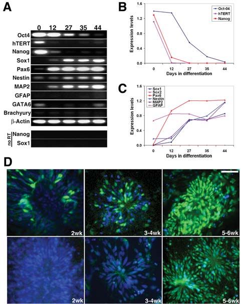
Gerrard, Rodgers, Cui                                                                                                                 1235

to directly visualize the process of neural conversion. In addi-          Immunocytochemistry
tion, because the process is in a chemically defined medium,              Cells were fixed at room temperature with 4% paraformaldehyde
it provides a good system in the future to investigate signaling          for 10 minutes, washed with PBS, and incubated with 10% goat
pathways in controlling the ESC neural differentiation. Here we           serum and 0.02%–0.1% Triton X-100 for 1 hour. The cells were
extend this study to hESCs, demonstrating that blocking bone              then incubated with primary antibodies at the appropriate dilu-
morphogenetic protein (BMP) signaling with its antagonist nog-            tion at 4°C overnight. Secondary antibody was applied for 30 min-
gin at the initial stage of the differentiation is sufficient for deri-   utes after washing with PBS. The cells were finally mounted with
vation of highly enriched and expandable populations of neural            Mowiol (Calbiochem, San Diego, http://www.emdbiosciences.
progenitors in adherent culture. Additionally, these progenitors          com) and then visualized and captured using Nikon TE2000-U
can be further differentiated into various types of neurons in vitro      (http://www.nikoninstruments.com) or Digital Pixel image anal-
but do not exhibit glial markers until long term in culture.              ysis system (Brighton, U.K., http://www.digitalpixel.co.uk).
                                                                               The following primary antibodies were used in this study:
Materials and Methods                                                     mouse monoclonal antibodies against nestin, polysialic acid–
                                                                          neural cell adhesion molecule (PSA-NCAM), and TH (1:200;
Derivation and Culture of Neural Progenitors                              all from Chemicon, Temecula, CA, http://www.chemicon.com),
hESCs were cultured on matrigel (BD Biosciences, San Jose,                β-tubulin III (1:1,000; Sigma), and α-fetoprotein (1:500; Sigma);
CA, http://www.bdbiosciences.com) in mouse embryonic fibro-               rabbit polyclonal antibodies against nestin and musashi (1:200),
blast–conditioned medium (MEF-CM) supplemented with                       GAD65 (1:500), MAP2 and Pax6 (1:1,000; all from Chemicon),
8 ng/ml basic fibroblast growth factor (bFGF) (Invitrogen,                GABA and Sox1 (1:500; Sigma), GATA6 (1:50; Santa Cruz Bio-
Carlsbad, CA, http://www.invitrogen.com) and propagated                   technology Inc., Santa Cruz, CA, http://www.scbt.com), and glial
routinely with collagenase in 1:3 ratios [6]. For neural differen-        fibrilliary acidic protein (GFAP) (1:500; DakoCytomation, Glos-
tiation, nearly confluent hESCs were split with 0.5 mM EDTA/              trup, Denmark, http://www.dakocytomation.com). Secondary
phosphate-buffered saline (PBS) in 1:5 ratios into culture dishes         antibodies used were goat anti-rabbit fluorescein isothiocyanate
coated with matrigel or poly-L-lysine/laminin (Sigma-Aldrich,             (FITC), goat anti-mouse FITC, and goat anti-rabbit Texas red (all
St. Louis, http://www.sigmaaldrich.com) and cultured in                   at 1:400; Jackson Laboratory, Bar Harbor, ME, http://www.jax.
N2B27 medium (1:1 mix of D-MEM/F12 supplemented with N2                   org), goat anti-mouse immunoglobulin G Alex Fluro 568, and
and Neurobasal medium supplemented with B27, all from Gibco               goat anti-rabbit Alex Fluro 488 (all at 1:500–1:1,000; Molecular
[Grand Island, NY, http://www.invitrogen.com], supplemented               Probes Inc., Eugene, OR, http://probes.invitrogen.com).
with 100 ng/ml mouse recombinant noggin (R&D Systems Inc.,
Minneapolis, http://www.rndsystems.com). At this stage, cells             RNA Isolation and Reverse Transcription–Polymerase
were defined as passage 1 (P1). In the neural differentiation, the        Chain Reaction
cells were consistently cultured in the same matrices-coated              Total RNA was extracted using QIAGEN RNeasy kit and DNase
culture plastics. Cells of P1 and P2 were split by collagenase            I treatment (Qiagen, Inc., Valencia, CA, http://www.qiagen.com).
into small clumps, similar to hESC culture, and continuously              First-strand cDNA was synthesized with 2 μg total RNA in 20–25
cultured in N2B27 medium plus noggin. From P3, cells were dis-            μl as described previously [8]. The cDNAs were then diluted to
associated into single cells by TrypLE express (Gibco), and nog-          225 μl with distilled H 2O. Diluted cDNA 3–10 μl was used for
gin could be withdrawn from N2B27 medium. Some of the cells               polymerase chain reaction (PCR) in 50-μl reactions. PCR condi-
were cultured in N2B27 medium supplemented with 20 ng/ml                  tions were optimized and linear amplification range was deter-
bFGF from P4-5.                                                           mined for each pair of primers by varying annealing temperature
     The growth curves were started at P5. The neural progenitors         and cycle numbers. Primer sequences and their PCR conditions
were plated at 1 × 10⁵ per cm2 in six-well culture plates and cultured    are provided upon request. The expression levels were analyzed
in N2B27 medium with or without 20 ng/ml bFGF. The cells were             using multianalysis software and standardized by β-actin house-
counted when splitting, and population doublings were calculated.         keeping gene expression.
     For tyrosine hydroxylase (TH) neuron differentiation, neu-
ral progenitors at P4 or P5 were seeded in poly-L-lysine/laminin          Results
(PLL/Lam)-coated culture dishes and induced with sonic hedge-
hog (300–400 ng/ml), FGF8 (100 ng/ml) [7], and ascorbic acid              Noggin Blocked hESC Commitment
(160 μM) for 1–2 weeks and then followed by brain-derived neu-            to Extraembryonic Endoderm and Enhanced
rotrophic factor (BDNF) (20 ng/ml), glial cell line–derived neu-          Neuroectoderm Formation
rotrophic factor (GDNF) (20 ng/ml), ascorbic acid (160 μM), and           It has been shown previously that culturing mouse ESCs in
laminin (0.5 μg/ml) for another 2 weeks.                                  modified N2B27 medium on gelatin-coated plastic can suffi-
1236 Neural Differentiation of hESCs in Adherent Culture
ciently differentiate them into neural precursors [5]. A modi- It has been reported that BMP2 plays a critical role in ESCs
fied version of this procedure was applied to hESCs (H1, H7, differentiation to extraembryonic endoderm [9] and inhibits
and H9) in which hESCs were dissociated with EDTA rather neuroectoderm differentiation [5, 13]. Therefore, we examined
than trypsin. EDTA-dissociated cells plated as small clumps whether blocking BMP signaling with noggin and/or follistatin
(~4 to 10 cells/clump), but trypsin generated single cells, might restrain the primitive endoderm formation and promote
which exhibited low seeding efficiency and spontaneous dif- neural differentiation. Both noggin and follistatin are well-char-
ferentiation. All three hESC lines showed very low attachment acterized BMP antagonists and have been shown as neural induc-
in gelatin-coated culture dishes and formed cell aggregates in ers in Xenopus embryos [14–16]. However, noggin is known to
suspension. Therefore, gelatin was replaced with matrigel or bind BMP 2, 4, and 7 with higher affinity, consequently prevent-
PLL/Lam because both matrices have been reported for hESC ing ligation of their receptors, whereas follistatin has high bind-
culture previously [6]. ing affinity for activin but lower affinity for BMPs and its antago-
On both matrigel and PLL/Lam, hESC plating efficiency nizing effects of BMPs through different mechanism rather than
was improved. After 4 days of culture in N2B27 medium, flat interfering with the bindings of receptors. Our results showed
cells started to emerge, which were very similar in appearance that hESCs cultured in N2B27 medium supplemented with 100
to BMP2-treated hESCs reported earlier by Pera et al. [9]. This ng/ml noggin significantly blocked extraembryonic endoderm
flat cell population expanded rapidly (Figs. 1A, 1B, 1D, 1E), formation in both matrigel and PLL/Lam-coated plastic and after
pushing other cell types into clumps, eventually forcing them to 10 days neural progenitor cells are clearly visible (Figs. 1C, 1F),
detach into the medium. This was most apparent in H9 cells and whereas follistatin did not show evident neuroectoderm forma-
then H1 and its clonal derivatives and less evident in H7 cells. tion, although it partially inhibited extraembryonic endoderm
These flat cells showed positive staining with GATA6 antibody formation. The hESCs in follistatin-treated cultures exhibited
(Fig. 1G) and sometimes formed fluid-filled cysts (Fig. 1B, mainly fibroblast-like morphology (Fig. 1I).
arrow) that expressed α-fetoprotein (AFP) (Fig. 1H). GATA6 Analysis of gene expression in H1, T5 (a clonal Oct4-GFP
has been implicated in mammalian yolk sac endoderm differ- cell line derived from H1 [17]), and H7 cell lines in the noggin-
entiation and function [10, 11]. Forced expression of GATA6 in treated as well as nontreated culture conditions revealed that at
mouse ESCs triggers differentiation to visceral endoderm [12]. day 6 of differentiation (Fig. 2A), noggin-treated cultures showed
Therefore, the results suggest that these cells are extraembry- apparent downregulation of GATA6, Id1, and Id3 transcripts in
onic endoderm [9]. all tested cell lines. Additionally, the downregulation of BMP4
was observed in most tested lines. Id proteins have been identi-
fied as targets of BMP signaling, and BMP2 induces expression
of Id1 and Id3 [18]. Both Id1 and Id3 repress the promoter activa-
tion induced by neurogenic bHLH factors and inhibit neurogen-
esis [18]. These results indicate that noggin inhibited BMP signal
pathways, and this may have been the mechanism that blocked
ESC differentiation to extraembryonic endoderm. However, the
levels of BMP2 expression exhibited upregulation in noggin-
treated cultures. This could be due to the feedback regulation of
downstream genes. There was no obvious expression of AFP at
day 6 of differentiation in both conditions. By day 13, however,
AFP expression was significantly upregulated in non–noggin-
treated cultures (Fig. 2B), whereas in noggin-treated cultures, no
or very low levels of AFP expression were detected.
Downregulation of Oct4 Expression Is a Relatively
Figure 1. Differentiation of human embryonic stem cells in N2B27 Slower Process During Neural Differentiation
medium with or without bone morphogenetic protein antagonists. H1
cells were cultured for 13 days in (A, B, D, E, G, H) N2B27 medium, It has been reported that Oct4, the POU-domain transcription
(C, F) N2B27 medium supplemented with 100 ng/ml noggin, or (I) factor encoded by Pou5f1, may be required for neurogenesis [19].
N2B27 medium supplemented with 50 ng/ml follistatin. (A–C): We have previously generated Oct4-GFP reporter hESC lines,
Cells cultured on matrigel. (D–F): Cells cultured on poly-L-lysine/ which retain hESC characteristics, and the expression of Oct4-
laminin. (A–F, I): Phase-contrast images. (G, H): Immunostaining
with GATA6 and α-fetoprotein antibodies, respectively. Arrow: Dif-
GFP reporter transgene mimics the expression of endogenous
ferentiated cells formed cyst. Scale bar = 250 μm in A and D, 75 μm in Oct4 gene [17]. The adherent differentiation culture protocol in
the others. Abbreviation: FC, flat cells. addition to these reporter cell lines provides a noninvasive andGerrard, Rodgers, Cui 1237
relatively direct model system to investigate Oct4 expression dur- neural differentiation than other hESC markers, such as Nanog
ing neural differentiation in vitro. Therefore, we took two Oct4- and human telomerase RT catalytic unit (hTERT). This is con-
EGFP clonal hESC lines through the differentiation protocol (as sistent with previous published reports [19, 4], suggesting that
described in the next section) and monitored the GFP expression transiently sustained Oct4 levels may be required to bypass the
regularly using a fluorescent microscope. initial differentiation to extraembryonic endoderm-like cells.
Oct4-GFP expression was readily visible in most cells
throughout the first week of hESC differentiation (Fig. 3Aa). Differentiation of Neural Progenitors and Neurons
After approximately 10 days of differentiation, downregula- from hESCs
tion of GFP expression was initiated. The areas where the dif- Our protocol of neural differentiation in adherent culture system
ferentiated cells became smaller and the nuclei were less visible comprises four stages (Fig. 5A). Stage 1 is hESC culture (Fig.
lost GFP expression, whereas the areas where the cells retained 5Ba). hESCs were cultured in matrigel-coated six-well culture
morphology of hESCs were still positive for GFP (Fig. 3Ab). plates with MEF-CM supplemented with bFGF as previously
After approximately 1 month of differentiation, cells exhib- described [6, 17]. When cells were almost confluent, they were
ited typical neural progenitor morphology, and almost no GFP disassociated with EDTA, seeded at a 1:5 ratio into new plates (~3
was visible (Fig. 3Ac). However, if the hESCs were cultured in × 10 4 per cm 2), coated with either matrigel or PLL/lam, and cul-
N2B27 medium without noggin, approximately 50% of cells lost tured in N2B27 medium supplemented with mouse recombinant
Oct4-GFP expression during the first week of differentiation noggin at 100 ng/ml. These cells were defined as passage 1 (P1).
and those cells exhibited extraembryonic endoderm-like mor- During the initial week of induction stage, the cells formed more
phology (Fig. 3B). The observations of GFP expression were compacted colonies, devoid of surrounding spontaneously dif-
compatible with the reverse transcription (RT)-PCR results ferentiated cells (Fig. 5Bb, stage 2, neural induction) compared
(Fig. 4A) in which Oct4 expression was retained longer during with hESCs grown in MEF-CM culture. Gradually the cells in
some areas started to look darker and smaller with less clearly vis-
ible nuclei, a sign of early neuroectoderm formation. These cells
Figure 3. Oct4-GFP expression in Oct4-GFP clonal cell line, T5,
during neural differentiation. (A): Phase-contrast and fluorescent
Figure 2. Comparison of gene-expression pattern during initial images of T5 human embryonic stem cell (hESC) differentiation
hESC differentiation in N2B27 with or without noggin treatment. in N2B27 medium supplemented with noggin for (a) 4 days, (b) 19
hESCs were differentiated for (A) 6 days and (B) 13 days. Reverse days, and (c) 30 days. The expression of Oct4-GFP was sustained
transcription–polymerase chain reaction analyses show that BMP during the first week of differentiation and then downregulated. No
target genes Id1 and Id3 are downregulated upon noggin treatment GFP was visible after approximately 1 month of differentiation. (B):
and expression of extraembryonic endoderm markers GATA6 and Phase-contrast and fluorescent images of T5 hESC differentiation in
AFP are inhibited. H1, H7, and T5 represent three hESC lines. Abbre- N2B27 medium for 4 days. The cells became flat, extraembryonic
viations: AFP, α-fetoprotein; BMP, bone morphogenetic protein; endoderm-like cells and the Oct4-GFP expression was dramatically
ECM, extracellular matrix; hESC, human embryonic stem cell; M, downregulated in the flat cells. Scale bar = 100 μm in Ac and 140 μm
matrigel; P/L, poly-L-lysine/laminin. in the others.1238 Neural Differentiation of hESCs in Adherent Culture
were split with collagenase at a 1:3 ratio (5 to 7 × 10 4 per cm 2) at
days 8 through 14 and continuously cultured in the same medium.
Collagenase was used rather than EDTA at this stage because the
seeding efficiency with EDTA was much lower even though there
were no significant morphological changes observed. The neural
tube–like structure became clearly visible at P2 (approximately
day 20), and some early rosette neural progenitors also started to
appear (Figs. 5Bc, 5Bd, stage 3, formation of neuroectoderm).
These early neural progenitors still prefer to be propagated in
small clumps to give a high yield of neural lineages. We found
that if we disassociated them into single cells at this stage, some
of these cells became other cell types rather than neural progeni-
tors. This could be due to three possibilities. One is that some cells
Figure 4. Alterations on expression levels of hESCs and neural
markers during neural differentiation. (A–C): Semiquantities
reverse transcription (RT)–polymerase chain reaction on hESCs
and neural markers. (D): Immunostaining with anti-Pax6 and anti-
Sox1 antibodies. Scale bar = 50 μm. Abbreviations: hESC, human
embryonic stem cell; hTERT, human telomerase reverse transcrip-
tion catalytic unit.
Figure 5. Differentiation of hESCs to neural progenitors in adherent culture system. (A): Schematic representation illustrating the sequential
steps in the neural differentiation of hESCs. The timeline was counted from the start of differentiation (see text for details). (B): Sequential
images during H1 cell differentiation in the matrigel-adherent culture as described in the text and Materials and Methods. (a): hESCs grown in
CM with bFGF. (b): The first week of differentiation (P1), hESCs still exhibit ESC-like morphology. (c, d): Neural tube–like structures (c) and
rosette (d) appeared at approximately 2–3 weeks of differentiation (P2). (e, f): Typical neural progenitors clearly emerged after 4 weeks of dif-
ferentiation (P3-4). (g-h): Neural progenitors at P5 cultured in N2B27 without or with bFGF, respectively. Scale bar = 100 μm in a–d; 75 μm in
e–h. (C): Growth curves of H1-T5 (triangle) and H7 (circle) neural progenitors (P5-P10) cultured in N2B27 with (solid symbol) or without (open
symbol) bFGF supplement show that the cells proliferated faster in response to bFGF. Abbreviations: bFGF, basic fibroblast growth factor; CM,
conditioned medium; hESC, human embryonic stem cell; PLL/Lam, poly-L-lysine/laminin.Gerrard, Rodgers, Cui 1239
were still undifferentiated at this stage so they could respond to 6F). However, no GFAP-positive cells were observed (Fig. 6K),
the change in the microenvironment to become other cell types. although they started to emerge after more than 80 days of differ-
Alternatively, the early neuroectoderm cells retained the capacity entiation in culture (Fig. 6L).
to generate other cell types in response to environment, or it could hESC-derived neural progenitors with this protocol were
be a simple in vitro selection for non-neural cells. expandable by seeding them no less than 1 × 10⁵ per cm 2 with-
At P3 (about 30 days of differentiation), the cells exhibited out addition of bFGF or epidermal growth factor, but the prolif-
typical rosette neural progenitor cell morphology (Fig. 5Be) and eration gradually slowed after six passages. However, the neural
showed robust staining with nestin antibody (Fig. 6A), indicating progenitors could be maintained at a relatively high dividing rate
their neural progenitor properties. From P3, the hESC-derived for much longer when bFGF was supplemented (Figs. 5Bg, 5Bh,
neural progenitor cells could be disassociated and seeded effi- 5C). At P3 without bFGF, cells in the center of the rosette retained
ciently with TrypLE express at approximately 1 × 10⁵ per cm 2 . strong proliferative potential, as shown by BrdU staining, and the
Most cells exhibited a homogenous appearance of neural pre- surrounding cells positive for MAP2 antibody showed less BrdU
cursor/early neurons, as shown in Figure 5Bf, and showed posi- incorporation (Fig. 6J). Some of the hESC-derived cells have been
tive staining for neural precursor markers, nestin, musashi, and cultured for longer than 4 months and still proliferate vigorously
PSA-NCAM (Figs. 6B–6D). By counting the cells positive for with the addition of bFGF. We also found that although addition
these markers over the number of cells positive for DAPI, 97.7% ± of bFGF from the beginning of differentiation enhanced the cell
0.55%, 96.3% ± 2.87%, and 92.3% ± 0.96% were positive for nes- proliferation and slightly delayed the time of differentiation, it did
tin, mushashi, and PSA-NCAM, respectively. We have repeated not affect the overall neural differentiation outcome.
the experiments several times on H1, H7, and T5 cell lines, and
the results are similar. The data were from three independent dif- Characterization of hESC-Derived Neural
ferentiation experiments of T5 cells. At P3, cells at the edge of the Progenitors and Neurons
rosette also stained positive for MAP2 and β-tublin III, and by P4, Expression analysis of the hESCs and their derived neural pro-
even more cells were positive for MAP2 and β-tublin III (Figs. 6E, genitors at various stages of differentiation confirmed morpho-
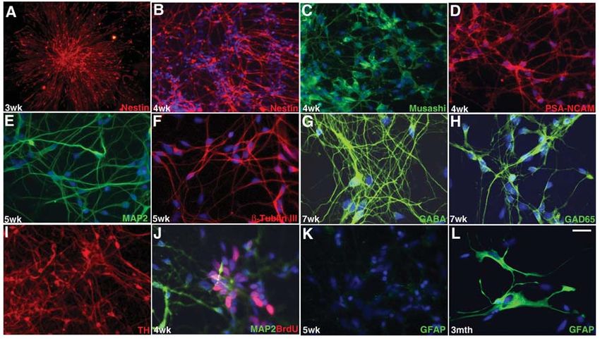
Figure 6. Characterization of hESC-derived neural progenitors and neurons by immunocytochemistry. (A): Typical rosette-neural progenitor
appeared at approximately 3 weeks of differentiation and is positive with nestin antibody staining. After disassociation, the neural progenitors
are also positive for other neural progenitor markers, such as (C) musashi and (D) PSA-NCAM, in addition to (B) nestin. These neural progeni-
tors are expandable, and the cells in the center of the rosette are positive for BrdU labeling (J). At P4, the cells are positive for early neuronal
markers as well such as (E) MAP2 and (F) β-tubulin III. Most of the neurons differentiated from hESCs in the N2B27 medium are GABA neu-
rons, indicated by their positive staining for (G) GABA and (H) GAD65 antibodies. TH neurons can be induced by exposure of neural progeni-
tors at 4 weeks of differentiation to SHH and FGF8 (I). No GFAP-positive cells were detected (K) until long term in culture (L). Blue staining
represents DAPI counterstain. Timelines are indicated at lower left corner. Scale bar = 75 μm in A, B, and I and 25 μm in the others. Abbrevia-
tions: DAPI, 4,6-diamidino-2-phenylindole; GABA, γ-aminobutyric acid; GFAP, glial fibrilliary acidic protein; hESC, human embryonic stem
cell; TH, tyrosine hydroxylase.1240 Neural Differentiation of hESCs in Adherent Culture
logical observations and immunostaining results. The semi- Mouse ESCs have been successfully converted to neuroec-
quantitative RT-PCR showed a time-dependent reduction in todermal precursors in adherent monoculture [5]. However, due
expression of ESC markers, including Oct4, hTERT, and nanog to the differences between mouse ESCs and hESCs, we failed to
(Figs. 4A, 4B), and the reduction of Oct4 was slower than hTERT differentiate hESCs efficiently into neural lineage using the same
and nanog. Furthermore, the reduction in expression of ESC protocol. First, mouse ESCs can grow in culture after disassocia-
markers was associated with increased expression of neural pro- tion into single cells and seeding at low density, whereas hESCs
genitor markers, such as Sox1, nestin, Pax6, and the neuronal are prone to spontaneous differentiation into flat or fibroblast-like
marker MAP2 (Figs. 4A, 4C). The glial marker GFAP, however, cells if plated at low density [6, 9], indicating that hESCs require
was not detected, which corresponded to the immunostain- cell-cell contacts to control their fate. Second, there is no problem
ing results. The mesodermal and endodermal markers AFP, to culture mouse ESCs in gelatin-coated culture plastics, whereas
GATA6, and brachyury showed almost undetectable expression hESCs are unable to efficiently attach in these dishes. This may
without consistent increase or decrease during differentiation reflect the differences of cell-surface molecules, including adhe-
(Fig. 4A). Pax6 antibody staining was observed by the second sion molecules, between these two ESCs. For example, mouse
week of differentiation and became more homogenous along the ESCs express SSEA1 cell-surface marker, but hESCs express
progress of differentiation (Fig. 4D, upper panel). No clear Sox1 SSEA3 and SSEA4. Therefore, instead of gelatin, matrigel and
antibody staining was observed until after 4 weeks of differen- poly-lysine/laminin were used. Similar to mouse ESCs, maybe
tiation (Fig. 4D, lower panel). The sequence of Pax6 and Sox1 even more prevalent, hESCs produced large numbers of flattened
expression is similar to the neural differentiation with multi- cells when plated on matrigel or laminin. These flat cells were
cellular aggregates [20]. Musashi showed a similar timeline as positive for GATA6 and AFP, indicating that they are extraem-
Sox1 (Fig. 6C). Together, these results suggest that hESCs cul- bryonic endoderm [21, 22]. Addition of BMP antagonist noggin
tured with this serum-free adherent protocol mainly differenti- but not follistatin into the culture medium of hESCs effectively
ated into the neural lineage, particularly neuronal cells. blocked the extraembryonic endoderm formation and promoted
Most neurons generated with this differentiation protocol by neural development. Such an effect of noggin has been observed
passage 5 (~60 days in differentiation) are immunopositive for in Xenopus laevis [23].
γ-aminobutyric acid (GABA) (Fig. 6G) and GAD65, an enzyme The neural progenitors generated in this study were mainly
required for GABA synthesis (Fig. 6H), with very few TH-posi- neuronal precursors. There were no detectable GFAP- and O4-
tive neurons. No glutamatergic neurons were detected, as indi- positive cells until late passage in long-term cultures. These
cated by negative staining with GluRI and GLAST antibodies. results differ from the neural progenitors produced by multicel-
However, exposing the cells to SHH, FGF8, and ascorbic acid for lular aggregates in the presence of serum. In the latter culture
1–2 weeks at passages 4 through 5 of differentiation (~40–50 days system, GFAP-positive glial cells are visible in less than 1 month
of differentiation) followed by treatment with BDNF, GDNF, of differentiation [2, 24]. This could be due to the application of
ascorbic acid, and laminin for another 1–2 weeks resulted in a sig- BMP antagonist because BMP has been indicated as an important
nificant increase in the numbers of TH-positive neurons (Fig. 6I). factor for glial cell formation [25]. The addition of noggin into
These results indicate that the hESC-derived neural progenitors the medium might have blocked the BMP signaling and inhibited
with this protocol have the potential for terminal differentiation the glial cell differentiation; withdrawing noggin at passage 3
into other neuronal types other than GABA neurons if optimal or 4 may permit glial development in the long-term culture. It is
culture conditions for a particular neuronal type are provided. also possible that cells in multicellular aggregates are at various
In the current protocol, neither GFAP-positive nor gal C–posi- developmental stages due to their complex microenvironments;
tive and O4-positive cells were detected by passage 5. However, some cells are more developmentally advanced than others, so
GFAP-positive cells were detectable after long-term culturing they may develop into glial cells when most of the cells are still
(>80 days). neuronal cells.
The adherent culture protocol facilitates visualization of
Discussion the process of neural conversion. This process seemed to reca-
The main finding of this study is the development of an adher- pitulate early steps of the nervous system development in vivo
ent culture system using either matrigel or laminin as matrix. in that undifferentiated ESCs become more compact and then
This culture system can efficiently differentiate hESCs to neural neural tube–like structures are formed. This could provide an
fate, particularly neuronal progenitors, without using multicel- experimental tool to study factors affecting human neural tube
lular aggregates or coculture. The progenitors generated by this formation under controlled conditions. We have used this system
method can be further differentiated to dopaminergic neurons, to investigate Oct4 expression during neural differentiation and
GABA neurons, and possibly other neural cell types if correct found that Oct4 expression is temporally retained before down-
culture conditions are provided. regulation to become neural progenitor type of cells. The cells thatGerrard, Rodgers, Cui 1241
lost Oct4 expression rapidly did not commit to neural fate, instead tem for differentiation of hESCs to neural lineages, particularly
turning into the flattened extraembryonic cells. Our observations neurons. This system will be useful to study factors for the deter-
support previous findings that forced rapid downregulation of mination of neural and other lineages.
Oct4 in ESCs pushes them into extraembryonic cells [26, 27]. Our
results and others [4, 19] suggest that transiently sustained levels Acknowledgments
of Oct4 expression may be required for in vitro differentiation of We would like to dedicate this paper to Prof. John Clark in mem-
human ESCs into neural lineages because rapid downregulation ory of his support to this work. We also thank Diana Wylie for her
of Oct4 expression in ESCs might have promoted the formation of technical assistance and the other members of the laboratory. This
primitive endoderm. work was supported by the Biotechnology and Biological Science
Understanding how to direct hESCs toward a specific lin- Research Council and the Geron Corporation.
eage pathway and generate appropriate cell types robustly is very
important not only for the study of developmental biology but Disclosures
also for potentially using these cells to treat human diseases. The The authors indicate no potential conflicts of interest.
results reported here provide a simple and relatively defined sys-
References 14 Balemans W, Van HW. Extracellular regulation of BMP signaling in ver-
tebrates: a cocktail of modulators. Dev Biol 2002;250:231–250.
1 Thomson JA, Itskovitz-Eldor J, Shapiro SS et al. Embryonic stem cell
lines derived from human blastocysts. Science 1998;282:1145–1147. 15 Bachiller D, Klingensmith J, Kemp C et al. The organizer factors Chor-
din and Noggin are required for mouse forebrain development. Nature
2 Zhang SC, Wernig M, Duncan ID et al. In vitro differentiation of trans- 2000;403:658–661.
plantable neural precursors from human embryonic stem cells. Nat Bio-
technol 2001;19:1129–1133. 16 Wang XP, Suomalainen M, Jorgez CJ et al. Follistatin regulates enamel
patterning in mouse incisors by asymmetrically inhibiting BMP signaling
3 Carpenter MK, Inokuma MS, Denham J et al. Enrichment of neurons and ameloblast differentiation. Dev Cell 2004;7:719–730.
and neural precursors from human embryonic stem cells. Exp Neurol
2001;172:383–397. 17 Gerrard G, Zhao D, Clark AJ et al. Stably transfected human embryonic
stem cell clones express OCT4-specific green fluorescent protein and
4 Perrier AL, Tabar V, Barberi T et al. Derivation of midbrain dopamine maintain self-renewal and pluripotency. Stem Cells 2005;23:124–133.
neurons from human embryonic stem cells. Proc Natl Acad Sci U S A
2004;101:12543–12548. 18 Miyazono K, Miyazawa K. Id: a target of BMP signaling. Sci STKE
2002;E40.
5 Ying QL, Stavridis M, Griffiths D et al. Conversion of embryonic stem
cells into neuroectodermal precursors in adherent monoculture. Nat Bio- 19 Shimozaki K, Nakashima K, Niwa H et al. Involvement of Oct3/4 in the
technol 2003;21:183–186. enhancement of neuronal differentiation of ES cells in neurogenesis-
inducing cultures. Development 2003;130:2505–2512.
6 Xu C, Inokuma MS, Denham J et al. Feeder-free growth of undifferenti-
ated human embryonic stem cells. Nat Biotechnol 2001;19:971–974. 20 Li XJ, Du ZW, Zarnowska ED et al. Specification of motoneurons from
human embryonic stem cells. Nat Biotechnol 2005;23:215–221.
7 Lee SH, Lumelsky N, Studer L et al. Efficient generation of midbrain and
hindbrain neurons from mouse embryonic stem cells. Nat Biotechnol 21 Morrisey EE, Ip HS, Lu MM et al. GATA-6: a zinc finger transcription
2000;18:675–679. factor that is expressed in multiple cell lineages derived from lateral meso-
derm. Dev Biol 1996;177:309–322.
8 Cui W, Aslam S, Fletcher J et al. Stabilization of telomere length and
karyotypic stability are directly correlated with the level of hTERT gene 22 Krumlauf R, Hammer RE, Tilghman SM et al. Developmental regulation
expression in primary fibroblasts. J Biol Chem 2002;277:38531–38539. of alpha-fetoprotein genes in transgenic mice. Mol Cell Biol 1985;5:1639–
1648.
9 Pera MF, Andrade J, Houssami S et al. Regulation of human embryonic
stem cell differentiation by BMP-2 and its antagonist noggin. J Cell Sci 23 Munoz-Sanjuan I, Brivanlou AH. Neural induction, the default model and
2004;117:1269–1280. embryonic stem cells. Nat Rev Neurosci 2002;3:271–280.
10 Morrisey EE, Tang Z, Sigrist K et al. GATA6 regulates HNF4 and is 24 Reubinoff BE, Itsykson P, Turetsky T et al. Neural progenitors from
required for differentiation of visceral endoderm in the mouse embryo. human embryonic stem cells. Nat Biotechnol 2001;19:1134–1140.
Genes Dev 1998;12:3579–3590.
25 Rajan P, Panchision DM, Newell LF et al. BMPs signal alternately through
11 Koutsourakis M, Langeveld A, Patient R et al. The transcription factor a SMAD or FRAP-STAT pathway to regulate fate choice in CNS stem
GATA6 is essential for early extraembryonic development. Development cells. J Cell Biol 2003;161:911–921.
1999;126:723–732.
26 Niwa H, Miyazaki J, Smith AG. Quantitative expression of Oct-3/4 defines
12 Fujikura J, Yamato E, Yonemura S et al. Differentiation of embryonic differentiation, dedifferentiation or self-renewal of ES cells. Nat Genet
stem cells is induced by GATA factors. Genes Dev 2002;16:784–789. 2000;24:372–376.
13 Tropepe V, Hitoshi S, Sirard C et al. Direct neural fate specification from 27 Hay DC, Sutherland L, Clark J et al. Oct-4 knockdown induces similar
embryonic stem cells: a primitive mammalian neural stem cell stage patterns of endoderm and trophoblast differentiation markers in human
acquired through a default mechanism. Neuron 2001;30:65–78. and mouse embryonic stem cells. Stem Cells 2004;22:225–235.You can also read