Epigallocatechin-3-Gallate, the Main Polyphenol in Green Tea, Inhibits Porcine Epidemic Diarrhea Virus In Vitro - Frontiers
←
→
Page content transcription
If your browser does not render page correctly, please read the page content below
ORIGINAL RESEARCH
                                                                                                                                            published: 22 February 2021
                                                                                                                                        doi: 10.3389/fphar.2021.628526

                                           Epigallocatechin-3-Gallate, the Main
                                           Polyphenol in Green Tea, Inhibits
                                           Porcine Epidemic Diarrhea Virus In
                                           Vitro
                                           Changchao Huan 1,2,3, Weiyin Xu 1,2,3, Bo Ni 4, Tingting Guo 5, Haochun Pan 1,2,3,
                                           Luyao Jiang 1,2,3, Lin Li 1,2,3, Jingting Yao 1,2,3 and Song Gao 1,2,3*
                                           1
                                             Institutes of Agricultural Science and Technology Development, College of Veterinary Medicine, Yangzhou University, Yangzhou,
                                           China, 2Jiangsu Co-Innovation Center for Prevention and Control of Important Animal Infectious Diseases and Zoonoses,
                                           Yangzhou, China, 3Key Laboratory of Avian Bioproduct Development, Ministry of Agriculture and Rural Affairs, Yangzhou, China,
                                           4
                                             China Animal Health And Epidemiology Center, Qingdao, China, 5College of Medicine, Yangzhou University, Yangzhou, China

                                           There are currently no licensed drugs against porcine epidemic diarrhea virus (PEDV), but
                                           vaccines are available. We identified a natural molecule, epigallocatechin-3-gallate
                                           (EGCG), the main polyphenol in green tea, which is effective against infection with
                                           PEDV. We used a variety of methods to test its effects on PEDV in Vero cells. Our
                                           experiments show that EGCG can effectively inhibit PEDV infections (with HLJBY and
                            Edited by:
                    Apostolos Zarros,
                                           CV777 strains) at different time points in the infection using western blot analysis. We found
                University of Glasgow,     that EGCG inhibited PEDV infection in a dose-dependent manner 24 h after the infection
                      United Kingdom
                                           commenced using western blotting, plaque formation assays, immunofluorescence
                        Reviewed by:
                                           assays (IFAs), and quantitative reverse-transcriptase PCR (qRT-PCR). We discovered
                    Barbara Schnierle,
        Paul-Ehrlich-Institut, Germany     that EGCG treatment of Vero cells decreased PEDV attachment and entry into them by the
                   Pritom Chowdhury,       same method analysis. Western blotting also showed that PEDV replication was inhibited
      Tea Research Association, India
                                           by EGCG treatment. Whereas EGCG treatment was found to inhibit PEDV assembly, it had
                  *Correspondence:
                          Song Gao
                                           no effect on PEDV release. In summary, EGCG acts against PEDV infection by inhibiting
                   gsong@yzu.edu.cn        PEDV attachment, entry, replication, and assembly.
                                           Keywords: porcine epidemic diarrhea virus, epigallocatechin-3-gallate, green tea polyphenol, drug, virus inhibition
                   Specialty section:
        This article was submitted to
Experimental Pharmacology and Drug
                           Discovery,      INTRODUCTION
              a section of the journal
           Frontiers in Pharmacology       Porcine epidemic diarrhea (PED) is characterized by acute villus atrophy and congestion, severe
       Received: 12 November 2020          watery diarrhea, dehydration and death. PED can infect pigs of all ages and causes high mortality in
         Accepted: 20 January 2021         newborn piglets (mortality rate is nearly 100%) (Li, et al., 2012; Zhang, et al., 2019). Its causative
        Published: 22 February 2021        agent is porcine epidemic diarrhea virus (PEDV). PEDV was first recognized in 1971 in England
                             Citation:     (Wood, 1977; Pensaert and de Bouck, 1978). Although PED has been reported in Asia since the
  Huan C, Xu W, Ni B, Guo T, Pan H,        1980s, its prevalence has been comparatively low (Takahashi, et al., 1983; Puranaveja, et al., 2009; Lin,
Jiang L, Li L, Yao J and Gao S (2021)      et al., 2014; Jung, et al., 2016; Sun, et al., 2016). Since late 2010, the new PED strains with high
  Epigallocatechin-3-Gallate, the Main
                                           pathogenicity in China have been regarded as pandemic strains (Sun, et al., 2016). In 2013, highly
     Polyphenol in Green Tea, Inhibits
      Porcine Epidemic Diarrhea Virus
                                           pathogenic PEDV was first seen in the USA where it quickly spread to the neighboring countries such
                               In Vitro.   as Canada and Mexico (Mole, 2013; Stevenson, et al., 2013; Chen, et al., 2014; Lowe, et al., 2014;
        Front. Pharmacol. 12:628526.       Vlasova, et al., 2014). To date, PED reoccurrence has become more common in pigs immunized with
     doi: 10.3389/fphar.2021.628526        a commercial vaccine (Li, et al., 2012; Sun, et al., 2012; Tian, et al., 2013). Therefore, PEDV continues

Frontiers in Pharmacology | www.frontiersin.org                                    1                                         February 2021 | Volume 12 | Article 628526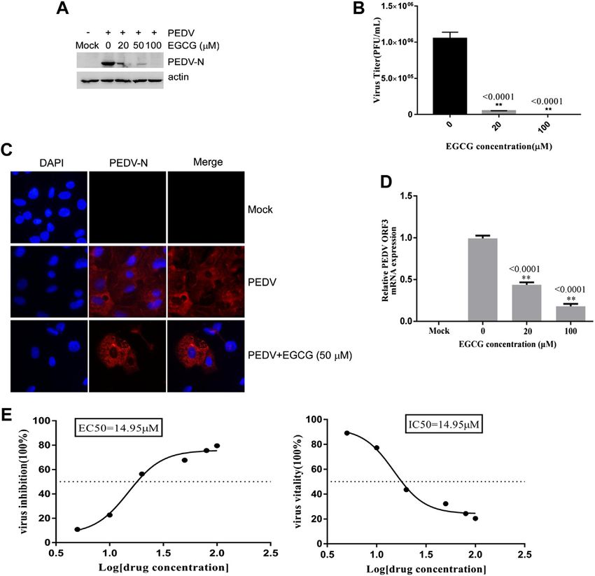
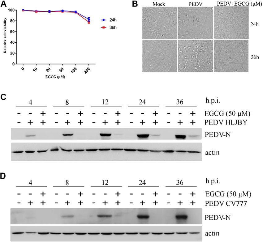
Huan et al.                                                                                                                              EGCG Inhibits PEDV In Vitro

 FIGURE 1 | EGCG inhibition of PEDV at different time points. (A) Cytotoxicity assay using soluble EGCG. EGCG concentrations (0, 10, 20, 50, 100 and 200 μm)
 were added to Vero cells and the cells were cultured for 24 h–36 h. Cell viability was evaluated by the CCK8 assay and calculated as (A450 compound/A450 mock) ×
 100%. (B–D) EGCG inhibits PEDV infection. Vero cells were pretreated with different concentrations of EGCG for 1 h and then infected with PEDV HLJBY (0.1 MOI) or
 PEDV CV777 (0.1 MOI) for 4, 8, 12, 24 and 36 h in the presence of EGCG. (B) EGCG treatment decreased the cytopathic effect on the cells at 24 and 36 h. (C,D)
 PEDV N protein expression levels were detected by western blotting at different time points.

to spread widely and cause huge economic losses to the swine                         the number of hydroxyl groups on its B-ring. EGCG displays
industry. With the prevention and control of PED now very                            potent inhibitory effects toward human immunodeficiency virus,
urgent, we have focused our attention on the potential use of                        influenza virus, hepatitis virus, hepatitis B virus, porcine
traditional Chinese medicine against PEDV.                                           reproductive and respiratory syndrome virus and porcine
   PEDV is a member of alpha coronavirus in the Coronaviridae                        circovirus type 2 (Fassina, et al., 2002; Yamaguchi, et al., 2002;
family, and it possesses a single-stranded positive-sense RNA                        Jariwalla, et al., 2007; Roomi, et al., 2008; Fukazawa, et al., 2012;
genome about 28 kb (Kocherhans, et al., 2001). PEDV size ranges                      Calland, et al., 2012; Chen, et al., 2012; Ge, et al., 2018; Li, et al.,
in diameter from 95 nm to 190 nm. PEDV genome includes a 5’                          2020). However, whether EGCG has an inhibitory effect on
untranslated region (UTR),two overlapping open reading                               PEDV has not been reported. Therefore, we evaluated whether
frames (ORFs) encoding two polyproteins (ORF1a and                                   EGCG inhibited PEDV infection.
ORF1b), ORFs 2–6 encoding four major structural proteins
(spike (S), ORF3, envelope (E), membrane (M), and
nucleocapsid (N)) and one accessory ORF3 protein (Duarte,                            RESULTS
et al., 1993; Lee and Lee, 2010).
   Green tea is consumed as a drink in worldwide (Hayat, et al.,                     EGCG Inhibits PEDV Infection
2015). In 2017, the production of global tea was 5.686 million                       To assess the antiviral activity of EGCG against PEDV, the
tons, while the production of China’s tea was 2.609 million tons.                    cytotoxicity of EGCG in Vero cells was first evaluated using a
Epigallocatechin-3-gallate (EGCG) is a natural compound in                           CCK8 assay. The results revealed that when used for 24 and
green tea, and EGCG accounts for about 59% of the total                              36 h at 100 μM, EGCG was not cytotoxic to Vero cells, whereas
catechin in it. Other catechins in green tea include epicatechin                     it was cytotoxic at 200 μm (Figure 1A). Therefore, the
(EC) (6.4%), epicatechin gallate (ECG) (13.6%), and                                  maximum concentration of EGCG used in the experiments
epigallocatechin (EGC) (19%) (McKay and Blumberg, 2002).                             was 100 μm. Vero cells were pretreated with different
Structurally and functionally, the properties of catechin are                        concentrations of EGCG for 1 h and then infected with
attributed to the presence or absence of a galloyl moiety and                        PEDV HLJBY (0.1 MOI) or PEDV CV777 (0.1 MOI) for

Frontiers in Pharmacology | www.frontiersin.org                                  2                                        February 2021 | Volume 12 | Article 628526
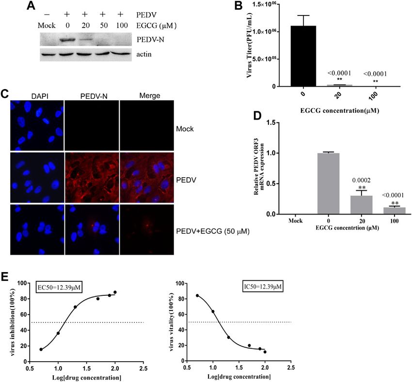
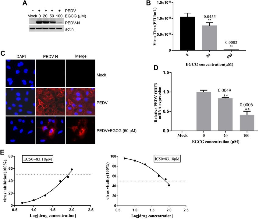
Huan et al.                                                                                                                                   EGCG Inhibits PEDV In Vitro

 FIGURE 2 | EGCG exhibits an anti-viral effect on PEDV infection in Vero cells. Cells were pre-treated with different concentrations of EGCG for 1 h before being
 infected with PEDV HLJBY (0.1 MOI) and were then treated with different concentrations of EGCG for 24 h. Intact cells and supernatants were collected at 24 h.p.i. (A)
 PEDV N and actin proteins were detected by western blotting. (B) Viral titers were determined using a plaque formation assay. (C) IFA detection of PEDV infected cells.
 (D) PEDV mRNA was quantified by qRT-PCR. (E) EC50 or IC50 of EGCG against PEDV infection was checked.

different time periods with EGCG present. EGCG treatment                                results confirm that EGCG displays marked antiviral activity
decreased the cytopathic effect of PEDV HLJBY on the cells                              against PEDV infections.
(Figure 1B). In addition, western blotting showed that PEDV
HLJBY and PEDV CV777 N protein expression levels
decreased significantly at the different time points (Figures                            EGCG Impairs PEDV Attachment to Vero
1C, D). Therefore, we determined the inhibitory effect of                               Cells
PEDV HLJBY infection on the cells for the 24 h period by                                To investigate the stage at which EGCG exerts its antiviral effect
western blotting, plaque formation assays, IFAs and qRT-PCR.                            in the PEDV infection process, we first explored the effect of
Western blotting showed that the expression level of the PEDV                           EGCG treatment on PEDV attachment to Vero cells. A binding
HLJBY N protein decreased significantly under different                                  assay was performed that involved pretreating Vero cells with
concentrations of EGCG, and the decrease in the protein                                 EGCG for 1 h before infecting them with PEDV HLJBY at 4°C for
level was approximately 99.6 and 99.9% at 50 μm and                                     1 h with EGCG present. The cells were then washed with PBS
100 μm of EGCG, respectively (Figure 2A). The inhibition                                3 times and cultured at 37°C for 23 h. The western blotting results
of PEDV HLJBY infection was approximately 99.9% at 100 μm                               showed that EGCG inhibited the expression level of PEDV N
of EGCG, as demonstrated by the decreased viral titers                                  protein, with an inhibition rate of 68.5–99.4% in an EGCG dose-
detected by the plaque formation assay (Figure 2B). The                                 dependent manner (Figure 3A). EGCG treatment also decreased
number of cells infected with PEDV HLJBY was clearly                                    the numbers of cells infected with PEDV, as determined by IFA
lower than that of the controls in the IFA (Figure 2C), and                             (Figure 3C). Supernatants were collected for assaying the viral
the reduction of the PEDV HLJBY ORF3 was about 93.2% at                                 titers, which revealed that the PEDV titer fell by approximately
100 μm EGCG by qRT-PCR (Figure 2D). In addition, the                                    99.3% at 100 μM EGCG, as determined by the plaque formation
EC50 or IC50 of the antiviral effect of EGCG on PEDV HLJBY                              assays (Figure 3B). Cells were also collected to determine the level
was further determined to be 12.39 µm (Figure 2E). These                                of PEDV ORF3 mRNA by qRT-PCR, which showed that EGCG

Frontiers in Pharmacology | www.frontiersin.org                                     3                                         February 2021 | Volume 12 | Article 628526
Huan et al.                                                                                                                                EGCG Inhibits PEDV In Vitro

 FIGURE 3 | EGCG prevents PEDV binding to Vero cells. (A–C) Vero cells were pretreated with EGCG for 1 h and then infected with PEDV HLJBY at 4°C for 1 h with
 EGCG present. The cells were washed three times with PBS and then cultured at 37°C for 23 h. (A) PEDV N protein levels were detected by western blotting. (B)
 Supernatant-specific PEDV titers were assayed by a plaque formation assay. (C) The number of cells infected with PEDV was evaluated by IFA. (D) Vero cells were pre-
 treated with different concentrations of EGCG for 1 h before being infected with PEDV (0.1MOI) at 4°C for 1 h with EGCG present. The cells were collected to assay
 the level of PEDV ORF3 mRNA by qRT-PCR at 1 h.p.i. (E) EC50 or IC50 of EGCG against PEDV binding was checked.

treatment significantly impaired the level of PEDV ORF3 mRNA                           respectively (Figure 4D). In addition, the EC50 or IC50 of EGCG
(by about 53 and 79.2% at 20 and 100 μm, respectively)                                inhibited entry was 83.18 µm (Figure 4E). These results confirm
(Figure 3D). In addition, the EC50 or IC50 of EGCG                                    that EGCG can block PEDV entry into Vero cells.
inhibited attachment was 14.95 µm (Figure 3E). These results
show that EGCG treatment reduced the attachment of PEDV to
the Vero cells such that PEDV was unable to infect them.                              The Effect of EGCG on PEDV Replication,
                                                                                      Assembly and Release in Vero Cells
EGCG Affects PEDV Entry Into Vero Cells                                               To clarify the effects of EGCG on PEDV infection, Vero cells were
To explore whether EGCG is able to inhibit PEDV entry, Vero                           infected with PEDV HLJBY for 1 h and then treated with EGCG
cells were infected with PEDV HLJBY in the presence of EGCG at                        for 3 and 5 h. Cells were collected to evaluate the expression levels
37°C for 1 h. After 1 h, the cells were washed three times in citric                  of the PEDV N protein at 4 and 6 h.p.i. Western blotting showed
acid and three times in PBS, and 2% DMEM was added. The                               that EGCG treatment decreased the level of the PEDV N protein
culture was allowed to continue for 23 h at 37°C. We found that                       (Figure 5A), suggesting that EGCG can prevent PEDV
EGCG reduced the level of PEDV N protein (by about 10–89.4%),                         replication.
as judged by western blotting (Figure 4A). The cells infected with                        To investigate whether EGCG treatment affected viral
PEDV were examined by IFA (Figure 4C). Supernatants were                              assembly, Vero cells were infected with PEDV HLJBY (0.1
also collected to assay the viral titers, and the plaque formation                    MOI) at 37°C for 1 h before adding EGCG. The cell
assays revealed that the PEDV titers fell significantly by about                       supernatants and intact cells were collected to determine
96.5% at 100 μm (Figure 4B). When the Vero cells were infected                        the copy numbers of the PEDV ORF3 mRNA at 24 h.p.i.
with PEDV with EGCG present at 37°C for 1 h, the PEDV ORF3                            The copy number ratio of the mRNA from the PEDV ORF3
mRNA levels fell by about 81.1 and 56.7% at 20 μm and 100 μm,                         gene in the supernatant to that of the PEDV ORF3 mRNA in

Frontiers in Pharmacology | www.frontiersin.org                                   4                                        February 2021 | Volume 12 | Article 628526
Huan et al. EGCG Inhibits PEDV In Vitro
FIGURE 4 | EGCG inhibition of PEDV entry into Vero cells. (A–C) Vero cells were infected with PEDV HLJBY with EGCG present at 37°C for 1 h. After 1 h the cells
were washed three times with citric acid and three times with PBS, and 2% DMEM was added. The culture was allowed to continue for 23 h at 37°C. (A) The level of
PEDV N protein was evaluated by western blotting. (B) Plaque formation assay for PEDV titers in the supernatants. (C) The number of cells infected with PEDV was
evaluated by IFA. (D) When the Vero cells were infected with PEDV with EGCG present at 37 C for 1 h, the PEDV ORF3 mRNA levels were detected by qRT-PCR.
(E) EC50 or IC50 of EGCG against PEDV entry was checked.
the cells showed that EGCG inhibited PEDV assembly by villous atrophy (Huan, et al., 2020). PEDV HLJBY and PEDV
about 99.6% at 100 μm (Figure 5B). In contrast, we found CV777 were stored at −80°C in the Yangzhou University
that the collected supernatant and intact cells that we used Infectious Diseases laboratory.
to determine the PEDV titer showed the opposite effect,
whereby the ratio of PEDV titer in the supernatant to that Reagents
of the PEDV titer in the cells showed that EGCG treatment had EGCG was purchased from Selleck (China) and was diluted
no effect on PEDV release, as evidenced by the results to stock solutions of 50 mm with PBS and stored at −80 °C
(Figure 5C). for all subsequent experiments. The purity of EGCG was
99.68% by HPLC assessed. The PEDV N antibody used
herein was generated in our laboratory. The anti-PEDV-N
MATERIALS AND METHODS polyclonal antibody was prepared in rabbits using PEDV-N
protein as antigen and western blot and IFA were diluted 1:
Cells and Virus 5,000 and 1:500, respectively (Gao, et al., 2020). The β-actin
Vero cells were cultured in Dulbecco’s modified Eagle’s medium antibody was purchased from TransGenBiotech (China).
(Sigma-Aldrich) supplemented with 8% fetal bovine serum HRP-labeled Goat Anti-Mouse IgG (H + L) and DAPI
(Lonsa) at 37°C with 5% CO2. PEDV HLJBY isolated from the (4′,6-diamidino-2-phenylindole) was purchased from
intestinal contents of a pig-let with diarrhea from Heilongjiang Beyotime Biotechnology (China). The citric acid solution
province, and PEDV reference strain CV777 was purchased from (PH 3.0) is composed of 40 mm citric acid, 10 mm
China Institute of Veterinary Drug Control. PEDV HLJBY and KCl, and135 mm NaCl, to remove un-internalized virus
PEDV CV777 caused pigs diarrhea, and acute viral enteritis with particles.
Frontiers in Pharmacology | www.frontiersin.org 5 February 2021 | Volume 12 | Article 628526Huan et al. EGCG Inhibits PEDV In Vitro
FIGURE 5 | EGCG treatment decreased the replication and assembly of PEDV. (A) Vero cells were infected with PEDV HLJBY for 1 h and then treated with EGCG
for 3 or 5 h. Western blotting analysis of PEDV N levels. (B,C) Vero cells were infected with PEDV HLJBY (0.1 MOI) at 37°C for 1 h before EGCG treatment. (B) The
supernatants from the cells and the intact cells were collected to determine the copy number of the PEDV ORF3 gene at 24 h.p.i., as well as the PEDV titer ratio in the
supernatant and in intact cells. (C) The collected supernatants and intact cells were used to determine the PEDV titers, from which the PEDV titer ratios in the
supernatants to those of the intact cells were calculated.
Cytotoxicity of EGCG Toward Vero Cells HLJBY (0.1 MOI) at 4°C with the corresponding concentrations
The cytotoxicity of EGCG toward Vero cells was evaluated of EGCG for 1 h. The cells were washed in cold PBS three times
following the manufacturer’s instructions from the CCK8 kit (Norkin, 2009; Huan, et al., 2015). The cells and cell supernatants
(Beyotime Biotechnology). Vero cells were seeded in a 96-well were then collected to evaluate PEDV N protein levels and
plate, and then treated with 10, 20, 50, 100, 200 μm determine the virus titer by western blotting and plaque
concentrations of EGCG for 24–36 h. After 24–36 h of culture, formation assays, respectively, at 24 h post-infection (h.p.i.).
10 μl of CCK-8 solution per well was added and the plate was To demonstrate the effect of EGCG on PEDV attachment, the
further incubated for 2 h at 37°C. Then we checked the cells were washed with cold PBS three times and then collected for
absorbance at 450 nm to determine the maximum measurement of the PEDV ORF3 mRNA expression levels by
concentration of EGCG that was not toxic to the Vero cells. qRT-PCR.
Cell-Based Assays Entry Assay
Infectivity Assay Vero cells were infected with PEDV HLJBY at 4°C for 1 h,
We checked the life cycle of PEDV (MOI 1) in Vero cells. We followed by washing with cold PBS three times. The cells were
collected supernatants to check viral titer at hourly intervals maintained in 2% DMEM containing different concentrations of
between 1 and 12 h. Vero cells were pre-treated with EGCG EGCG (0, 20, 50, 100 μm) at 37°C for 1 h. After washing three
(0, 20, 50,100 μm) for 1 h and then infected with PEDV (0.1 times with the citric acid solution (40 mm citric acid, 10 mm KCl,
multiplicity of infection; MOI) for 4, 8, 12, 24, and 36 h with 135 mm NaCl, pH 3.0) to remove un-internalized virus particles
EGCG present. Vero cells were also pre-treated with different (Hancock, et al., 2010; Wang, et al., 2014; Huan, et al., 2017). Vero
concentrations of EGCG for 1 h, followed by infection with cells were washed with PBS three times. Intracellular viral
PEDV (0.1 MOI) for 24 h with EGCG. Cells were collected to proteins and cell supernatants were collected at 24 h.p.i to
determine whether any changes had occurred in the N protein or detect any changes that occurred in PEDV N protein levels or
the ORF3 mRNA of PEDV by western blot analysis and virus titers using western blots and plaque formation assays,
quantitative reverse–transcriptase quantitative PCR (qRT- respectively. The same steps were used with the Vero cells when
PCR), respectively. Cell supernatants were collected to measuring the levels of PEDV ORF3 mRNA by qRT-PCR.
measure viral titers using a plaque formation assay.
Replication, Assembly and Release of PEDV
Attachment/Binding Assay Vero cells were infected with PEDV HLJBY at 37°C for 1 h,
Vero cells were pre-treated with different concentrations of followed by washing and incubation in 2% DMEM containing
EGCG (0, 20, 50,100 μm) for 1 h, followed by washing with EGCG. The cells were collected to check the level of PEDV N
cold PBS three times. The cells were then infected with PEDV protein on western blots at 4 and 6 h.p.i.
Frontiers in Pharmacology | www.frontiersin.org 6 February 2021 | Volume 12 | Article 628526Huan et al. EGCG Inhibits PEDV In Vitro
Vero cells were incubated with PEDV HLJBY at 37°C for analyzed by one-way ANOVA using the SPSS 17.0 software
1 h. The cells were washed three times in PBS and then package. When the p values were less than 0.05, the
incubated with 2% DMEM containing various differences were considered to be statistically significant (*p <
concentrations of EGCG at 37°C. At 24 h.p.i., cells or cell 0.05 and **p < 0.01).
supernatants were collected to determine the viral titers and
the number of viral RNA copies using a plaque formation assay
and qRT-PCR, respectively. DISCUSSION
Western Blotting EGCG is known to exert its anti-infective effect on bacteria,
The western blotting procedure followed previous descriptions viruses, and different fungal species (Steinmann, et al., 2013).
(Huan, et al., 2017; Gao, et al., 2020). PEDV N and actin were It does this with hepatitis C virus (HCV) by inhibiting its entry
detected using the above-mentioned primary antibody and a by interfering with its binding to the target cells (Ciesek, et al.,
secondary antibody. 2011; Calland, et al., 2012; Chen, et al., 2012). EGCG reduces
hepatitis B virus (HBV) infection by decreasing HBV antigen
Immunofluorescence Assays expression, and decreasing the levels of extracellular HBV
IFAs were performed as per a previous description (Gao, et al., DNA and covalently closed circular DNA (Xu, et al., 2008;
2020). Briefly, Vero cells were fixed with 4% paraformaldehyde, He, et al., 2011). EGCG possibly damages and inactivates the
permeabilized with 0.1% Triton X-100, blocked with 3% bovine virions of herpes simplex virus 1 and 2 by binding to their
serum albumin, and incubated with the anti-PEDV N antibody envelope proteins (Isaacs, et al., 2008; Isaacs, et al., 2011).
and an Alexa Fluor 5558-conjugated goat anti-rabbit secondary EGCG was found to inhibit transcription of the immediate-
antibody (Invitrogen, United States). All images were taken at early genes (Rta, Zta and EA-D) of Epstein–Barr virus (Chang,
×200 magnification. et al., 2003). But the effect of EGCG on PEDV has awaited
elucidation. Therefore, we explored the activity of EGCG
qRT-PCR against PEDV.
qRT-PCR was performed as described previously (Gao, et al., SARS coronavirus attaching and entering Vero E6 cells
2020). Total RNA was extracted, and gene expression was needs 30 –60 min, and extracellular virus particles were
assessed by qRT-PCR using specific primers (PEDV-ORF3-F: checked at 5 – 6 h.p.i. (Ng, et al., 2003a; Ng, et al., 2003b;
TTTGCACTGTTTAAAGCGTCT, PEDV-ORF3-R: AGTAAA Qinfen, et al., 2004; Hartenian, et al., 2020). Furthermore, one
AGCAGACTAAACAAAGCCT). replication cycle of Porcine deltacoronavirus (PDCoV) takes
5–6 h (Qin, et al., 2019). In addition, we checked the life cycle
Plaque Formation Assays of PEDV (MOI 1) in Vero cells. Infectious progeny virus was
Viral culture supernatants were diluted from 10−1 to 10−6 in detected in the supernatants at 6 h at a low titer (1.6 ×
DMEM and then used to infect Vero cells seeded in 6-well plates 102 PFU/ml), and progeny virus increased. The result
at 37°C for 2 h before the DMEM was added to each well. Vero revealed that the first generation of progeny virus were
cells were cultured at 37°C with 5% CO2 for 3 days, after which released at about 5 h.p.i., and one life cycle of PEDV takes
they were stained with 0.5% crystal violet. about 6 h to complete. In this study, we showed that EGCG
inhibited two different PEDV strains in Vero cells (Figures 1,
Calculation of EC50 and IC50 2), and went on to elucidate the anti-PEDV mechanism of
EGCG was serially diluted to 100, 80, 50, 20, 10 and 5 μm, EGCG. To investigate the effect of EGCG against PEDV
and was added to Vero cells which were infected with infection, we first explored its effect on PEDV HLJBY
PEDV (MOI 0.1). EGCG was present during the different binding. The results showed that EGCG reduced the
stages of PEDV infection. The infected cells without EGCG expression of the N protein of PEDV and also reduced
treatment were set as mock control. We determined ORF3 PEDV ORF3 mRNA levels and PEDV titers (Figure 3) in
mRNA levels at 24 h.p.i. by qRT-PCR and the values of the binding assay, revealing that EGCG significantly reduced
inhibition were calculated as (1-ORF3 mRNA (compound)/ PEDV HLJBY attachment to Vero cells. These results are
ORF3 mRNA (mock)) × 100%. The values of inhibition consistent with the findings from another study where EGCG
and EGCG concentration were used to establish a dose inhibited the binding of human immunodeficiency virus 1
response curve and further calculated the EC50 (Williamson, et al., 2006; Nance, et al., 2009; Jiang, et al., 2010;
(concentration for 50% of maximal effect). The values of Li, et al., 2011). EGCG potently inhibited PEDV infection by
vitality were calculated as ORF3 mRNA (compound)/ORF3 preventing PEDV attachment to Vero cells. Therefore, EGCG
mRNA (mock) × 100%. The values of vitality and EGCG may provide a new way of preventing PEDV infections. We
concentration were used to establish a dose response curve next explored the effect of EGCG on PEDV HLJBY entry into
and further calculate the IC50. Vero cells. We observed that EGCG decreased the levels of the
PEDV N protein, PEDV ORF3 mRNA and the PEDV titer
Statistical Analysis using an entry assay (Figure 4). Other studies have shown that
All experiments were independently repeated at least three times EGCG can block influenza virus entry of cells by binding to
and all data were presented as means ± SD. All the data were hemagglutinin (Nakayama, et al., 1993; Imanishi, et al., 2002;
Frontiers in Pharmacology | www.frontiersin.org 7 February 2021 | Volume 12 | Article 628526Huan et al. EGCG Inhibits PEDV In Vitro
Song, et al., 2005). Our results indicate that EGCG was able to DATA AVAILABILITY STATEMENT
inhibit PEDV infection by blocking PEDV entry into
Vero cells. The original contributions presented in the study are included in
EGCG has been reported to suppress enterovirus the article/Supplementary Material, further inquiries can be
replication (Ho, et al., 2009). Therefore, we investigated directed to the corresponding author.
the effect of EGCG on PEDV HLJBY replication. We
found that EGCG reduced PEDV replication, but this
effect was inferior to that seen with PEDV attachment and AUTHOR CONTRIBUTIONS
entry. This result shows that EGCG has potential to inhibit
PEDV replication. Although we discovered that EGCG All authors listed have made a substantial, direct, and intellectual
treatment was able to decrease PEDV assembly, it had no contribution to the work and approved it for publication.
effect on PEDV release.
In conclusion, we have provided evidence that EGCG
inhibited PEDV infection by blocking viral attachment, FUNDING
entry, replication and assembly. Although The
bioavailability of EGCG is usually poor (about 4.5–7.2%) This research was funded by the National Natural Science Foundation
(Del Rio, et al., 2010), EGCG contribute to the health of China (31902253), the Natural Science Foundation of Jiangsu
benefits (Chen, et al., 1997; Sabu, et al., 2002; Henning, Province (BK20180921), the Individual Technology Research and
et al., 2004; Yang and Wang, 2016). Chow HH. revealed Development of the Modern Agricultural Industry of Jiangsu Province
repeated intake of catechins in humans increased (CX (19)3024), the China Postdoctoral Science Foundation
bioavailability of EGCG (Chow, et al., 2003). In addition, (2018M632399), the Priority Academic Program Development of
the increase in EGCG bioavailability in mice fed the Jiangsu Higher Education Institutions, and the earmarked fund for
catechin diet for two weeks was reported (Ishii, et al., Jiangsu Agricultural Industry Technology System.
2019). In previous studies, EGCG had similar antiviral
activity against various viruses, EGCG against PRRSV with
an EC50 of 48.2–63.09 µm in vitro (Ge, et al., 2018), and EGCG ACKNOWLEDGMENTS
inhibited JEV with an IC50 of 4.9–20 µm in vitro (Wang, et al.,
2018). EGCG against PCV2 in vitro with EC50 was 37.79 ± We thank Sandra Cheesman, from Liwen Bianji, Edanz Group
1.64 µm (Li, et al., 2020). In our study, the EC50 or IC50 of China (www.liwenbianji.cn/ac) and Xiang Mao for editing the
EGCG against PEDV in vitro was12.39–83.18 µm. English text of a draft of this manuscript.
Duarte, M., Gelfi, J., Lambert, P., Rasschaert, D., and Laude, H. (1993). Genome
REFERENCES organization of porcine epidemic diarrhoea virus. Adv. Exp. Med. Biol. 342,
55–60. doi:10.1007/978-1-4615-2996-5_9
Calland, N., Albecka, A., Belouzard, S., Wychowski, C., Duverlie, G., Descamps, V., Fassina, G., Buffa, A., Benelli, R., Varnier, O. E., Noonan, D. M., and Albini, A.
et al. (2012). (-)-Epigallocatechin-3-gallate is a new inhibitor of hepatitis C virus (2002). Polyphenolic antioxidant (-)-epigallocatechin-3-gallate from green tea
entry. Hepatology 55, 720–729. doi:10.1002/hep.24803 as a candidate anti-HIV agent. AIDS 16, 939–941. doi:10.1097/00002030-
Chang, L. K., Wei, T. T., Chiu, Y. F., Tung, C. P., Chuang, J. Y., Hung, S. K., et al. 200204120-00020
(2003). Inhibition of Epstein-Barr virus lytic cycle by (-)-epigallocatechin Fukazawa, H., Suzuki, T., Wakita, T., and Murakami, Y. (2012). A cell-based,
gallate. Biochem. Biophys. Res. Commun. 301, 1062–1068. doi:10.1016/ microplate colorimetric screen identifies 7,8-benzoflavone and green tea gallate
s0006-291x(03)00067-6 catechins as inhibitors of the hepatitis C virus. Biol. Pharm. Bull. 35, 1320–1327.
Chen, C., Qiu, H., Gong, J., Liu, Q., Xiao, H., Chen, X. W., et al. (2012). doi:10.1248/bpb.b12-00251
(-)-Epigallocatechin-3-gallate inhibits the replication cycle of hepatitis C Gao, R., Zhang, Y., Kang, Y., Xu, W., Jiang, L., Guo, T., et al. (2020). Glycyrrhizin
virus. Arch. Virol. 157, 1301–1312. doi:10.1007/s00705-012-1304-0 inhibits PEDV infection and proinflammatory cytokine secretion via the
Chen, L., Lee, M. J., Li, H., and Yang, C. S. (1997). Absorption, distribution, HMGB1/TLR4-MAPK p38 pathway. Int. J. Mol. Sci. 21, 2961. doi:10.3390/
elimination of tea polyphenols in rats. Drug Metab. Dispos. 25, 1045–1050. ijms21082961
Chen, Q., Li, G., Stasko, J., Thomas, J. T., Stensland, W. R., Pillatzki, A. E., et al. Ge, M., Xiao, Y., Chen, H., Luo, F., Du, G., and Zeng, F. (2018). Multiple antiviral
(2014). Isolation and characterization of porcine epidemic diarrhea viruses approaches of (-)-epigallocatechin-3-gallate (EGCG) against porcine
associated with the 2013 disease outbreak among swine in the United States. reproductive and respiratory syndrome virus infection in vitro. Antiviral
J. Clin. Microbiol. 52, 234–243. doi:10.1128/JCM.02820-13 Res. 158, 52–62. doi:10.1016/j.antiviral.2018.07.012
Chow, H. H., Cai, Y., Hakim, I. A., Crowell, J. A., Shahi, F., Brooks, C. A., et al. Hancock, M. H., Cliffe, A. R., Knipe, D. M., and Smiley, J. R. (2010). Herpes simplex
(2003). Pharmacokinetics and safety of green tea polyphenols after multiple- virus VP16, but not ICP0, is required to reduce histone occupancy and enhance
dose administration of epigallocatechin gallate and polyphenon E in healthy histone acetylation on viral genomes in U2OS osteosarcoma cells. J. Virol. 84,
individuals Clin. Cancer Res. 9, 3312–3319. 1366–1375. doi:10.1128/JVI.01727-09
Ciesek, S., von Hahn, T., Colpitts, C. C., Schang, L. M., Friesland, M., Steinmann, J., Hartenian, E., Nandakumar, D., Lari, A., Ly, M., Tucker, J. M., and Glaunsinger, B.
et al. (2011). The green tea polyphenol, epigallocatechin-3-gallate, inhibits A. (2020). The molecular virology of coronaviruses. J. Biol. Chem. 295,
hepatitis C virus entry. Hepatology 54, 1947–1955. doi:10.1002/hep.24610 12910–12934. doi:10.1074/jbc.REV120.013930
Del Rio, D., Calani, L., Scazzina, F., Jechiu, L., Cordero, C., and Brighenti, F. (2010). Hayat, K., Iqbal, H., Malik, U., Bilal, U., and Mushtaq, S. (2015). Tea and its
Bioavailability of catechins from ready-to-drink tea. Nutrition 26, 528–533. consumption: benefits and risks. Crit. Rev. Food Sci. Nutr. 55, 939–954. doi:10.
doi:10.1016/j.nut.2009.06.013 1080/10408398.2012.678949
Frontiers in Pharmacology | www.frontiersin.org 8 February 2021 | Volume 12 | Article 628526Huan et al. EGCG Inhibits PEDV In Vitro
He, W., Li, L. X., Liao, Q. J., Liu, C. L., and Chen, X. L. (2011). Epigallocatechin Lowe, J., Gauger, P., Harmon, K., Zhang, J., Connor, J., Yeske, P., et al. (2014). Role
gallate inhibits HBV DNA synthesis in a viral replication - inducible cell line. of transportation in spread of porcine epidemic diarrhea virus infection,
World J. Gastroenterol. 17, 1507–1514. doi:10.3748/wjg.v17.i11.1507 United States. Emerging Infect. Dis. 20, 872–874. doi:10.3201/eid2005.131628
Henning, S. M., Niu, Y., Lee, N. H., Thames, G. D., Minutti, R. R., Wang, H., et al. McKay, D. L., and Blumberg, J. B. (2002). The role of tea in human health: an
(2004). Bioavailability and antioxidant activity of tea flavanols after update. J. Am. Coll. Nutr. 21, 1–13. doi:10.1080/07315724.2002.10719187
consumption of green tea, black tea, or a green tea extract supplement. Am. Mole, B. (2013). Deadly pig virus slips through US borders. Nature 499, 388. doi:10.
J. Clin. Nutr. 80, 1558–1564. doi:10.1093/ajcn/80.6.1558 1038/499388a
Ho, H. Y., Cheng, M. L., Weng, S. F., Leu, Y. L., and Chiu, D. T. (2009). Antiviral Nakayama, M., Suzuki, K., Toda, M., Okubo, S., Hara, Y., and Shimamura, T.
effect of epigallocatechin gallate on enterovirus 71. J. Agric. Food Chem. 57, (1993). Inhibition of the infectivity of influenza virus by tea polyphenols.
6140–6147. doi:10.1021/jf901128u Antiviral Res. 21, 289–299. doi:10.1016/0166-3542(93)90008-7
Huan, C. C., Pan, H. C., Fu, S. Y., Xu, W. Y., Gao, Q. Q., Wang, X. B., et al. (2020). Nance, C. L., Siwak, E. B., and Shearer, W. T. (2009). Preclinical development of the
Characterization and evolution of the coronavirus porcine epidemic diarrhoea green tea catechin, epigallocatechin gallate, as an HIV-1 therapy. J. Allergy Clin.
virus HLJBY isolated in China. Transbound. Emerg. Dis. 67, 65–79. doi:10.1111/ Immunol. 123, 459–465. doi:10.1016/j.jaci.2008.12.024
tbed.13321 Ng, M. L., Tan, S. H., See, E. E., Ooi, E. E., and Ling, A. E. (2003a). Early events of
Huan, C. C., Wang, H. X., Sheng, X. X., Wang, R., Wang, X., and Mao, X. (2017). SARS coronavirus infection in vero cells. J. Med. Virol. 71, 323–331. doi:10.
Glycyrrhizin inhibits porcine epidemic diarrhea virus infection and attenuates 1002/jmv.10499
the proinflammatory responses by inhibition of high mobility group box-1 Ng, M. L., Tan, S. H., See, E. E., Ooi, E. E., and Ling, A. E. (2003b). Proliferative
protein. Arch. Virol. 162, 1467–1476. doi:10.1007/s00705-017-3259-7 growth of SARS coronavirus in Vero E6 cells. J. Gen. Virol. 84, 3291–3303.
Huan, C. C., Wang, Y., Ni, B., Wang, R., Huang, L., Ren, X. F., et al. (2015). Porcine doi:10.1099/vir.0.19505-0
epidemic diarrhea virus uses cell-surface heparan sulfate as an attachment Norkin, L. C. (2009). Virology: molecular biology and pathogenesis. Washington,
factor. Arch. Virol. 160, 1621–1628. doi:10.1007/s00705-015-2408-0 DC: ASM Press.
Imanishi, N., Tuji, Y., Katada, Y., Maruhashi, M., Konosu, S., Mantani, N., et al. Pensaert, M. B., and de Bouck, P. (1978). A new coronavirus-like particle associated
(2002). Additional inhibitory effect of tea extract on the growth of influenza A with diarrhea in swine. Arch. Virol. 58, 243–247. doi:10.1007/BF01317606
and B viruses in MDCK cells. Microbiol. Immunol. 46, 491–494. doi:10.1111/j. Puranaveja, S., Poolperm, P., Lertwatcharasarakul, P., Kesdaengsakonwut, S.,
1348-0421.2002.tb02724.x Boonsoongnern, A., Urairong, K., et al. (2009). Chinese-like strain of
Isaacs, C. E., Wen, G. Y., Xu, W., Jia, J. H., Rohan, L., Corbo, C., et al. (2008). porcine epidemic diarrhea virus, Thailand. Emerging Infect. Dis. 15,
Epigallocatechin gallate inactivates clinical isolates of herpes simplex 1112–1115. doi:10.3201/eid1507.081256
virus. Antimicrob. Agents Chemother. 52, 962–970. doi:10.1128/AAC. Qin, P., Du, E. Z., Luo, W. T., Yang, Y. L., Zhang, Y. Q., Wang, B., et al. (2019).
00825-07 Characteristics of the life cycle of porcine deltacoronavirus (PDCoV) in vitro:
Isaacs, C. E., Xu, W., Merz, G., Hillier, S., Rohan, L., and Wen, G. Y. (2011). replication kinetics, cellular ultrastructure and virion morphology, and
Digallate dimers of (-)-epigallocatechin gallate inactivate herpes simplex virus. evidence of inducing autophagy. Viruses-Basel. 11, 455. doi:10.3390/v11050455
Antimicrob. Agents Chemother. 55, 5646–5653. doi:10.1128/AAC.05531-11 Qinfen, Z., Jinming, C., Xiaojun, H., Huanying, Z., Jicheng, H., Ling, F., et al.
Ishii, S., Kitazawa, H., Mori, T., Kirino, A., Nakamura, S., Osaki, N., et al. (2019). (2004). The life cycle of SARS coronavirus in Vero E6 cells. J. Med. Virol. 73,
Identification of the catechin uptake transporter responsible for intestinal 332–337. doi:10.1002/jmv.20095
absorption of epigallocatechin gallate in mice. Sci. Rep. 9, 11014. doi:10. Roomi, M. W., Jariwalla, R. J., Kalinovsky, T., Roomi, N., Niedzwiecki, A., and
1038/s41598-019-47214-4 Rath, M. (2008). Inhibition of cellular invasive parameters in influenza A virus-
Jariwalla, R. J., Roomi, M. W., Gangapurkar, B., Kalinovsky, T., Niedzwiecki, A., infected MDCK and Vero cells by a nutrient mixture. Biofactors 33, 61–75.
and Rath, M. (2007). Suppression of influenza A virus nuclear antigen doi:10.1002/biof.5520330106
production and neuraminidase activity by a nutrient mixture containing Sabu, M. C., Smitha, K., and Kuttan, R. (2002). Anti-diabetic activity of green tea
ascorbic acid, green tea extract and amino acids. Biofactors 31, 1–15. doi:10. polyphenols and their role in reducing oxidative stress in experimental diabetes.
1002/biof.5520310101 J. Ethnopharmacol. 83, 109–116. doi:10.1016/s0378-8741(02)00217-9
Jiang, F., Chen, W., Yi, K., Wu, Z., Si, Y., Han, W., et al. (2010). The evaluation of Song, J. M., Lee, K. H., and Seong, B. L. (2005). Antiviral effect of catechins in green
catechins that contain a galloyl moiety as potential HIV-1 integrase inhibitors. tea on influenza virus. Antiviral Res. 68, 66–74. doi:10.1016/j.antiviral.2005.
Clin. Immunol. 137, 347–356. doi:10.1016/j.clim.2010.08.007 06.010
Jung, K., Hu, H., and Saif, L. J. (2016). Porcine deltacoronavirus infection: etiology, Steinmann, J., Buer, J., Pietschmann, T., and Steinmann, E. (2013). Anti-infective
cell culture for virus isolation and propagation, molecular epidemiology and properties of epigallocatechin-3-gallate (EGCG), a component of green tea. Br.
pathogenesis. Virus Res. 226, 50–59. doi:10.1016/j.virusres.2016.04.009 J. Pharmacol. 168, 1059–1073. doi:10.1111/bph.12009
Kocherhans, R., Bridgen, A., Ackermann, M., and Tobler, K. (2001). Completion of Stevenson, G. W., Hoang, H., Schwartz, K. J., Burrough, E. R., Sun, D., Madson, D.,
the porcine epidemic diarrhoea coronavirus (PEDV) genome sequence. Virus et al. (2013). Emergence of Porcine epidemic diarrhea virus in the United States:
Genes 23, 137–144. doi:10.1023/a:1011831902219 clinical signs, lesions, and viral genomic sequences. J. Vet. Diagn. Invest. 25,
Lee, Y. J., and Lee, C. (2010). Porcine reproductive and respiratory syndrome virus 649–654. doi:10.1177/1040638713501675
replication is suppressed by inhibition of the extracellular signal-regulated Sun, D., Wang, X., Wei, S., Chen, J., and Feng, L. (2016). Epidemiology and vaccine
kinase (ERK) signaling pathway. Virus Res. 152, 50–58. doi:10.1016/j.virusres. of porcine epidemic diarrhea virus in China: a mini-review. J. Vet. Med. Sci. 78,
2010.06.002 355–363. doi:10.1292/jvms.15-0446
Li, J. R., Song, D. F., Wang, S. N., Dai, Y. D., Zhou, J. Y., and Gu, J. Y. (2020). Sun, R. Q., Cai, R. J., Chen, Y. Q., Liang, P. S., Chen, D. K., and Song, C. X. (2012).
Antiviral effect of epigallocatechin gallate via impairing porcine circovirus type Outbreak of porcine epidemic diarrhea in suckling piglets, China. Emerging
2 attachment to host cell receptor. Viruses-Basel. 12, 176. doi:10.3390/ Infect. Dis. 18, 161–163. doi:10.3201/eid1801.111259
v12020176 Takahashi, K., Okada, K., and Ohshima, K. (1983). An outbreak of swine diarrhea
Li, S., Hattori, T., and Kodama, E. N. (2011). Epigallocatechin gallate inhibits the of a new-type associated with coronavirus-like particles in Japan. Nippon
HIV reverse transcription step. Antivir. Chem. Chemother. 21, 239–243. doi:10. Juigaku Zasshi 45, 829–832. doi:10.1292/jvms1939.45.829
3851/IMP1774 Tian, Y., Yu, Z., Cheng, K., Liu, Y., Huang, J., Xin, Y., et al. (2013). Molecular
Li, W., Li, H., Liu, Y., Pan, Y., Deng, F., Song, Y., et al. (2012). New variants of characterization and phylogenetic analysis of new variants of the porcine
porcine epidemic diarrhea virus, China, 2011. Emerging Infect. Dis. 18, epidemic diarrhea virus in Gansu, China in 2012, Viruses 5, 1991–2004.
1350–1353. doi:10.3201/eid1808.120002 doi:10.3390/v5081991
Lin, C. N., Chung, W. B., Chang, S. W., Wen, C. C., Liu, H., Chien, C. H., et al. Vlasova, A. N., Marthaler, D., Wang, Q., Culhane, M. R., Rossow, K. D., Rovira, A.,
(2014). US-like strain of porcine epidemic diarrhea virus outbreaks in et al. (2014). Distinct characteristics and complex evolution of PEDV strains,
Taiwan, 2013-2014. J. Vet. Med. Sci. 76, 1297–1299. doi:10.1292/jvms.14- North America, May 2013-February 2014. Emerging Infect. Dis. 20, 1620–1628.
0098 doi:10.3201/eid2010.140491
Frontiers in Pharmacology | www.frontiersin.org 9 February 2021 | Volume 12 | Article 628526Huan et al. EGCG Inhibits PEDV In Vitro Wang, C. Y., Hour, M. J., Lai, H. C., Chen, C. H., Chang, P. J., Huang, S. H., et al. immunodeficiency virus type 1 (HIV-1). Antiviral Res. 53, 19–34. doi:10.1016/ (2018). Epigallocatechin-3-gallate inhibits the early stages of Japanese s0166-3542(01)00189-9 encephalitis virus infection. Virus Res. 253, 140–146. doi:10.1016/j.virusres. Yang, C. S., and Wang, H. (2016). Cancer preventive activities of tea catechins. 2018.06.009 Molecules 21, 1679. doi:10.3390/molecules21121679 Wang, Y., Li, J. R., Sun, M. X., Ni, B., Huan, C., Huang, L., et al. (2014). Triggering Zhang, L., Liu, X., Zhang, Q., Zhou, P., Fang, Y., Dong, Z., et al. (2019). Biological unfolded protein response by 2-Deoxy-D-glucose inhibits porcine epidemic characterization and pathogenicity of a newly isolated Chinese highly virulent diarrhea virus propagation. Antiviral Res. 106, 33–41. doi:10.1016/j.antiviral. genotype GIIa porcine epidemic diarrhea virus strain. Arch. Virol. 164, 2014.03.007 1287–1295. doi:10.1007/s00705-019-04167-3 Williamson, M. P., McCormick, T. G., Nance, C. L., and Shearer, W. T. (2006). Epigallocatechin gallate, the main polyphenol in green tea, binds to the T-cell Conflict of Interest: The authors declare that the research was conducted in the receptor, CD4: potential for HIV-1 therapy. J. Allergy Clin. Immunol. 118, absence of any commercial or financial relationships that could be construed as a 1369–1374. doi:10.1016/j.jaci.2006.08.016 potential conflict of interest. Wood, E. N. (1977). An apparently new syndrome of porcine epidemic diarrhoea. Vet. Rec. 100, 243–244. doi:10.1136/vr.100.12.243 Copyright © 2021 Huan, Xu, Ni, Guo, Pan, Jiang, Li, Yao and Gao. This is an open- Xu, J., Wang, J., Deng, F., Hu, Z., and Wang, H. (2008). Green tea extract access article distributed under the terms of the Creative Commons Attribution and its major component epigallocatechin gallate inhibits hepatitis License (CC BY). The use, distribution or reproduction in other forums is permitted, B virus in vitro. Antiviral Res. 78, 242–249. doi:10.1016/j.antiviral.2007. provided the original author(s) and the copyright owner(s) are credited and that the 11.011 original publication in this journal is cited, in accordance with accepted academic Yamaguchi, K., Honda, M., Ikigai, H., Hara, Y., and Shimamura, T. (2002). practice. No use, distribution or reproduction is permitted which does not comply Inhibitory effects of (-)-epigallocatechin gallate on the life cycle of human with these terms. Frontiers in Pharmacology | www.frontiersin.org 10 February 2021 | Volume 12 | Article 628526
You can also read