EVIDENCE OF SARS-COV2 ENTRY PROTEIN ACE2 IN THE HUMAN NOSE AND OLFACTORY BULB
←
→
Page content transcription
If your browser does not render page correctly, please read the page content below
Developmental Biology / Research Article

                                                   Cells Tissues Organs 2020;209:155–164                                 Received: September 7, 2020
                                                                                                                         Accepted: November 12, 2020
                                                   DOI: 10.1159/000513040                                                Published online: January 22, 2021

Evidence of SARS-CoV2 Entry Protein
ACE2 in the Human Nose and Olfactory
Bulb
Moritz Klingenstein a Stefanie Klingenstein a Peter H. Neckel b
Andreas F. Mack b Andreas P. Wagner b Alexander Kleger c Stefan Liebau a
Alfio Milazzo a
aInstitute
        of Neuroanatomy and Developmental Biology, Eberhard Karls University Tübingen, Tübingen, Germany;
bInstitute
        of Clinical Anatomy and Cell Analysis, Eberhard Karls University Tübingen, Tübingen, Germany;
cDepartment of Internal Medicine I, University Medical Center Ulm, Ulm, Germany

Keywords                                                                 hardly accessible human olfactory bulb. ACE2 can be detect-
SARS-CoV2 · ACE2 · Human · Olfactory epithelium ·                        ed in the olfactory epithelium as well as in the respiratory
Olfactory bulb                                                           epithelium of the nasal septum, the nasal conchae, and the
                                                                         paranasal sinuses. ACE2 is located in the sustentacular cells
                                                                         and in the glandular cells in the olfactory epithelium as well
Abstract                                                                 as in the basal cells, glandular cells, and epithelial cells of the
Usually, pandemic COVID-19 disease, caused by SARS-CoV2,                 respiratory epithelium. Intriguingly, ACE2 is not expressed in
presents with mild respiratory symptoms such as fever,                   mature or immature olfactory receptor neurons and basal
cough, but frequently also with anosmia and neurological                 cells in the olfactory epithelium. Similarly, ACE2 is not local-
symptoms. Virus-cell fusion is mediated by angiotensin-con-              ized in the olfactory receptor neurons albeit the olfactory
verting enzyme 2 (ACE2) and transmembrane serine prote-                  bulb is positive. Vice versa, TMPRSS2 can also be detected in
ase 2 (TMPRSS2) with their organ expression pattern deter-               the sustentacular cells and the glandular cells of the olfac-
mining viral tropism. Clinical presentation suggests rapid vi-           tory epithelium. Our findings provide the basic anatomical
ral dissemination to the central nervous system leading                  evidence for the expression of ACE2 and TMPRSS2 in the hu-
frequently to severe symptoms including viral meningitis.                man nose, olfactory epithelium, and olfactory bulb. Thus,
Here, we provide a comprehensive expression landscape of                 they are substantial for future studies that aim to elucidate
ACE2 and TMPRSS2 proteins across human postmortem na-                    the symptom of SARS-CoV2 induced anosmia via the olfac-
sal and olfactory tissue. Sagittal sections through the human            tory pathway.                                 © 2021 S. Karger AG, Basel
nose complemented with immunolabelling of respective
cell types represent different anatomically defined regions
including olfactory epithelium, respiratory epithelium of the
nasal conchae and the paranasal sinuses along with the                   M.K. and S.K. contributed equally to this work.

karger@karger.com     © 2021 S. Karger AG, Basel                         Stefanie Klingenstein
www.karger.com/cto                                                       Institute of Neuroanatomy and Developmental Biology
                                                                         Eberhard Karls University Tübingen
                                                                         Oesterbergstr. 3, DE–72074 Tübingen (Germany)
                                                                         stefanie.klingenstein @ uni-tuebingen.de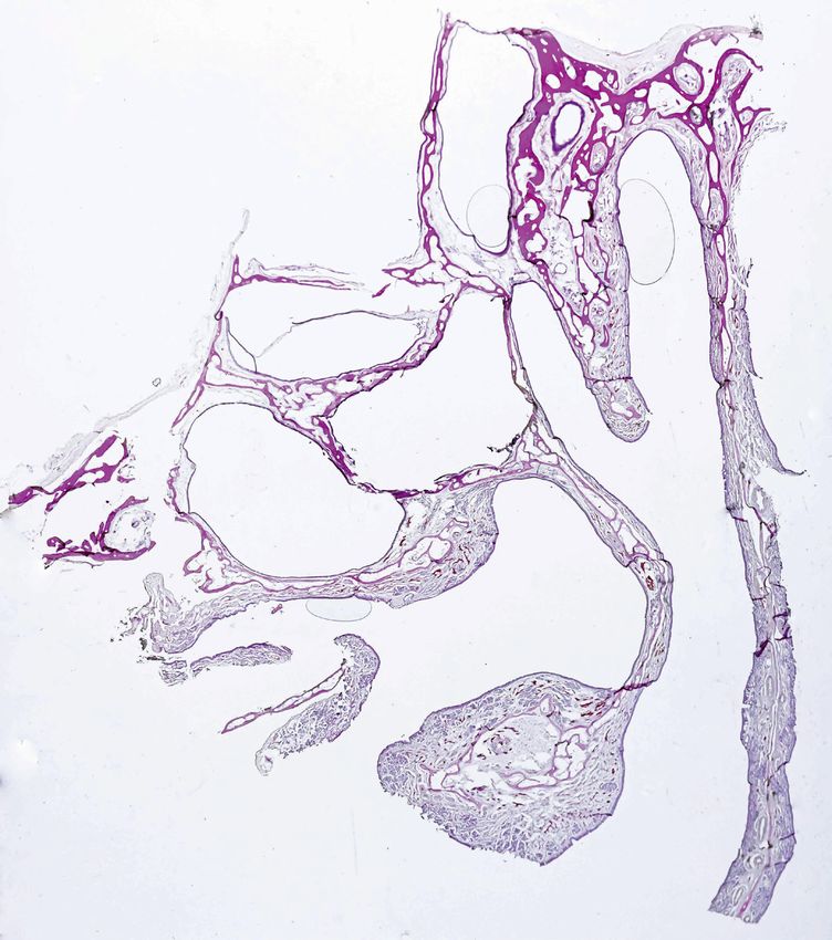
Introduction leads to different symptoms with varying severity. Ac-
cording to clinical studies worldwide, the most prevalent
The Coronavirus Disease 2019 (COVID-19) emerged symptoms are fever, cough, fatigue, headache, dyspnea,
from East Asia and quickly spread all over the world sputum production, arthralgia, diarrhea, rhinorrhea, and
reaching a pandemic scale [Guan et al., 2020]. The infil- sore throat [Krajewska et al., 2020; Zhou et al., 2020].
tration of the virus SARS-CoV2 into different cell types Since many patients also report olfactory and gustatory
a
OB
b
b CP
OE
IV
CE
I SNC
III
NS
INC
II
a b
a a
IV IV
I I
III
III
II
ACE2 II
c d TUBB3
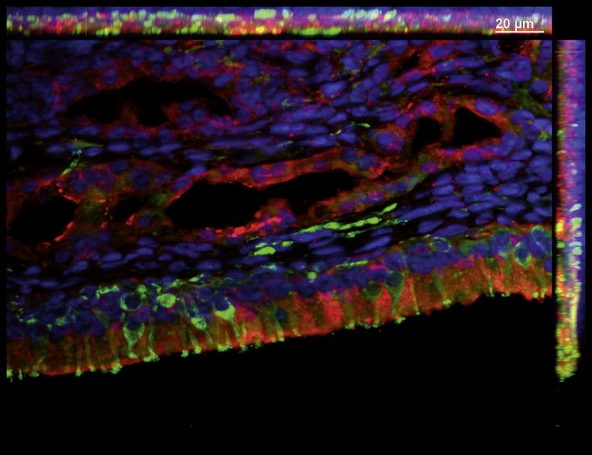
Fig. 1. a Schematic illustration of a human head. Frontal section (II), the cellulae ethmoidales (CE) (III) and the olfactory epitheli-
highlights the nose specimen (a). Sagittal view of the olfactory bulb um (OE) (IV). CP, cribriform plate; SNC, superior nasal conchae.
section (b). Olfactory epithelium and olfactory bulb, both marked c. Hematoxylin eosin staining of the right half of the nose speci-
in green, are located at the upper nasal cavity and above the crib- men. Dashed boxes show the same areas found in the schematic
riform plate. b Schematic representation of the nose specimen picture in b. d Immunofluorescence staining of the right half of
from the frontal section through the head (a). The olfactory epi- human nose specimen. TUBB3 is shown in green, ACE2 in red,
thelium (OE) and the olfactory bulb (OB) are shown in green. Sec- nuclei in blue. Dashed boxes show the same areas found in the
tion plane for better comprehensibility of the olfactory bulb (b). schematic picture in b. More details for area I, II, III of the respira-
The dashed boxes show the section of the respiratory epithelium tory epithelium can be found in Fig. 2. More details for area IV
of the nasal septum (NS) (l), the intermediate nasal conchae (INC) olfactory epithelium can be found in Fig. 3. Scale bar, 2.5 mm.
156 Cells Tissues Organs 2020;209:155–164 Klingenstein/Klingenstein/Neckel/Mack/
DOI: 10.1159/000513040 Wagner/Kleger/Liebau/Milazzo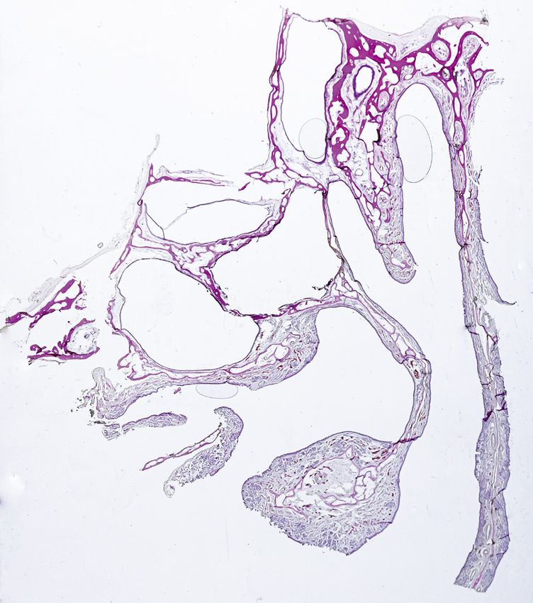
II               Ciliated Cell

                 I
                                                       Goblet Cell

                                                       Basal Cell

                                                       Gll. nasales
      a

                                                                        I                                   I

                     I                                                                                               I
                                            Respiratory Epithelium
                                                Nasal Septum

                                                                                                                ACE2
                                                                        II                                 II

                           II               Respiratory Epithelium
                                                                                                                     II
                                         Intermediate Nasal Conchae

                                                                                                                ACE2
                                                                       III                                III

                 III
                                            Respiratory Epithelium
                                             Cellulae Ethmoidale

       III                                                                                                      ACE2
      b                                                                      c

Fig. 2. a Schematic illustration of the pseudostratified respiratory         thelium of the nasal septum as well as positive apical staining of
epithelium with ciliated cells, goblet cells, basal cells, and nasal         respiratory epithelial cells and nasal submucosal glands (l). ACE2
submucosal glands. b Hematoxylin eosin stainings of the respira-             (red) protein expression is located in the ciliated epithelium cells
tory epithelium in the area of the nasal septum (l), the intermediate        and the basal cells of the respiratory epithelium of intermediate
nasal conchae (II), and the cellulae ethmoidales (III). c Immuno-            nasal conchae as well as in the underlying nasal submucosal glands
fluorescent stainings of the respiratory epithelium verified by ab-          (II). ACE2 (red) expression in the cellulae ethmoidales can be
sent TUBB3 expression in the epithelium from dashed boxes I, II              found in epithelial cells and basal cells (III). Nuclei are shown in
and III. TUBB3 is a highly specific marker for mature and imma-              blue. Scale bar, 20 μm.
ture ORN. ACE2 (red) positive basal cells of the respiratory epi-

Detection of ACE2 in Human Olfactory                                         Cells Tissues Organs 2020;209:155–164                           157
Tissue                                                                       DOI: 10.1159/000513040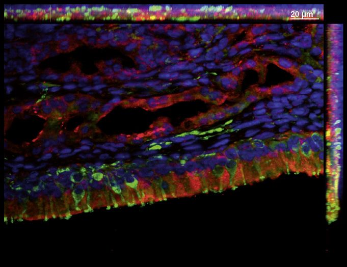
dysfunctions, these symptoms are considered typical of                   epithelium and murine olfactory epithelium [Bilinska et
the SARS-CoV2 infection [Lechien et al., 2020; Lee et al.,               al., 2020]. Similarly, experiments performed in the mu-
2020]. There are many steps involved in the perception of                rine olfactory bulb showed partly contradictory results
smell where the infection with SARS-CoV2 could poten-                    for ACE2 staining. Until now, there are no protein verifi-
tially be the cause of anosmia, starting with the transport              cations in human olfactory bulb due to its difficult acces-
of the odorants to the receptors in the olfactory neurons                sibility.
extending to the signal transduction to different olfactory                  Here, we employ a unique human postmortem tissue
cortex areas.                                                            resource to thoroughly study the ACE2 and TMPRSS2
    In the olfactory epithelium, a variety of histological               protein expression patterns in the human olfactory sys-
target structures, including the olfactory receptor neu-                 tem, including olfactory epithelium, respiratory epithe-
rons (ORN) with their ensheathed axons, sustentacular,                   lium of the nasal septum, the nasal conchae, and the pa-
microvillar or glandular cells could serve as a viral target             ranasal sinuses as well as the olfactory bulb. These find-
and therefore influence olfactory function. Another area                 ings will help to explain the symptom of anosmia as well
of viral attack could be the olfactory bulb. Here, the fila              as frequent dissemination to the central nervous system
olfactoria or projection neurons in different layers of the              in COVID-19 patients and give a starting point to further
olfactory bulb could be targeted by the virus, causing dis-              investigations on how SARS-CoV2 can affect the olfac-
ruption in olfactory perception.                                         tory system.
    Infection of host cells with SARS-CoV-2 is preceded
by a complex process of virus attaching, receptor recogni-
                                                                            Materials and Methods
tion and proteolytic cleavage of the transmembrane spike
glycoprotein to promote virus-cell fusion mediated by                        Tissue Processing
angiotensin-converting enzyme 2 (ACE2) and trans-                            The tissue was obtained from human body donors. Surgery was
membrane serine protease 2 (TMPRSS2) [Hoffmann et                        performed transcranial with particular attention to remove the
al., 2020].                                                              cribriform plate together with the olfactory bulb as well as the na-
                                                                         sal septum and the nasal conchae. Fixation of the whole specimen
    Up to date, there is no clear evidence which cell types              was performed for 4 days with daily changes of Roti Histofix (Carl
of the olfactory and respiratory epithelium express ACE2                 Roth, P087.1) fixation media, followed by 1-day washing step with
and TMPRSS2. Transcriptional but also data from mu-                      PBS. Decalcification was achieved with 10% EDTA for 70 days
rine and human olfactory tissue show as of now report                    with medium change twice a week. After a washing step for 1 day
conflicting data [Bilinska et al., 2020; Brann et al., 2020;             with PBS, the specimen was incubated for another day in 30% su-
                                                                         crose, followed by embedding with Tissue-Tek (Thermo Fisher,
Hou et al., 2020; Ueha et al., 2020]. In addition, the re-               12351753) for frozen sections.
ports on localization of the viral entry proteins in differ-
ent cell types from the epithelia vary [Bilinska et al., 2020;               Immunohistological Staining
Ueha et al., 2020]. Protein expression was found in sus-                     For hematoxylin-eosin staining, 12 μm frozen sections were
                                                                         treated with filtered hematoxylin (Sigma, H9627) for 10 min, fol-
tentacular cells in all publications, but only one paper                 lowed by a washing step with tap water for 2–5 min. Eosin (Sigma,
claims a low ACE2 expression in ORN [Ueha et al., 2020].                 230251) counterstaining was performed for 7 min, followed by
TMPRSS2 was found in murine and human respiratory                        careful rinses with distilled H2O and dehydration with increasing

Fig. 3. a Schematic structure of human olfactory epithelium. The         and mucosa showing ACE2 positive sustentacular cells and posi-
mature olfactory receptor neurons are highlighted in green and are       tive Bowman glands. Upper dashed box shows an enlarged picture
surrounded by sustentacular cells. The sensory neurons arise via         of ACE2 positive sustentacular cell, marked with dashed white
an olfactory precursor step from basal cells. There are 2 types of       lines, but negative for TUBB3. TUBB3 positive olfactory sensory
basal cells: horizontal and globose basal cells. Bowman glands are       neuron marked with dashed yellow lines. Lower dashed box shows
secretory glands that are found exclusively in the olfactory epithe-     ACE2 positive staining in Bowman glands in the olfactory submu-
lium. b Hematoxylin eosin staining of the olfactory epithelium in        cosa. One ACE2 positive Bowman gland cell is marked with a
the area of the nasal septum (IV). c Immunofluorescent staining          dashed white line. e Immunofluorescent picture with TMPRSS2
of the olfactory epithelium from dashed box (IV). Merged picture         (green) and OMP (red) expression in the olfactory epithelium
shows ACE2 in red, OMP, a marker for mature ORN, in white and            (IV). TMPRSS2 is mainly located in the sustentacular cells and
TUBB3, a marker for mature and immature ORN, in green. Posi-             shows a minor expression in Bowman glands, but no expression in
tive ACE2 staining is exclusively located to the region of the sus-      basal cell or OMP-positive mature ORN. Nuclei are shown in blue.
tentacular cells, no appearance in basal cells or co-localization with   Scale bar, 20 μm.
OMP or TUBB3. d Enlarged picture from the olfactory epithelium                                                  (For figure see next page.)

158                     Cells Tissues Organs 2020;209:155–164                                   Klingenstein/Klingenstein/Neckel/Mack/
                        DOI: 10.1159/000513040                                                  Wagner/Kleger/Liebau/Milazzo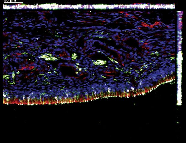
Sustentacular Cell                                                       IV
                                                       Olfactory Receptor Neuron
                                                       Olfactory Precursor Cell
                                                       Globose Basal Cell
                                                       Horizontal Basal Cell               IV                Olfactory Epithelium
                                                                                                                Nasal Septum
                                                       Bowman‘s Gland
                          a                                                         b

                                          IV

                                                                                               II
                              c
                                               TUBB3                  ACE2                 OMP
                                                                 IV            IV

                                                                                            Sustentacular Cell

                                    IV                                                      Olfactory Receptor Neuron

                                                                                           Bowman‘s Gland Cell

                                                                                        III

                              d   TUBB3                               ACE2
                                          IV

                              e                TMPRSS2                OMP

                  3

Detection of ACE2 in Human Olfactory                           Cells Tissues Organs 2020;209:155–164                                 159
Tissue                                                         DOI: 10.1159/000513040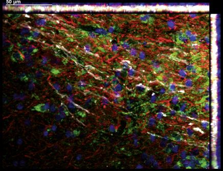
alcohol concentrations (70, 95, and 100%). After washing 2 times        TMPRSS2 are found in the respiratory and olfactory mu-
in xylene, sections were mounted with DPX mounting media                cosa. First, a frontal section of human postmortem tissue
(VWR, 13,514).
                                                                        through the nose with distinct anatomical regions (shown
    Immunofluorescence Staining                                         in Fig. 1a) is examined. The olfactory epithelium (IV) is
    For immunofluorescence staining, 12 μm frozen sections were         located at the roof of the nasal cavity between the nasal
rehydrated for 5 min with PBS (Thermo Fisher, 10010056) fol-            septum and the superior nasal conchae. The paranasal
lowed by an ethanol gradient each 30 s (70, 95, 99, 95, and 70%).       sinuses, in particular the cellulae ethmoidales (III), are
The frozen sections were blocked in skimmed milk blocking buffer
(TBS + 10% NDS +1.25% BSA, 4% skimmed milk, 0.1% Triton X)              located next to the nasal turbinate. In addition, the nasal
for 30 min at room temperature. The primary antibody was di-            septum (I), the superior nasal conchae, and the interme-
luted in skimmed milk blocking solution and incubated over night        diate nasal conchae (II), all of which covered with respira-
at 4°C. The following primary antibodies were used: ACE2 rb (Ab-        tory epithelium, are present in this section (shown in
cam, ab15348 1:100), glial fibrillary acidic protein (GFAP) ms          Fig. 1b). With hematoxylin-eosin staining, these different
(Merck, MAB360, 1:100), TMPRSS2 ms (Santa Cruz, Sc515727,
1:50), ßIII-tubulin (TUBB3) ms (BioLegend, 802002, 1:1000), and         regions of the nasal cavity were identified and visualized
olfactory marker protein (OMP) gt (WAKO, 544-1001 1:500).               (shown in Fig. 1c, 2b, 3b). Overview of ACE2 and TUBB3
Synaptophysin rb (Abcam, 14,692-100, 1:100). All primary anti-          immunolabelling in postmortem nasal tissue (shown in
bodies were verified in appropriate tissue (online suppl. Fig 1; see    Fig. 1d). TUBB3, a marker for mature and immature ol-
www.karger.com/doi/10.1159/513040). After several washing               factory sensory neurons allows distinguishing the olfac-
steps, secondary antibodies were diluted 1:100 in PBS together
with DAPI (Abcam, ab228549) and incubated for 45 min in room            tory epithelium from the other non-neural areas within
temperature away from daylight. The following secondary anti-           the nose specimen (Fig. 1d).
bodies were used: Dαrb 488 (Invitrogen, A32790), Dαms 488 (In-
vitrogen, A32766), Dαrb 546 (Invitrogen, A10040), Dαgt 546 (In-             Identification of ACE2 in Different Areas of the
vitrogen, A11056), and Dαms 647 (Abcam, ab150107). Sections                 Respiratory Epithelium
were embedded with Mowiol (Carl Roth, 0713). Immunofluores-
cence stainings were analyzed using the Axio Imager. M2 micro-              Schematic illustration and hematoxylin-eosin staining
scope with the AxioVision software (Zeiss).                             show the anatomical stratification of the analyzed regions
    Scans of immunolabelled nasal specimen were done by order           of the nasal cavity (Fig. 1c, 2a, b). The different areas of
of Zeiss, pictures were analyzed with ZEN Blue software (Zeiss).        the respiratory epithelium, namely the nasal septum (I),
                                                                        the intermediate nasal conchae (II), and the cellulae eth-
                                                                        moidales (III) show pseudostratified ciliated epithelial
   Results                                                              cells arising from a basal cell layer. Mucus-producing
                                                                        goblet cells can be found in the respiratory epithelium of
   Anatomical Regions Analyzed in Human Postmortem                      all 3 areas (Fig. 2b). In the respiratory epithelium of the
   Nasal and Olfactory Tissue                                           nasal septum, the intermediate nasal conchae and the pa-
   High ACE2 and TMPRSS2 expressions have been doc-                     ranasal sinus, ACE2 expression was located in epithelial
umented in the respiratory, gastrointestinal, and repro-                cells as well as in basal cells. High expression was found
ductive system [Fan et al., 2020; Ren et al., 2020]. Focus-             in the nasal submucosal glands underneath the respira-
ing on the upper respiratory tract, increased ACE2 and                  tory epithelium. The ACE2 expression can be found uni-

Fig. 4. a Schematic illustration of the human olfactory bulb with       merular layer; EPL, external plexiform layer; MCL, mitral cell lay-
its layers: Under most is the outer nerve layer with the axons of the   er; IPL, internal plexiform layer; GCL, glomerular cell layer.
olfactory receptor neurons followed by the glomerular layer with        d Overview picture with immunofluorescence stainings for ACE2
olfactory glomerula connecting the axons with interneurons. Syn-        (green), TUBB3 (red), and OMP (white) illustrating the different
aptic processing between the glomerular layer and the mitral cell       layers of the olfactory bulb. The white dashed box indicates the
layer occurs in the external plexiform cell layer. In the mitral cell   magnified area illustrated in the next image, showing representa-
layer, the mitral cells are located. They form synapses in the inner    tive ACE2 positive cells, not co-localizing with TUBB3 or OMP.
plexiform layer with the granular cells of the granular cell layer.     High protein expression of TUBB3 and OMP can be found in the
b Hematoxylin eosin staining of a sagittal section of the human         outer nerve layer, whereas ACE2 is mainly located in the glomeru-
olfactory bulb (b). Dashed box shows an enlarged picture in (c).        lar layer. e Picture of the olfactory bulb showing ACE2 (green) and
Scale bar, 2.5 mm. c Immunofluorescence staining of human olfac-        glial marker GFAP (red) positive cells. Some GFAP positive glia
tory bulb. Synaptophysin is shown in green. The specific layers of      cells co-localize with ACE2. Nuclei are shown in blue. Scale bar, 20
the olfactory bulb are marked. ONL, outer nerve layer; GL, glo-         μm.                                          (For figure see next page.)

160                     Cells Tissues Organs 2020;209:155–164                                    Klingenstein/Klingenstein/Neckel/Mack/
                        DOI: 10.1159/000513040                                                   Wagner/Kleger/Liebau/Milazzo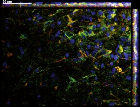
formly in the gland cells in the submucosa as well as in the                                        show primary sensory neurons that are embedded be-
excretory part of the nasal glands, localized within the                                            tween the supporting cells. These sustentacular cells pro-
epithelium (Fig. 2c).                                                                               vide mechanical strength to the epithelium, generate the
                                                                                                    olfactory binding protein, and support the other cells
   Identification of ACE2 and TMPRSS2 in the Olfactory                                              with nutrients [Choi and Goldstein, 2018]. Additionally,
   Epithelium                                                                                       sustentacular cells are responsible for the maintenance of
   Schematic illustration of the olfactory epithelium                                               the ion and water balance within the olfactory epithelium
(Fig. 3a) and histological staining of the same area (Fig. 3b)                                      [Suzuki et al., 2000]. ORN arise from basal stem cells via

                                                                                                                          Granular Cell Layer
                                                                                                                          Inner Plexiform Layer
                                                                                                                          Mitral Cell Layer
                                                                                                                          External Plexiform Layer
                                                                                                                          Glomerular Layer
                                                                                                                          Outer Nerve Layer
        a

    b                                                         dorsal
                                                                                                                                                     GCL
                                                    frontal             occipital                                                                    IPL
                                                                                                                                                     MCL
                                                                                    SYNAPTOPHYSIN

                                                              ventral

                                                                                                                                                     EPL

                                                                                                                                                     GL

                                                                                        c                                                            ONL
    b                                                                                                                          nuclei

    EPL

    GL

    d ONL                                            ACE2                                                       TUBB3                        OMP

    e                       ACE2                     GFAP
                                                                                                                                                                 4

Detection of ACE2 in Human Olfactory                                                                Cells Tissues Organs 2020;209:155–164                  161
Tissue                                                                                              DOI: 10.1159/000513040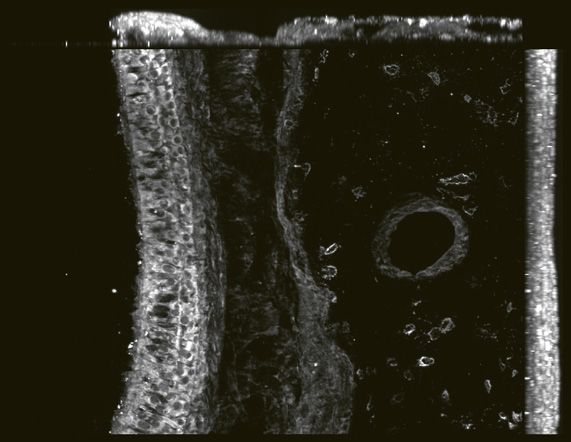
immature and intermediate steps [Caggiano et al., 1994;         localized in some glial fibrillary acidic protein (GFAP)-
Fletcher et al., 2017]. Moreover, microvillar cells and ex-     positive glia cells (Fig. 4e), which is consistent with the
cretory ducts from the specialized Bowman glands be-            findings in other brain regions [Deffner et al., 2020].
neath the epithelium lie scattered in the epithelium
[Getchell and Getchell, 1992; Miller et al., 1995]. In addi-
tion, the data confirmed the expression of ACE2 in the             Discussion and Conclusion
olfactory epithelium (Fig. 3c, d). The ACE2 staining was
mainly located in the supporting cells and could not be             In our present work, we show that both virus entry pro-
found in basal cells, immature ORN, marked with ßIII-           teins, ACE2 and TMPRSS2, are found in the sustentacular
tubulin (TUBB3) nor in mature ORN, marked with olfac-           cells and Bowman glands of the olfactory epithelium (IV),
tory marker protein (OMP) (Fig. 3c). In the underlying          but not in the primary sensory neurons. In the respiratory
lamina propria, high concentrations of ACE2 were found          epithelium from the nasal septum (I), the intermediate na-
in the Bowman gland cells. ACE2 expression is mainly            sal conchae (II), and the cellulae ethmoidales (III), ACE2
limited to the sustentacular cells and shows no co-local-       expression was detected in the basal cell layer and in the
ization with adjacent TUBB3 positive sensory neurons            apical part of the respiratory epithelial cells as well as in
(Fig. 3d). TMPRSS2 was also expressed in the supporting         the nasal submucosal glands. We conclude, based on our
cells of the olfactory epithelium and in the glandular cells    findings, that SARS-CoV2 may bind to ACE2 and
of both epithelia, but no expression in the other cell types    TMPRSS2 in epithelial cells of the respiratory, olfactory,
(Fig. 3e).                                                      and paranasal sinus epithelium and can thus penetrate the
                                                                upper respiratory system. In the olfactory bulb, ACE2 was
    Identification of ACE2 in the Olfactory Bulb                widely distributed with the highest expression in the glo-
    To complete our view of ACE2 expression in the olfac-       merular layer, but was not co-localized with OMP positive
tory system, stainings of the human olfactory bulb were         neurons or other neuronal cell types.
performed. Axons from the ORN pervade through the                   Taken all findings together, this leads to the question,
lamina cribrosa to the olfactory bulb. Here, the first syn-     how the symptoms of anosmia can be explained in CO-
aptic interconnections to the downstream secondary              VID-19 patients. It could be hypothesized that anosmia
neurons appear [Nagayama et al., 2014]. These defined           in COVID-19 is caused by viral infection of ORN, which
interconnections are located in 6 distinct layers of the ol-    further leads to their damage. Corrupted ORN cannot
factory bulb (Fig. 4a, c). With hematoxylin-eosin stain-        process and project odorant information to the brain via
ing, these different regions of the olfactory bulb were         the olfactory bulb and odorant information cannot be
identified and visualized (Fig. 4b). The processed infor-       processed and projected to the brain via the olfactory
mation projects along the olfactory tract to different re-      bulb. However, in our work we can clearly demonstrate
gions of the olfactory cortex [Scott et al., 1993]. The hall-   that there is no expression of ACE2 in the primary neu-
mark feature of the outer nerve layer is the ensheathed fila    rons, which supports results from previous work [Brann
olfactoria coming from the olfactory epithelium, pene-          et al., 2020; Chen et al., 2020]. Probably, the sustentacular
trate the bone plate and enter the olfactory bulb. Different    cells are affected by the SARS-CoV2 virus, as these cells
cell types and their interconnections, like interneurons        express both proteins ACE2 and TMPRSS2. Sustentacu-
and projection neurons are located in deeper layers of the      lar cells are essential for the olfactory system. They do not
olfactory bulb and lead to processed and fine-tuned sen-        only provide structural stability comparable to glia cells,
sory information (Fig. 4a) [Au et al., 2002]. The olfactory     but also support all other cells of the epithelium in a nu-
bulb has been analyzed on RNA and on protein level with         tritious and metabolic way as they are connected via tight
mouse tissue so far [Brann et al., 2020; Ueha et al., 2020].    and adherens junctions with the other cell types [Suzuki
In the human olfactory bulb, ACE2 could be found wide-          et al., 2000; Steinke et al., 2008; Bilinska et al., 2020].
ly distributed, with high expression in the glomerular lay-     Moreover, sustentacular cells perform phagocytosis and
er (Fig. 4d). Faint stainings of ACE2 in the mitral cell lay-   are probably involved in protective mechanisms by ex-
er were also detected. However, no co-localization of           pressing antiviral and antibacterial proteins [Suzuki et al.,
ACE2 and OMP or TUBB3 could be detected, suggesting             1996]. Sustentacular cells are in close contact to the ORN,
that ACE2 is not expressed in the axons of the ORN or           forming intercellular connections. Potentially, infected
other neurons (Fig. 4d). While neurons in the olfactory         sustentacular cells may also invade ORN across these
bulb lack the expression of ACE2, the protein could be          bridges, unassisted by the virus entry genes.

162                  Cells Tissues Organs 2020;209:155–164                           Klingenstein/Klingenstein/Neckel/Mack/
                     DOI: 10.1159/000513040                                          Wagner/Kleger/Liebau/Milazzo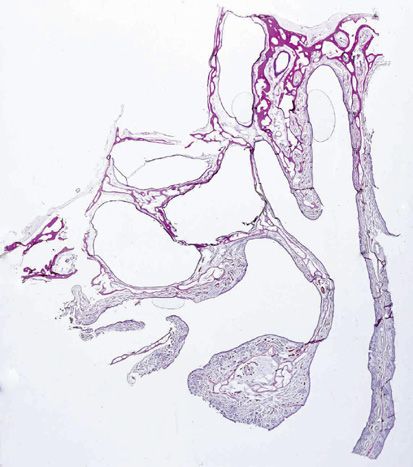
How does the localization of ACE2 and TMPRSS2 in            together with the underling Bowman gland cells may lead
Bowman gland cells and nasal submucosal glands of the           to altered mucus production, metabolism and structural
respiratory epithelium fit to our hypothesis? Glandular         instability in the olfactory epithelium. In addition, infec-
cells in the nasal cavity, together with goblet cells, which    tion may result in the inability of the ORNs to connect to
are found in respiratory epithelium, are necessary for          odorants via OBPs. Nevertheless, most patients regain
the production of mucus. Mucus is a viscous solution,           their ability of smell perception, due to the fact that the
which contains mainly water, ions, proteins and mucins          basal cells are presumably not affected by the virus and
and covers the apical side of the nasal epithelium              can therefore replace destroyed cells of the olfactory epi-
[Escada et al., 2009]. It helps to maintain the physiolog-      thelium.
ical barrier of the epithelium against foreign substances
from the air. In particular, the Bowman glands are sup-
posed to produce enzymes providing xenobiotic-me-                  Statement of Ethics
tabolizing functions [Renne et al., 2007]. Due to the
                                                                   Sampling of human material from body donors and all follow-
virus entry protein expression in the Bowman gland
                                                                ing experiments were made in accordance to local laws and regula-
and nasal submucosal glands, SARS-CoV2 could de-                tions approved by the responsible ethical committee at the Medical
stroy many glandular cells which would probably result          Department of the University of Tübingen (Project Nr.
in a reduced or incorrect mucus production, leading to          284/2020BO2). The body donors gave their informed consent in
dysfunction or even damage of ORN. In addition, sus-            concert with the declaration of Helsinki to use the cadaver for re-
                                                                search purposes. The procedure was approved by the ethics com-
tentacular cells and Bowman glands are supposed to
                                                                mission at the Medical Department of the University of Tübingen
produce odorant-binding proteins (OBPs), which are              (Project Nr. 237/2007 BO1).
indispensable for the perception of odorants [Vogt et
al., 2002; Nagnan-Le Meillour et al., 2019]. Without
OBPs in the mucus of the olfactory epithelium, the                 Conflict of Interest Statement
binding of odorants to the olfactory receptors is ham-
pered.                                                             The authors have no conflicts of interest to declare.
    That in mind, we hypothesize that a disruption of high
numbers of sustentacular cells as well as mucus produc-
ing glandular cells could lead to a decreased perception of        Funding Sources
smell and leave the olfactory epithelium less protected
                                                                   No specific funding was received for this study.
against other viral or bacterial threats. The stem cells of
the olfactory epithelium, the basal cells, are most proba-
bly not affected by COVID-19 infections, maintaining the
                                                                   Author Contributions
potential of reproducing ORN, sustentacular cells as well
as Bowman glands [Maddux et al., 1993]. This could ex-              M.K., S.K., A.M. invented and designed the project. A.M.,
plain the relatively fast recovery in most patients suffering   P.H.N., and A.W. performed the surgeries and extraction of hu-
from COVID-19 triggered anosmia [Hopkins et al., 2020;          man tissues. A.M. and M.K. performed the tissue processing pro-
Lee et al., 2020]. However, some cases have been reported       cedures and generated the frozen sections. A.M. performed the
                                                                histological stainings. S.K. optimized and performed the immuno-
with very slow or nearly no recovery of anosmia [Kosugi         fluorescence stainings. M.K. took and edited the microscope pic-
et al., 2020]. Depending on the dimension of destruction        tures and created the Figures and schemata. S.K. and M.K. wrote
of sustentacular and glandular cells, reproduction from         the manuscript. A.W., A.F.M., P.H.N. helped in discussion of the
basal cells followed by recovery of the nasal mucus may         project and performed proofreading of the manuscript. M.K., A.K.
be prolonged or, as in severe cases, the destruction of sus-    and S.L. were proofreading of the manuscript. Financial support
                                                                was provided by S.L.
tentacular cells may also affect the basal cells leading to
extended symptoms. In this context, it is noteworthy that
we also found ACE2 expression in the basal cells of the
                                                                   Acknowledgment
respiratory epithelium.
    Based on our findings, we presume that SARS-CoV2                This study has been published on pre-print server [Klingen-
can enter the cells from the upper respiratory system via       stein et al., 2020].
the viral entry proteins ACE2 and TMPRSS2. The infec-
tion of the sustentacular cells of the olfactory epithelium

Detection of ACE2 in Human Olfactory                            Cells Tissues Organs 2020;209:155–164                          163
Tissue                                                          DOI: 10.1159/000513040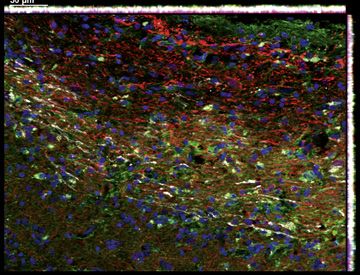
References
Au WW, Treloar HB, Greer CA. Sublaminar or-           Hoffmann M, Kleine-Weber H, Schroeder S,               Nagayama S, Homma R, Imamura F. Neuronal
    ganization of the mouse olfactory bulb nerve          Krüger N, Herrler T, Erichsen S, et al. SARS-          organization of olfactory bulb circuits. Front
    layer. J Comp Neurol. 2002;446(1):68–80.              CoV-2 cell entry depends on ACE2 and TM-               Neural Circuits. 2014;8:98.
Bilinska K, Jakubowska P, Von Bartheld CS, Bu-            PRSS2 and is blocked by a clinically proven pro-   Nagnan-Le Meillour P, Joly A, Le Danvic C, Marie
    towt R. Expression of the SARS-CoV-2 Entry            tease inhibitor. Cell. 2020;182(2):271–280e.8.         A, Zirah S, Cornard JP. Binding Specificity of
    Proteins, ACE2 and TMPRSS2, in Cells of the       Hopkins C, Surda P, Whitehead E, Kumar BN.                 Native Odorant-Binding Protein Isoforms Is
    Olfactory Epithelium: Identification of Cell          Early recovery following new onset anosmia             Driven by Phosphorylation and O-N-Acetyl-
    Types and Trends with Age. ACS Chem Neu-              during the COVID-19 pandemic - an obser-               glucosaminylation in the Pig Sus scrofa. Front
    rosci. 2020;11(11):1555–62.                           vational cohort study. J Otolaryngol Head              Endocrinol (Lausanne). 2019;9:816.
Brann DH, Tsukahara T, Weinreb C, Lipovsek M,             Neck Surg. 2020;49(1):26.                          Ren X, Wang S, Chen X, Wei X, Li G, Ren S, et al.
    Van den Berge K, Gong B, et al. Non-neuronal      Hou YJ, Okuda K, Edwards CE, Martinez DR,                  Multiple expression assessments of ACE2 and
    expression of SARS-CoV-2 entry genes in the           Asakura T, Dinnon KH, et al. SARS-CoV-2                TMPRSS2 SARS-CoV-2 entry molecules in
    olfactory system suggests mechanisms under-           Reverse Genetics Reveals a Variable Infection          the urinary tract and their associations with
    lying COVID-19-associated anosmia. Sci                Gradient in the Respiratory Tract. Cell. 2020;         clinical manifestations of COVID-19. Infect
    Adv.2020;6(31):eabc5801.                              182(2):429–446e.14.                                    Drug Resist. 2020;13:3977–90.
Caggiano M, Kauer JS, Hunter DD. Globose basal        Klingenstein M, Klingenstein S, Neckel PH, Mack        Renne RA, Gideon KM, Harbo SJ, Staska LM,
    cells are neuronal progenitors in the olfactory       AF, Wagner A, Kleger A, et al. Evidence of             Grumbein SL. Upper respiratory tract lesions
    epithelium: a lineage analysis using a replica-       SARS-CoV2 entry protein ACE2 in the hu-                in inhalation toxicology. Toxicol Pathol.
    tion-incompetent retrovirus. Neuron. 1994;            man nose and olfactory bulb. BioRxiv. 2020;            2007;35(1):163–9.
    13(2):339–52.                                         204602.                                            Scott JW, Wellis DP, Riggott MJ, Buonviso N.
Chen M, Shen W, Rowan NR, Kulaga H, Hillel A,         Kosugi EM, Lavinsky J, Romano FR, Fornazieri               Functional organization of the main olfactory
    Ramanathan M, et al. Elevated ACE2 expres-            MA, Luz-Matsumoto GR, Lessa MM, et al. In-             bulb. Microsc Res Tech. 1993;24(2):142–56.
    sion in the olfactory neuroepithelium: impli-         complete and late recovery of sudden olfac-        Steinke A, Meier-Stiegen S, Drenckhahn D, Asan
    cations for anosmia and upper respiratory             tory dysfunction in COVID-19. Braz J Oto-              E. Molecular composition of tight and adher-
    SARS-CoV-2 entry and replication. Eur                 rhinolaryngol. 2020;86(4):490–96.                      ens junctions in the rat olfactory epithelium
    Respir J. 2020;56:2001948.                        Krajewska J, Krajewski W, Zub K, Zatoński T.               and fila. Histochem Cell Biol. 2008; 130(2):
Choi R, Goldstein BJ. Olfactory epithelium: Cells,        COVID-19 in otolaryngologist practice: a re-           339–61.
    clinical disorders, and insights from an adult        view of current knowledge. Eur Arch Otorhi-        Suzuki Y, Takeda M, Farbman AI. Supporting
    stem cell niche. Laryngoscope Investig Oto-           nolaryngol. 2020;277(7):1885–97.                       cells as phagocytes in the olfactory epithelium
    laryngol. 2018;3(1):35–42.                        Lechien JR, Chiesa-Estomba CM, De Siati DR,                after bulbectomy. J Comp Neurol. 1996;
Deffner F, Scharr M, Klingenstein S, Klingenstein         Horoi M, Le Bon SD, Rodriguez A, et al. Ol-            376(4):509–17.
    M, Milazzo A, Scherer S, et al. Histological          factory and gustatory dysfunctions as a clini-     Suzuki Y, Takeda M, Obara N, Suzuki N, Takeichi
    Evidence for the Enteric Nervous System and           cal presentation of mild-to-moderate forms             N. Olfactory epithelium consisting of sup-
    the Choroid Plexus as Alternative Routes of           of the coronavirus disease (COVID-19): a               porting cells and horizontal basal cells in the
    Neuroinvasion by SARS-CoV2. Front Neuro-              multicenter European study. Eur Arch Oto-              posterior nasal cavity of mice. Cell Tissue Res.
    anat. 2020;14(74):596439.                             rhinolaryngol. 2020;277(8):2251–61.                    2000;299(3):313–25.
Escada PA, Lima C, da Silva JM. The human olfac-      Lee DJ, Lockwood J, Das P, Wang R, Grinspun E,         Ueha R, Kondo K, Kagoya R, Shichino S, Ueha S,
    tory mucosa. Eur Arch Otorhinolaryngol.               Lee JM. Self-reported anosmia and dysgeusia            Yamasoba T. Background mechanisms of ol-
    2009;266(11):1675–80.                                 as key symptoms of coronavirus disease 2019.           factory dysfunction in COVID-19: expression
Fan C, Li K, Ding Y, Lu WL, Wang J. ACE2 Ex-              CJEM. 2020;22(5):595–602.                              of ACE2, TMPRSS2, and Furin in the nose
    pression in Kidney and Testis May Cause Kid-      Lee Y, Min P, Lee S, Kim S-W. Prevalence and               and olfactory bulb in human and mice. bioRx-
    ney and Testis Damage After 2019-nCoV In-             Duration of Acute Loss of Smell or Taste in            iv. 2020:097352.
    fection. medRxiv. 2020, 20022418.                     COVID-19 Patients. J Korean Med Sci. 2020;         Vogt RG, Rogers ME, Franco MD, Sun M. A com-
Fletcher RB, Das D, Gadye L, Street KN, Baud-             35(18):e174.                                           parative study of odorant binding protein
    huin A, Wagner A, et al. Deconstructing Ol-       Maddux JF, Vogtsberger KN, Desmond DP, Es-                 genes: differential expression of the PBP1-
    factory Stem Cell Trajectories at Single-Cell         quivel M. Program changes and retention on             GOBP2 gene cluster in Manduca sexta (Lepi-
    Resolution. Cell Stem Cell. 2017; 20(6): 817–         methadone. J Subst Abuse Treat. 1993; 10(6):           doptera) and the organization of OBP genes
    830e8.                                                585–8.                                                 in Drosophila melanogaster (Diptera). J Exp
Getchell ML, Getchell TV. Fine structural aspects     Miller ML, Andringa A, Evans JE, Hastings L. Mi-           Biol. 2002;205(Pt 6):719–44.
    of secretion and extrinsic innervation in the         crovillar cells of the olfactory epithelium:       Zhou P, Yang XL, Wang XG, Hu B, Zhang L,
    olfactory mucosa. Microsc Res Tech. 1992;             morphology and regeneration following ex-              Zhang W, et al. A pneumonia outbreak asso-
    23(2):111–27.                                         posure to toxic compounds. Brain Res. 1995;            ciated with a new coronavirus of probable bat
Guan WJ, Ni ZY, Hu Y, Liang WH, Ou CQ, He                 669(1):1–9.                                            origin. Nature. 2020;579(7798):270–3.
    JX, China Medical Treatment Expert Group
    for, C. Clinical Characteristics of Coronavirus
    Disease 2019 in China. N Engl J Med. 2020;
    382(18):1708–20.

164                        Cells Tissues Organs 2020;209:155–164                                             Klingenstein/Klingenstein/Neckel/Mack/
                           DOI: 10.1159/000513040                                                            Wagner/Kleger/Liebau/Milazzo
You can also read