LY 294002 enhances the chemosensitivity of liver cancer to oxaliplatin by blocking the PI3K/AKT/HIF 1α pathway
←
→
Page content transcription
If your browser does not render page correctly, please read the page content below
MOLECULAR MEDICINE REPORTS 24: 508, 2021
LY‑294002 enhances the chemosensitivity of liver cancer to
oxaliplatin by blocking the PI3K/AKT/HIF‑1α pathway
RUYUE XU1,2*, YINCI ZHANG1,2*, AMIN LI1,2*, YONGFANG MA1,2*, WENPENG CAI1,
LI SONG1,2, YINGHAI XIE2, SHUPING ZHOU2, WEIYA CAO1,2 and XIAOLONG TANG1,2
1
Medical School, Anhui University of Science and Technology, Huainan, Anhui 232001; 2Institute of Environmentally
Friendly Materials and Occupational Health, Anhui University of Science and Technology, Wuhu, Anhui 241000, P.R. China
Received September 4, 2020; Accepted April 12, 2021
DOI: 10.3892/mmr.2021.12147
Abstract. Liver cancer remains one of the leading causes of signaling pathway, which may be related to the inhibition
cancer deaths worldwide. The therapeutic effect of oxaliplatin of HIF‑1α expression. These findings may have clinical
on liver cancer is often limited by acquired resistance of the significance for the treatment of oxaliplatin‑resistant liver
cancer cells. Abnormal activation of the PI3K/AKT pathway cancer.
plays an important role in the acquired resistance of oxaliplatin.
The present study investigated the effects of the PI3K inhibitor Introduction
LY‑294002 and AKT inhibitor MK2206 on the chemosensitivity
of oxaliplatin‑resistant liver cancer cells and the molecular Liver cancer is a malignant tumor with high morbidity and
mechanism involved. An oxaliplatin‑resistant liver cancer mortality rate. Liver cancer is the sixth commonest cancer;
cell line HepG2R was developed. MTT assay, clone formation its 2018 global mortality rate was ~8.2%, ranking fourth
experiments, flow cytometry and Annexin V‑FITC/PI staining among all types of cancer mortality (1‑3). Most liver cancer
were used to determine the proliferation, cycle and apoptosis cases reach middle or late stages before they are diagnosed,
of HepG2 R cells when oxaliplatin was combined with and the best time for surgical treatment is lost (4,5). Therefore,
LY‑294002 or MK2206 treatment. The effects of LY‑294002 chemotherapy is the main treatment for liver cancer. However,
and MK‑2206 on the abnormal activation of PI3K/AKT chemotherapy is prone to drug resistance, which has become
pathway and hypoxia inducible factor (HIF)‑1α protein level in a major problem faced by chemotherapy for liver cancer.
HepG2R cells were detected using western blotting. The results Oxaliplatin has been approved for systemic chemotherapy
indicated that the PI3K/AKT pathway is stably activated in in patients with liver cancer, but its efficacy is often reduced
HepG2R cells. Compared with the AKT inhibitor MK2206, the because of drug resistance (6,7). Therefore, it is necessary to
PI3K inhibitor LY‑294002 more effectively downregulated the explore the resistance mechanisms and find a more effective
phosphorylation levels of p85, p110α, p110β, p110γ and AKT in treatment strategy.
the PI3K/AKT pathway in HepG2R cells, and more effectively Resistance of liver cancer to chemotherapy is related to
inhibited the proliferation of the cells. LY‑294002 enhanced abnormal activation of the PI3K/AKT/hypoxia inducible factor
the chemotherapy sensitivity of HepG2R cells to oxaliplatin (HIF)‑1α signaling pathway (8‑10). PI3K catalyzes the production
by inducing G0/G1 phase arrest and increasing the proportion ofphosphatidylinositol‑3,4,5‑triphosphatethroughphosphorylation
of apoptotic cells. In addition, LY‑294002 reduced the level of phosphatidy‑linositol, phosphatidylinositol‑4‑phosphate and
of HIF‑1α, which is highly expressed in HepG2R cells. It was phosphatidylinositol‑4,5‑bisphosphate (11,12). Chemotherapy
concluded that LY‑294002 enhanced the chemosensitivity of drugs trigger this phosphorylation event, which in turn regulates
liver cancer cells to oxaliplatin by inhibiting the PI3K/AKT cell proliferation, cell‑cycle progression, cell migration and
cell survival (13). In addition, several chemotherapy drugs
can activate the serine/threonine kinase AKT (also known
as protein kinase B). As a proto‑oncogene, AKT plays an
important role in regulating various cell functions, including
Correspondence to: Dr Xiaolong Tang, Medical School, Anhui
metabolism, proliferation, survival, transcription and protein
University of Science and Technology, 168 Taifeng Street, Huainan,
Anhui 232001, P.R. China synthesis (14‑17). HIF‑1α, a functional subunit of HIF‑1, can
E‑mail: txljd2006@126.com regulate angiogenesis, red blood cell production, cell cycle,
metabolism and apoptosis. More importantly, HIF‑1α can be
*
Contributed equally induced by a variety of cytokines (including PTEN, SRC and
P53) in an oxygen‑independent manner through the PI3K/AKT
Key words: hepatocellular carcinoma, oxaliplatin, LY‑294002, pathway (18,19). However, whether the combined inhibition of
MK‑2206, PI3K/AKT, hypoxia inducible factor‑1α the PI3K/AKT pathway and HIF‑1α expression can increase
the sensitivity of liver cancer cells to chemotherapy is not
known.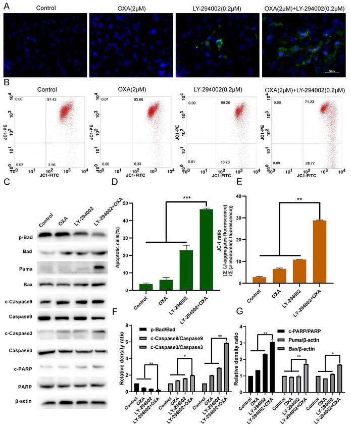
2          XU et al: LY-294002 ENHANCES THE CHEMOSENSITIVITY OF HEPATOCELLULAR CARCINOMA TO OXALIPLATIN

    LY294002 is a protein kinase inhibitor that can block the      (cat. no. BM4141) and eukaryotic translation initiation factor
cell signaling pathway of phosphotidylinsitol‑3‑kinase and         4E‑binding protein 1 (eIF4EBP1) antibodies (cat. no. BM4851)
inhibit the expression of PI3Kα, PI3Kδ and PI3Kβ. LY294002         were from Wuhan Boster Biological Technology, Ltd.
can enter cells and inhibit the PI3K and PI3K/Akt signaling        The p‑eIF4EBP1 antibody (cat. no. bs‑14550R) was from
pathways, including inhibition of Akt phosphorylation,             BIOSS. β ‑actin antibody (cat. no. AF5003) and BCA‑200
thereby inhibiting cell division and induce G1 arrest of           protein detection kits were from Biosharp Life Sciences.
pancreatic cancer and other cells (20). MK‑2206 is a highly        All antibodies were diluted 1:1,000 before use. Phosphatase
selective AKT1/2/3 inhibitor, activated by the pleckstrin          inhibitor mixture were provided by Beyotime Institute of
homology domain. MK‑2206 inhibits the autophosphorylation          Biotechnology.
of AKT at threonine 308 and serine 473 (21). Preclinical
studies have shown that MK‑2206 has an inhibitory effect           Western blot analysis. HepG2 and HepG2R [Oxaliplatin
on human cancer cell lines, such as breast, lung, colon and        (2 µM) treated 0, 6, 12, 24, 48, 72 and 96 h] cells (5.0x10⁶)
liver cancer (22‑25). PI3K/AKT/HIF‑1α reportedly induces           were added with lysis buffer (phosphatase inhibitor mixture)
chemoresistance in liver cancer cells (26,27), and LY‑294002       to extract protein. SDS‑PAGE gels (10%) were placed at
can enhance the chemosensitivity of liver cancer (28).             room temperature for 30 min, and 20 µl of protein samples
Therefore, the present study used LY‑294002 in combination         were added to each well. The protein samples, separated
with oxaliplatin.                                                  by SDS‑PAGE, were sequentially transferred on PVDF
    The present study explored the effect of LY‑294002 or          membrane (EMD Millipore) according to molecular weight.
MK‑2206 in combination with oxaliplatin on the abnormal            After being blocked with 5% skimmed milk at 37˚C for 1 h,
activation of the PI3K/AKT pathway in oxaliplatin‑resistant        the corresponding primary antibodies were incubated at 4˚C
liver cancer. The focus of the study was the more significant      overnight After blocking with 5% skimmed milk at 37˚C for 1 h,
inhibitory effect of LY‑294002 and whether its effect is           the corresponding primary antibody was incubated overnight
due to the inhibition of HIF‑1α expression. It was proposed        at 4˚C, and β‑actin was used as a load control. The membrane
that HIF‑1α may be a target to improve the sensitivity of          was washed three times with TBST [10 M Tris, 150 mM
chemotherapeutics, and that the combination of LY‑294002           NaCl, 0.05% Tween 20 (pH 8.3)], and secondary antibody
and oxaliplatin will be beneficial in the treatment of liver       was added and incubated at 37˚C for 1 h. The membrane was
cancer and provide a basis for the development of targeted         washed again three times with TBST, luminescent solution
therapeutic strategies against liver cancer.                       (ECL Luminescence kit; EMD Millipore) was added for color
                                                                   development. ImageJ 1.44p software (National Institutes of
Materials and methods                                              Health) was used for immunoblotting quantitative analysis.
                                                                   The gray value of each imprinted signal was compared with
Cell source and culture. The human liver cancer cell line          the control group to analyze the expression of the target gene
HepG2 (control HepG2 cells) was purchased from Shanghai            protein.
Mingjin Biological Technology Co, Ltd. The oxaliplatin‑resis‑
tant HepG2 cell line, HepG2 R, was induced in medium               MTT assay. Cells were seeded in 96‑well plates at a density
containing increasing concentration of oxaliplatin (0‑40 µM)       of 5x10³ cells/well and placed in an incubator for 24 h, and
for a total of seven months. The two cell lines were cultured in   drugs were added. After 24 h, the medium was discarded,
RPMI‑1640 medium (Hyclone; Cyvita) containing 10% fetal            10 µl of MTT medium was added to each well, and the plates
bovine serum (Hangzhou Sijiqing Biological Engineering             were placed in an incubator. After culturing for 4 h, the culture
Materials Co., Ltd.) and maintained in a 5% CO2 incubator          solution was discarded, 100 µl of DMSO was added to each
at 37˚C.                                                           well and shaken for 10 min on a shaker. The absorbance
                                                                   at 490 nm was detected with an enzyme‑linked immunoassay
Chemicals, antibodies and reagents. LY‑294002 (0.2 µM) and         detector (ELx800; Omega Bio‑Tek, Inc.).
MK‑2206 (50 nmol/l; MedChem Express) were dissolved in
dimethyl sulfoxide (DMSO) to prepare a 2 mM stock solution.        Colony formation assay. The cells [HepG2R, plus oxaliplatin
Oxaliplatin (Sigma‑Aldrich; Merck KGaA) was dissolved in           (2 µM) and/or LY‑294002(0.2 µM)] were seeded in a six‑well
water to produce a 2 mM stock solution and stored at ‑20˚C.        plate at a density of 1x10³ cells/well. After the cells had
Phosphorylated (p‑) retinoblastoma [Rb; anti‑Rb (phospho           adhered to the wall, drug was added. After 24 h, the drug‑free
S807) antibody [EPR17732] (cat. no. ab184796)] and cyclin D1       medium was replaced, and the culture was continued for
[anti‑Cyclin D1 antibody (SP4) cat. no. ab16663] antibodies        ~3 weeks. When the cell clones were clearly visible, the cell
were purchased from Abcam. Antibodies against caspase‑3            colonies were washed twice with pre‑cooled physiological
(cat. no. 9662), cleaved caspase‑3 (cat. no. 9661), caspase‑9      saline and fixed with 4% paraformaldehyde for 15 min at room
(cat. no. 9502), cleaved caspase‑9 (cat. no. 20750), PARP          temperature. After the fixing solution was removed, the cells
(cat. no. 9532), cleaved RARP (cat. no. 5625), PI3Kp110 α          were stained with crystal violet staining solution for 15 min at
(cat. no. 4249), PI3Kp110 β (cat. no. 3011), PI3Kp110 γ            room temperature. The number of clones formed was counted
(cat. no. 5405), PI3Kp85 (cat. no. 4292), Akt (cat. no. 4691),     to reflect the proliferation ability of cells under the action of
p‑Akt (Ser473; cat. no. 4060), Bad (cat. no. 9268), p‑Bad          each drug.
(cat. no. 5284), Bax (cat. no. 14796) and Puma (cat. no. 24633)
were purchased from Cell Signaling Technology, Inc.                Cell cycle analysis. The cells were seeded in a six‑well plate at
The p70S6K (cat. no. BM4240), p‑S6K1 (T421+S424)                   a density of 1x10⁵ cells/well and placed in an incubator for 24 h.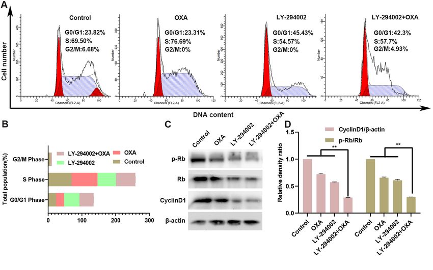
MOLECULAR MEDICINE REPORTS 24: 508, 2021                                                 3

Drugs were added for 24 h, and the cells were digested with         Results
trypsin for collection of the cells, which were fixed with 70%
ethanol at room temperature for 1 h. Pre‑cooled physiological       Stable activation of PI3K/AKT pathway in HepG2R cells
saline (1 ml) was added to the cells, the cells were centrifuged    induced by oxaliplatin. HepG2 and HepG2 R cells were
(562.5 x g; room temperature; 5 min) and 100 µl of staining         treated with 2 µM oxaliplatin. Protein was extracted after
solution from the AnnexinV‑FITC/PI staining kit (Jiangsu KGI        0, 6, 12, 24, 48, 72 and 96 h and measured using western
Biotechnology Co., Ltd.) was added and incubated at 4˚C in          blotting. p‑AKT (Ser473), PI3Kp85, PI3Kp110α, PI3Kp110β
the dark for 30 min (staining solution is a mixture of 50 µg/ml     were found all strongly expressed in HepG2R but weakly
PI, 100 µg/ml RNaseA and 0.2% Triton X‑100 solution). In            expressed in HepG2 (Fig. 1A‑F). However, the expression
total, 1 ml of pre‑cooled physiological saline was added to the     level of PI3Kp110γ was not different between the HepG2 and
staining solution, and the cells were centrifuged (562.5 x g;       HepG2R cells; it is possible that PI3Kp110γ is not as effective
room temperature; 5 min) to obtain a cell pellet. Finally, the      as PI3Kp110 α and PI3Kp110 β in the PI3K/AKT signaling
cells were resuspended in 300 µl of physiological saline, resus‑    pathway. These results indicated that the PI3K/AKT signaling
pended and analyzed with a flow cytometer (BD FACSCalibur;          pathway activation level was higher in HepG2R compared with
BD Biosciences). The results were analyzed with ModiFit             in HepG2 cells. The HepG2 and HepG2R cells were treated
LT 3.0 (Verity Software House, Inc.).                               with various concentrations of oxaliplatin for 24 or 48 h,
                                                                    and cell viability of was analyzed using an MTT assay. As
Annexin V FITC/PI and DAPI staining. The cells were                 shown in Fig. 1G‑H, oxaliplatin inhibited the cell viability of
seeded in a 24‑well plate containing slides at a density of         HepG2 in a time‑ and concentration‑dependent manner, but
1x10⁴ cells/well, placed in an incubator for 24 h, and treated      the proliferation of HepG2R was almost unaffected. HepG2
with drug. After 24 h, the slides were taken out, washed twice      and HepG2R cells were treated with 2 µmol/l oxaliplatin for
with normal saline, and stained with Annexin V FITC/PI              the same time periods, and cell viability was determined. The
(Jiangsu KGI Biotechnology Co., Ltd.) and DAPI (Shanghai            viability of HepG2 cells was found gradually decreased over
Biyuntian Biotechnology Co., Ltd.) for 15 min at room               time, but the viability of HepG2R did not change significantly
temperature in the dark. Apoptotic rate was determined with         (Fig. 1I). Since the abnormal activation of the PI3K/AKT
a fluorescence microscope; 10 fields of view were evaluated         signaling pathway promoted the proliferation of HepG2R,
at x200 magnification. Apoptosis rate=(early apoptotic              it was hypothesized that the PI3K/AKT pathway inhibitors
cells + late apoptotic cells)/total number of cells.                (LY‑294002 and MK‑2206) enhanced the sensitivity of liver
                                                                    cancer to oxaliplatin by targeting the PI3K/AKT pathway.
Measurement of mitochondrial membrane potential (JC‑1).
The cells were inoculated into the well plates at a density of      LY‑294002 can more effectively inhibit the PI3K/AKT pathway
1x10 4 cells/well, placed in an incubator for 24 h, and treated     in the HepG2R compared with HepG2 cell line. To determine
with drug for another 24 h. The cells were rinsed twice with        the effect of the PI3K site‑specific inhibitor (LY‑294002) or the
saline, 500 µl JC‑1 working solution was added to each well,        AKT site‑specific inhibitor (MK‑2206) in combination with
and the plates were placed in an incubator (5% CO2, 37˚C)           2 µmol/l oxaliplatin on the PI3K/AKT pathway in HepG2R, the
and incubated for an additional 20 min. The staining solu‑          expression levels of PI3Kp110α, PI3Kp110β, PI3Kp110γ, AKT,
tion was discarded, and the cells were rinsed with saline           p‑AKT (Ser473) and PI3Kp85 were measured. It was found
twice, then stained with DAPI solution at room temperature          that LY‑294002 (0.2 µM) and MK‑2206 (50 nmol/l) down‑
in dark for 15 min. As JC‑1 aggregates in the mitochondria          regulated the activation of the PI3K/AKT signaling pathway.
when the mitochondrial membrane potential is high, red              The inhibitory effect for p‑AKT(Ser473) was similar, but for
fluorescence is emitted; conversely, when mitochondria              PI3Kp110α, PI3Kp110β and PI3Kp110γ, the inhibitory effect
are depolarized, JC‑1 is in a monomer state and green               of LY‑294002 was greater (Fig. 2A‑D). MTT assays were also
fluorescence is emitted. Thus, the mitochondrial membrane           conducted. As shown in Fig. 2E‑H, the combination treatment
potential of the cell can be judged according to the ratio of       more effectively inhibited HepG2R cell viability compared
red and green fluorescence, and change of JC‑1 fluorescence         with single‑drug treatment, and LY‑294002 combined with
color can indicate change of the mitochondrial membrane             oxaliplatin had a stronger inhibitory effect compared with
potential. A decrease in mitochondrial membrane poten‑              MK‑2206 with oxaliplatin. These results demonstrated that
tial is considered a sign of early apoptosis (29). Red and          LY‑294002 targeting the PI3K/AKT pathway can enhance the
green fluorescence were analyzed with a flow cytometer              chemosensitivity of oxaliplatin.
(BD FACSCalibur; BD Biosciences). The results were
analyzed with CellQuest Pro 5.1.1 (Becton, Dickinson and            LY‑294002 combined with oxaliplatin effectively inhibits the
Company).                                                           proliferation of HepG2R cells. To improve our understanding
                                                                    of the molecular mechanism by which LY‑294002 inhibits
Statistical analysis. The experimental data were obtained           cell proliferation, HepG2R cells were treated with 2 µmol/l
through at least three independent experiments. The data            oxaliplatin and/or 0.2 µmol/l LY‑294002. As illustrated in
are expressed as mean ± SD, one‑way ANOVA to measure                Fig. 3A and B, the combination of LY‑294002 and oxaliplatin
significant differences between the means followed by Tukey's       significantly downregulated the phosphorylation levels of
post hoc tests. P
4 XU et al: LY-294002 ENHANCES THE CHEMOSENSITIVITY OF HEPATOCELLULAR CARCINOMA TO OXALIPLATIN Figure 1. In liver cancer cells, the PI3K/AKT signaling pathway in HepG2R cell line is abnormally activated compared with that in HepG2 cell line. (A) Cell lysates were collected and the designated protein detected using western blotting. Expression of (B) P13Kp85, (C) p‑AKT(Ser473)/AKT, (D) PI3Kp110α, (E) PI3Kp110β and (F) PI3Kp110γ in HepG2 and HepG2R cells incubated with oxaliplatin (2 µmol/l) at various time points. (G) MTT analysis was used to determine the cell viability of HepG2 and HepG2R cells at different concentrations of oxaliplatin at 24 h. (H). MTT analysis was used to determine the cell viability of HepG2 and HepG2R cells at different concentrations of oxaliplatin at 48 h. (I) MTT analysis was used to determine the cell viability of HepG2 and HepG2R cells at 2 µM oxaliplatin at different time points. *P
MOLECULAR MEDICINE REPORTS 24: 508, 2021                                                                 5

Figure 2. In the HepG2R cell line, PI3K/AKT pathway inhibitors downregulate the activation level of PI3K/AKT. (A‑D) HepG2R cells were incubated with
oxaliplatin (2 µmol/l) and/or LY‑294002 (0.2 µmol/l) and MK‑2206 (50 nmol/l) for 24 h. Cell lysates were collected, and the designated proteins were detected
using western blotting. (E‑H) Cytotoxicity of oxaliplatin and/or LY‑294002 and MK‑2206 to HepG2R by MTT analysis. *P
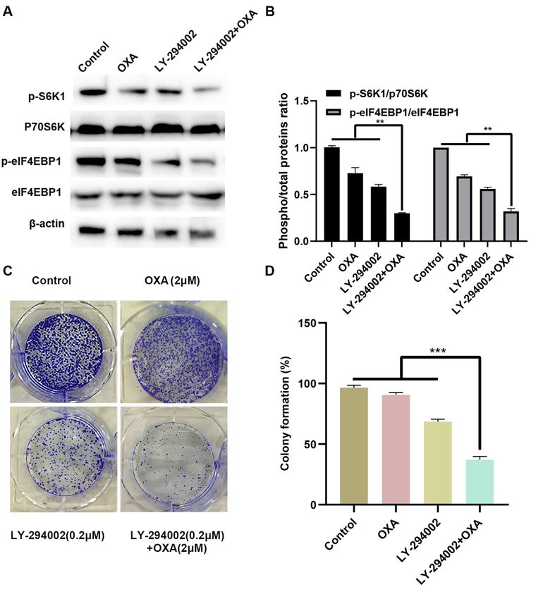
6           XU et al: LY-294002 ENHANCES THE CHEMOSENSITIVITY OF HEPATOCELLULAR CARCINOMA TO OXALIPLATIN

Figure 3. LY‑294002 enhances oxaliplatin induced cytotoxicity by blocking the PI3K/mTOR/S6K1/4EBP1 signaling pathway in HepG2 R cells.
(A and B) Treatment of HepG2R cells with oxaliplatin, LY‑294002, or LY‑294002 combined with oxaliplatin for 24 h. Direct effector response (S6K1 and
eIF4EBP1) downstream of the PI3K/AKT pathway was illustrated. Cell lysates were collected, and the designated proteins were detected using western
blotting. (C and D) Representative images of the colony formation assay in HepG2R cells. **P
MOLECULAR MEDICINE REPORTS 24: 508, 2021                                                               7

Figure 4. LY‑294002 inhibits oxaliplatin‑induced S phase arrest and enhances G 0/G1 phase cell arrest in HepG2R cells. (A and B) After 24 h of treatment
with LY‑294002, oxaliplatin, or LY‑294002 combined with oxaliplatin, the cell cycle distribution of HepG2R cells was determined using flow cytometry.
(C and D) Expression of cyclin D1 and p‑Rb in HePG2R cells was determined using western blotting analysis. **P
8           XU et al: LY-294002 ENHANCES THE CHEMOSENSITIVITY OF HEPATOCELLULAR CARCINOMA TO OXALIPLATIN

Figure 5. LY‑294002 promotes HepG2R apoptosis induced by oxaliplatin. (A and D) LY‑294002, oxaliplatin or LY‑294002 combined with oxaliplatin for
24 h. Effect of LY‑294002 on the apoptosis of liver cancer cells induced by oxaliplatin was determined with Annexin V FITC/PI staining. Magnification,
x400. (B and E) After 24 h treatment with LY‑294002, oxaliplatin, or LY‑294002 combined with oxaliplatin, the mitochondrial membrane potential was
measured using JC‑1 staining and flow cytometry. (C and F, G) After 24 h of treatment with LY‑294002, oxaliplatin or LY‑294002 combined with oxaliplatin,
western blot analysis was used to determine the effect of LY‑294002 on oxaliplatin‑induced apoptosis. *P
MOLECULAR MEDICINE REPORTS 24: 508, 2021                                                                 9

Figure 6. LY‑294002 downregulates the expression level of HIF‑1α through the PI3K/AKT signaling pathway. (A and D, E) HepG2 and HepG2R cells were
incubated with various concentrations of oxaliplatin for 24 h. Cell lysates were collected, and the designated proteins were detected using western blotting.
(B and F) After 24‑h treatment with LY‑294002, oxaliplatin or LY‑294002 combined with oxaliplatin, western blot analysis was performed to determine the
expression level of HIF‑1α. (C and G) After 24 h treatment with MK‑2206, oxaliplatin, or MK‑2206 combined with oxaliplatin, western blot analysis was used
to determine that MK‑2206 downregulated the expression level of HIF‑1α combined with oxaliplatin. *P
10          XU et al: LY-294002 ENHANCES THE CHEMOSENSITIVITY OF HEPATOCELLULAR CARCINOMA TO OXALIPLATIN

Authors' contributions                                                   14. Grosbois J and Demeestere I: Dynamics of PI3K and Hippo
                                                                             signaling pathways during in vitro human follicle activation.
                                                                             Hum Reprod 33: 1705‑1714, 2018.
RYX performed the experiments, analyzed the data and was                 15. Chang L, Graham PH, Ni J, Hao J, Bucci J, Cozzi PJ and Li Y:
the main contributor to writing the manuscript. YCZ analyzed                 Targeting PI3K/Akt/mTOR signaling pathway in the treatment
                                                                             of prostate cancer radioresistance. Crit Rev Oncol Hematol 96:
the data. AML was responsible for the English revision work                  507‑517, 2015.
and performed some of the experiments. YFM, WPC, LS,                     16. Brown JS and Banerji U: Maximising the potential of AKT
YHX, SPZ and WYC performed the experiments. XLT was the                      inhibitors as anti‑cancer treatments. Pharmacol Ther 172:
                                                                             101‑115, 2017.
project leader and was responsible for the design of the project,        17. Mehal W: NASH and HCC are driven by different signaling
the revision of the manuscript and performed some of the                     pathways with a common regulator. Cell Metab 29: 3‑4, 2019.
experiments. RYX and XLT confirm the authenticity of all the             18. Lee JH, Lee HJ, Sim DY, Jung JH, Kim KR and Kim SH:
                                                                             Apoptotic effect of lambertianic acid through AMPK/FOXM1
raw data. All authors read and approved the final manuscript.                signaling in MDA‑MB231 breast cancer cells. Phytother Res 32:
                                                                             1755‑1763, 2018.
Ethics approval and consent to participate                               19. Laughner E, Taghavi P, Chiles K, Mahon PC and Semenza GL:
                                                                             HER2 (neu) signaling increases the rate of hypoxia‑inducible
                                                                             factor 1alpha (HIF‑1alpha) synthesis: Novel mechanism for
Not applicable.                                                              HIF‑1‑mediated vascular endothelial growth factor expression.
                                                                             Mol Cell Biol 21: 3995‑4004, 2001.
                                                                         20. Qu D, Weygant N, Yao J, Chandrakesan P, Berry WL, May R,
Patient consent for publication                                              Pitts K, Husain S, Lightfoot S, Li M, et al: Overexpression of
                                                                             DCLK1‑AL increases tumor cell invasion, drug resistance, and
Not applicable.                                                              KRAS activation and can be targeted to inhibit tumorigenesis in
                                                                             pancreatic cancer. J Oncol 2019: 6402925, 2019.
                                                                         21. Simioni C, Martelli AM, Cani A, Cetin‑Atalay R, McCubrey JA,
Competing interests                                                          Capitani S and Neri LM: The AKT inhibitor MK‑2206 is
                                                                             cytotoxic in hepatocarcinoma cells displaying hyperphosphory‑
                                                                             lated AKT‑1 and synergizes with conventional chemotherapy.
The authors declare that they have no competing interests.                   Oncotarget 4: 1496‑1506, 2013.
                                                                         22. Yu X, Liu J, Qiu H, Hao H, Zhu J and Peng S: Combined inhibi‑
References                                                                   tion of ACK1 and AKT shows potential toward targeted therapy
                                                                             against KRAS‑mutant non‑small‑cell lung cancer. Bosn J Basic
                                                                             Med Sci 21: 198‑207, 2021.
 1. Feng RM, Zong YN, Cao SM and Xu RH: Current cancer                   23. Li YL, Weng HC, Hsu JL, Lin SW, Guh JH and Hsu LC: The
    situation in China: Good or bad news from the 2018 Global                combination of MK‑2206 and WZB117 exerts a synergistic
    cancer statistics? Cancer Commun (Lond) 39: 22, 2019.                    cytotoxic effect against breast cancer cells. Front Pharmacol 10:
 2. Miller KD, Goding Sauer A, Ortiz AP, Fedewa SA, Pinheiro PS,             1311, 2019.
    Tortolero‑Luna G, Martinez‑Tyson D, Jemal A and Siegel RL:           24. Zhu Y, Zhong Y, Long X, Zhu Z, Zhou Y, Ye H, Zeng X and
    Cancer statistics for Hispanics/Latinos, 2018. CA Cancer                 Zheng X: Deoxyshikonin isolated from Arnebia euchroma inhibits
    J Clin 68: 425‑445, 2018.                                                colorectal cancer by down‑regulating the PI3K/Akt/mTOR
 3. Li A, Zhang R, Zhang Y, Liu X, Wang R, Liu J, Liu X, Xie Y,              pathway. Pharm Biol 57: 412‑423, 2019.
    Cao W, Xu R, et al: BEZ235 increases sorafenib inhibition of hepa‑   25. Wang X, Wang X, Xu Y, Yan M, Li W, Chen J and Chen T: Effect
    tocellular carcinoma cells by suppressing the PI3K/AKT/mTOR              of nicastrin on hepatocellular carcinoma proliferation and apop‑
    pathway. Am J Transl Res 11: 5573‑5585, 2019.                            tosis through PI3K/AKT signalling pathway modulation. Cancer
 4. Smith DK and Murphy BA: Lower levels of education and                    Cell Int 20: 91, 2020.
    household income mediate lower dental care utilization among         26. Jiao M and Nan KJ: Activation of PI3 kinase/Akt/HIF‑1α
    survivors of early life cancers. Prev Med Rep 14: 100868, 2019.          pathway contributes to hypoxia‑induced epithelial‑mesenchymal
 5. Liu X, Xie C, Li A, Zhang Y, Liu X, Zhou S, Shen J, Huo Z,               transition and chemoresistance in hepatocellular carcinoma. Int
    Cao W, Ma Y, et al: BEZ235 enhances chemosensitivity of                  J Oncol 40: 461‑468, 2012.
    paclitaxel in hepatocellular carcinoma through inhibiting the        27. Ling S, Li J, Shan Q, Dai H, Lu D, Wen X, Song P, Xie H,
    PI3K/Akt/mTOR pathway. Am J Transl Res 11: 7255‑7271, 2019.              Zhou L, Liu J, et al: USP22 mediates the multidrug resistance of
 6. Yang Y, Yao JH, Du QY, Zhou YC, Yao TJ, Wu Q, Liu J and                  hepatocellular carcinoma via the SIRT1/AKT/MRP1 signaling
    Ou YR: Connexin 32 downregulation is critical for chemoresis‑            pathway. Mol Oncol 11: 682‑695, 2017.
    tance in oxaliplatin‑resistant HCC cells associated with EMT.        28. Ma J, Xie SL, Geng YJ, Jin S, Wang GY and Lv GY: In vitro
    Cancer Manag Res 11: 5133‑5146, 2019.                                    regulation of hepatocellular carcinoma cell viability, apoptosis,
 7. Ye JZ, Yan SM, Yuan CL, Wu HN, Zhang JY, Liu ZH, Li YQ,                  invasion, and AEG‑1 expression by LY294002. Clin Res Hepatol
    Luo XL, Lin Y and Liang R: GP73 level determines chemo‑                  Gastroenterol 38: 73‑80, 2014.
    therapeutic resistance in human hepatocellular carcinoma cells.      29. Elefantova K, Lakatos B, Kubickova J, Sulova Z and Breier A:
    J Cancer 9: 415‑423, 2018.                                               Detection of the mitochondrial membrane potential by the
 8. Zhu YJ, Zheng B, Wang HY and Chen L: New knowledge of                    cationic Dye JC‑1 in L1210 cells with massive overexpression of
    the mechanisms of sorafenib resistance in liver cancer. Acta             the plasma membrane ABCB1 drug transporter. Int J Mol Sci 19:
    Pharmacol Sin 38: 614‑622, 2017.                                         1985, 2018.
 9. Zhang Y, Liu X, Zhang J, Xu Y, Shao J, Hu Y, Shu P and               30. Tang X, Li A, Xie C, Zhang Y, Liu X, Xie Y, Wu B, Zhou S,
    Cheng H: Inhibition of miR‑19a partially reversed the resistance         Huang X, Ma Y, et al: The PI3K/mTOR dual inhibitor BEZ235
    of colorectal cancer to oxaliplatin via PTEN/PI3K/AKT pathway.           nanoparticles improve radiosensitization of hepatoma cells
    Aging (Albany NY) 12: 5640‑5650, 2020.                                   through apoptosis and regulation DNA repair pathway. Nanoscale
10. Kang HG, Wang BZ, Zhang J, Liu MR and Li YX: Combination                 Res Lett 15: 63, 2020.
    of temsirolimus and Adriamycin exhibits an enhanced anti‑            31. Devanabanda B and Kasi A: Oxaliplatin. StatPearls. Treasure
    tumor effect in hepatocellular carcinoma. Clin Res Hepatol               Island (FL), StatPearls Publishing Copyright© 2020, StatPearls
    Gastroenterol 41: 197‑203, 2017.                                         Publishing LLC, 2020.
11. Sun X, Su Y, He Y, Zhang J, Liu W, Zhang H, Hou Z, Liu J and         32. Tang X, Chen L, Li A, Cai S, Zhang Y, Liu X, Jiang Z,
    Li J: New strategy for in vitro activation of primordial follicles       Liu X, Liang Y and Ma D: Anti‑GPC3 antibody‑modified
    with mTOR and PI3K stimulators. Cell Cycle 14: 721‑731, 2015.            sorafenib‑loaded nanoparticles significantly inhibited HepG2
12. Fruman DA, Chiu H, Hopkins BD, Bagrodia S, Cantley LC and                hepatocellular carcinoma. Drug Deliv 25: 1484‑1494, 2018.
    Abraham RT: The PI3K pathway in human disease. Cell 170:             33. Shen JH, Chen PH, Liu HD, Huang DA, Li MM and Guo K:
    605‑635, 2017.                                                           HSF1/AMPKα2 mediated alteration of metabolic phenotypes
13. Aoki M and Fujishita T: Oncogenic roles of the PI3K/AKT/mTOR             confers increased oxaliplatin resistance in HCC cells. Am
    axis. Curr Top Microbiol Immunol 407: 153‑189, 2017.                     J Cancer Res 9: 2349‑2363, 2019.
MOLECULAR MEDICINE REPORTS 24: 508, 2021 11
34. Liao X, Song G, Xu Z, Bu Y, Chang F, Jia F, Xiao X, Ren X, 48. Fang X, Yang D, Luo H, Wu S, Dong W, Xiao J, Yuan S, Ni A,
Zhang M and Jia Q: Oxaliplatin resistance is enhanced Zhang KJ, Liu XY and Chu L: SNORD126 promotes HCC and
by saracatinib via upregulation Wnt‑ABCG1 signaling in CRC cell growth by activating the PI3K‑AKT pathway through
hepatocellular carcinoma. BMC Cancer 20: 31, 2020. FGFR2. J Mol Cell Biol 9: 243‑255, 2017.
35. He J, Ma J, Ren B and Liu A: Advances in systemic lupus 49. Zhou M, Zhang Q, Zhao J, Liao M, Wen S and Yang M:
erythematosus pathogenesis via mTOR signaling pathway. Phosphorylation of Bcl‑2 plays an important role in glycoche‑
Semin Arthritis Rheum 50: 314‑320, 2020. nodeoxycholate‑induced survival and chemoresistance in HCC.
36. Mammana S, Bramanti P, Mazzon E, Cavalli E, Basile MS, Oncol Rep 38: 1742‑1750, 2017.
Fagone P, Petralia MC, McCubrey JA, Nicoletti F and Mangano K: 50. Pan W, Li W, Zhao J, Huang Z, Zhao J, Chen S, Wang C, Xue Y,
Preclinical evaluation of the PI3K/Akt/mTOR pathway in animal Huang F, Fang Q, et al: lncRNA‑PDPK2P promotes hepatocel‑
models of multiple sclerosis. Oncotarget 9: 8263‑8277, 2018. lular carcinoma progression through the PDK1/AKT/Caspase 3
37. Nicoletti F, Fagone P, Meroni P, McCubrey J and Bendtzen K: pathway. Mol Oncol 13: 2246‑2258, 2019.
mTOR as a multifunctional therapeutic target in HIV infection. 51. Jiang S, Wang Q, Feng M, Li J, Guan Z, An D, Dong M, Peng Y,
Drug Discov Today 16: 715‑721, 2011. Kuerban K and Ye L: C2‑ceramide enhances sorafenib‑induced
38. Fagone P, Ciurleo R, Lombardo SD, Iacobello C, Palermo CI, caspase‑dependent apoptosis via PI3K/AKT/mTOR and Erk
Shoenfeld Y, Bendtzen K, Bramanti P and Nicoletti F: signaling pathways in HCC cells. Appl Microbiol Biotechnol 101:
Transcriptional landscape of SARS‑CoV‑2 infection dismantles 1535‑1546, 2017.
pathogenic pathways activated by the virus, proposes unique 52. Sui Y, Zheng X and Zhao D: Rab31 promoted hepatocellular
sex‑specific differences and predicts tailored therapeutic strate‑ carcinoma (HCC) progression via inhibition of cell apoptosis
gies. Autoimmun Rev 19: 102571, 2020. induced by PI3K/AKT/Bcl‑2/BAX pathway. Tumour Biol 36:
39. Gong C, Ai J, Fan Y, Gao J, Liu W, Feng Q, Liao W and Wu L: 8661‑8670, 2015.
NCAPG promotes the proliferation of hepatocellular carcinoma 53. Mendez‑Bla nco C, Fondevi la F, Ga rcia‑Pa lomo A,
through PI3K/AKT signaling. Onco Targets Ther 12: 8537‑8552, Gonzalez‑Gallego J and Mauriz JL: Sorafenib resistance in
2019. hepatocarcinoma: Role of hypoxia‑inducible factors. Exp Mol
40. Xie Y and Zhong DW: AEG‑1 is associated with hypoxia‑induced Med 50: 1‑9, 2018.
hepatocellular carcinoma chemoresistance via regulating 54. Gong J, Zhou S and Yang S: Vanillic Acid Suppresses HIF‑1α
PI3K/AKT/HIF‑1alpha/MDR‑1 pathway. EXCLI J 15: 745‑757, expression via inhibition of mTOR/p70S6K/4E‑BP1 and
2016. Raf/MEK/ERK pathways in human colon cancer HCT116 cells.
41. Li XZ, Sun YL, Cao LQ and Li MJ: Oxaliplatin‑rapamycin Int J Mol Sci 20: 465, 2019.
combination was superior to mono‑drug in treatment of hepa‑ 55. Xu LF, Ni JY, Sun HL, Chen YT and Wu YD: Effects of
tocellular carcinoma both in vitro and in vivo. Neoplasma 63: hypoxia‑inducible factor‑1α silencing on the proliferation of
880‑887, 2016. CBRH‑7919 hepatoma cells. World J Gastroenterol 19: 1749‑1759,
42. Liao B, Zhang Y, Sun Q and Jiang P: Vorinostat enhances the 2013.
anticancer effect of oxaliplatin on hepatocellular carcinoma 56. Chen J, Bai M, Ning C, Xie B, Zhang J, Liao H, Xiong J, Tao X,
cells. Cancer Med 7: 196‑207, 2018. Yan D, Xi X, et al: Gankyrin facilitates follicle‑stimulating
43. Fu X, Wen H, Jing L, Yang Y, Wang W, Liang X, Nan K, Yao Y hormone‑driven ovarian cancer cell proliferation through the
and Tian T: MicroRNA‑155‑5p promotes hepatocellular carci‑ PI3K/AKT/HIF‑1α/cyclin D1 pathway. Oncogene 35: 2506‑2517,
noma progression by suppressing PTEN through the PI3K/Akt 2016.
pathway. Cancer Sci 108: 620‑631, 2017. 57. Ferrin G, Guerrero M, Amado V, Rodriguez‑Peralvarez M
44. Zhang Y, Xie C, Li A, Liu X, Xing Y, Shen J, Huo Z, Zhou S, and De la Mata M: Activation of mTOR signaling pathway in
Liu X, Xie Y, et al: PKI‑587 enhances chemosensitivity of oxali‑ hepatocellular carcinoma. Int J Mol Sci 21: 1266, 2020.
platin in hepatocellular carcinoma through suppressing DNA 58. Duan Y, Haybaeck J and Yang Z: Therapeutic potential of
damage repair pathway (NHEJ and HR) and PI3K/AKT/mTOR PI3K/AKT/mTOR pathway in gastrointestinal stromal tumors:
pathway. Am J Transl Res 11: 5134‑5149, 2019. Rationale and progress. Cancers (Basel) 12: 2972, 2020.
45. Wang Y, Nie H, Zhao X, Qin Y and Gong X: Bicyclol induces 59. Steelman LS, Martelli AM, Cocco L, Libra M, Nicoletti F,
cell cycle arrest and autophagy in HepG2 human hepatocellular Abrams SL and McCubrey JA: The therapeutic potential of
carcinoma cells through the PI3K/AKT and Ras/Raf/MEK/ERK mTOR inhibitors in breast cancer. Br J Clin Pharmacol 82:
pathways. BMC Cancer 16: 742, 2016. 1189‑1212, 2016.
46. Li TT, Zhu D, Mou T, Guo Z, Pu JL, Chen QS, Wei XF and Wu ZJ:
IL‑37 induces autophagy in hepatocellular carcinoma cells by This work is licensed under a Creative Commons
inhibiting the PI3K/AKT/mTOR pathway. Mol Immunol 87: Attribution-NonCommercial-NoDerivatives 4.0
132‑140, 2017. International (CC BY-NC-ND 4.0) License.
47. Zhu F, Jiang D, Zhang M and Zhao B: 2,4‑Dihydroxy‑
3'‑methoxy‑4'‑ethoxychalcone suppresses cell proliferation and
induces apoptosis of multiple myeloma via the PI3K/akt/mTOR
signaling pathway. Pharm Biol 57: 641‑648, 2019.You can also read