Bioscience Research - Innovative Scientific Information & Services ...
←
→
Page content transcription
If your browser does not render page correctly, please read the page content below
Available online freely at www.isisn.org

                           Bioscience Research
                                 Print ISSN: 1811-9506 Online ISSN: 2218-3973
                              Journal by Innovative Scientific Information & Services Network

RESEARCH ARTICLE                   BIOSCIENCE RESEARCH, 2020 17(2): 1274-1288.                     OPEN ACCESS

Morphology, histology, histochemistry and fine
structure of venom apparatus of the medically
relevant Scorpion, Leiurus quinquestriatus
Mahmoud Abd El-Atti1, Jihad A El-Qassas1, Ali Gamal Gadel-Rab2, Moustafa
Sarhan2 and Mahmoud Desouky1*
1Department   of Zoology, Faculty of Science, Zagazig University, Zagazig, Egypt
2Department   of Zoology, Faculty of Science, Al-Azhar University (Assuit Branch), Assuit, Egypt

*Correspondence: mdesouky0@yahoo.com Received 17-05-2020, Revised: 08-06-2020, Accepted: 10-06-2020 e-Published:
14-06-2020
Leiurus quinquestriatus is one of the dangerous scorpions all over the world and represents a health
hazards in Egypt. It has garnered a lot of attention after the isolation of a peptide called chlorotoxin from
its venom which used for identification and treatment of gliomas. The present study was carried out to
critically describe the morphology, histology, histochemistry and ultrastructure of its venom gland before
and after milking to understand the mode of venom formation and secretion in this medically important
scorpion. Microscopical examination revealed that the venom apparatus of L. quinquestriatus is
composed of paired venom glands situated inside the telson’s vesicle. Two venom ducts extend from
these glands to open at the subterminal sides of the stinger. SEM revealed the presence of many
sensory setae over the cuticle of venom apparatus. The venom epithelium is highly folded and has three
types of cells: venom-producing cells, mucous (goblet) cells and supporting cells. Three types of distinct
secretory granules were found in venom-producing cells that gave a variable positive response with
histological and histochemical stains reflecting their constituent’s richness. The herein results revealed
also that these granules are distinct types and no transitional stages reflecting that they are at a
maturation stage .The mode of venom secretion from venom glands of the studied species were found
to be of apocrine type. Ultrastructural architecture of these cells and their granules were described.
Keywords: L. quinquestriatus, Venom apparatus, Morphology, Histology, Histochemistry, Ultrastructure

INTRODUCTION                                                   In addition, recent researches proved that these
     Scorpions are very successful arachnids that              toxins are effective in cancer therapy (Nafie MS,
inhabit all major terrestrial habitats (Possani LD, et         et al., 2020) including; glioma (Perumal SR, et al.,
al., 1999). Scorpion’s venoms are known for their              2017), breast adenocarcinoma (Crusca EJR, et
harmful effects and may cause severe health                    al., 2018), prostate cancer (Ben Aissa R, et al.,
problems (Veiga ABG, et al., 2009). In spite of                2020).
their negative effects, scorpion venoms are a rich                  Most of the previous studies on Egyptian
source of bioactive ingredients that are                       scorpions were focused on their morphology and
extensively used as anti-parasitic (Adade CM, and              taxonomy (e.g. Kaltsas D, et al., 2008; El-
Souto-Padrón T, 2015), insecticidal (Juichi H, et              Hennawy HK, 2014; Badry A, et al., 2018). In
al., 2019), anti-bacterial (Amorim-Carmo B, 2019)              recent years, some peptides of medical
and anti-viral agents (El-Bitar AMH, et al., 2019).            importance were isolated from Egyptian scorpions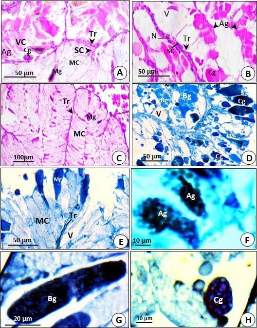
El-Atti et al.                   Venom Apparatus of the Medically Relevant Scorpion, Leiurus quinquestriatus

(El-Bitar AMH, et al., 2019; Elrayess, R.A. et. al.      provide proper ventilation. Scorpions were fed
2019). However, works on the morphological,              with grasshoppers.
histological and fine structures of Egyptian
                                                         Ethical Approval
scorpion venom glands are very rare (Soliman BA,
                                                              All protocols and experimental procedures
et al., 2013).
     Leiurus quinquestriatus represents one of the       were performed as per the norms of the
                                                         Institutional Animal Care and Ethics Committee,
most dangerous species of scorpions all over the
                                                         Zagazig University, (ZU-IACUC) Zagazig, Egypt
world and it is encountered in Egypt, especially in
                                                         (Research          protocol      No.,      ZU-
Upper Egypt and Sinai (El-Hennawy HK, 2014). It
                                                         IACUC/1/F/178/2019).
represents a health hazards in Egypt and was
reported to be more toxic than Androctonus
                                                         Venom Extraction
crassicauda (Abd El-Aziz FE, et al., 2019). It has
                                                              For this purpose, Electrical method (Yaqoob
garnered a lot of attention after the isolation of a     RHM, et al., 2016) was used. Scorpion was
36-amino acid peptide called chlorotoxin (CTX)           placed on a Petri plate with sticky tape. With the
from its venom by DeBin JA, et al., (1993). CTX          help of pointed electrode rinsed in saline solution,
has been used as a potential agent for diagnosis         electric current (20 V) was applied at the base of
and treatment of gliomas (Lyons SA, et al., 2002;        telson for 5 s until the venom was released.
Biswas A, et al., 2012). Moreover, several
bioactive toxins were isolated from the venom of         Histological and Histochemical preparations
this     scorpion.    For    example,     Bradykinin          The telson was cut at its articulation with the
Potentiating Factor (BPF) normalized the hepatic         last abdominal segment and quickly fixed in 10%
injury induced by CCl4 (Salman MMA, 2018).               neutral buffered formalin (pH 7.4) with 2% calcium
Furthermore, the crude venom of this scorpion            acetate. Bouin’s fluid, Gendre's fluid and Zenker's
induced cytotoxicity, elevated the reactive oxygen       fluid were also used for histochemical
species and enhanced apoptotic pathways in               preparations. Specimens were then treated with
some cancer cell lines (Salama W, and Geasa N,           formic acid for 2h for softening of their hard
2014; Al-Asmari AK, et al., 2018). The venom of          cuticle. Tissue samples were then dehydrated
this species has also antimicrobial activity             through ascending series of ethanol (60- 100%),
against different types of pathogenic bacteria           cleared in xylol and finally embedded in paraffin
(Alajami R, et al., 2020). However, sufficient gaps      wax (Gabe M, and Saint-Girons H, 1969).
exist in the literatures regarding the structure and     Sections were then cut at 5-7 µm with a rotary
function of venom glands of this species.                microtome. Sections were stained by H&E (Drury
     For all the above aforementioned information,       RA, and Wallington EA, 1980). Periodic Acid
the present study was carried out in order to            Schiff (PAS) technique (Gurr E, 1962) was used
describe        the      morphology,       histology,    for recognition and differentiation of carbohydrates
histochemistry and ultrastructure of venom glands        while mercuric bromophenol blue method (Mazia
of L. quinquestriatus. In this study venom gland         D, et al., 1953) was used for recognition of
was observed before and after milking to                 proteins.
understand the mode of venom formation and
secretion in this medically important scorpion.          Scanning Electron Microscopy (SEM)
                                                             The telson was fixed in 3% glutaraldehyde in
MATERIALS AND METHODS                                    0.1 ml sodium phosphate buffer (pH 7.2) for three
                                                         hours. Specimens were then washed two times in
Collections and Maintenance of Scorpions                 sodium phosphate buffer, post fixed in 1%
    L. quinquestriatus specimens were collected          osmium tetroxide (OSO4) in the same buffer for 2
by hunters during the months (May-September              hours at +4 ºC and then washed four times in
2018) from different geographical wild infested          sodium phosphate buffer. Telsons were then
area in Egypt. Specimens were brought to                 dehydrated in a graded series of ethanol (40%–
Invertebrate Lab., Zoology Department, Faculty of        100%). The last stages of dehydration were
Science, Zagazig University, Zagazig, Egypt.             performed with propylene oxide. The specimens
They were placed in fairly small transparent             were dried and coated in a Polaron SC 500
plastic boxes (20 × 10 × 10 cm) with 5–6 cm of           sputter coater with a thin layer of gold (Hayat MA,
sand at the bottom. The lid was perforated to            2000). The materials were examined with a Joel
                                                         JSM 5800 Scanning Electron Microscope (EM

                           Bioscience Research, 2020 volume 17(2): 1274-1288                           1275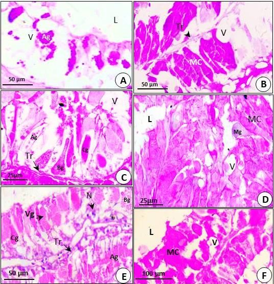
El-Atti et al.                    Venom Apparatus of the Medically Relevant Scorpion, Leiurus quinquestriatus

Unit, Faculty of        Agriculture,    El-Mansoura      Histology and Histochemistry            of   Venom
University, Egypt).                                      Glands of L. quinquestriatus

Transmission Electron Microscopy (TEM)                   Before milking
     The    specimens     were    fixed  in   3%               Light microscopical examination of the venom
glutaraldehyde for 3 hours at 4°C with 0.1 ml            apparatus revealed that two completely separated
sodium phosphate buffer (pH 7.2) and then                bilateral venom glands are found extended one on
washed three times with the same buffer.                 each side of the midline in the vesicle (ampulla) of
Specimens were then post fixed in 1% OSO4 for            telson (Plate II: A). The telson is covered by a
one hour at room temperature and then washed             hard, thick cuticle that composed of two layers: an
three times in sodium phosphate buffer to remove         outer homogeneous thin exocuticle and an inner
OSO4. They were then dehydrated in ascending             thick lamellar endocuticle (Plate II: B). Each gland
series of ethanol (40%–100%). After dehydration,         is surrounded by a layer of striated muscular
the specimens were embedded in Araldite CY 212           tissue covers the gland from its mesal and mid-
(Agar Scientific Ltd., UK) (Hayat MA, 1981). Thin        ventral sides (Plate II: C). The muscle fibrils
sections (60 - 70-nm-thick) were cut with glass          appear striated with alternating light (I) and dark
knife on RMC MT-X ultra-microtome (Boeckeler             (A) bands and have oval nuclei (Plate II: C). A
Instruments, USA) and mounted on 100-mesh                layer of dense connective tissue appeared purple
copper grids. Sections were stained with uranyl          in colour underlying the cuticle and the muscular
acetate followed by lead citrate and examined            tissue with trabeculea that extends inside the
under a Joel JEM 100 SX TEM (Jeol Ltd., Japan)           gland dividing it into lobules (Plate II: A,B&D).
at 80 kV (EM Unit, Faculty of Agriculture, El-                 Each venom gland comprises a sac-like
Mansoura University, Egypt).                             structure with lumen (Plate II: A). The glandular
                                                         epithelium lies between the connective tissue and
RESULTS                                                  lumen and thrown into a multitude of irregular
                                                         folds and finger-like structures (Plate II: D, E&F).
Morphology of Venom Apparatus of L.                      Three types of cells can be recognized: the goblet
quinquestriatus                                          (mucous) cells, the venom-producing cells and
     The venom apparatus of L. quinquestriatus is        supporting cells. The mucous cells are found in
composed of a bulbus vesicle and a stinger or            separate folded epithelium and occupy most of
aculeus. The vesicle is yellowish and globose            the stroma on the dorsal side of the gland while
while the brownish aculeus is longer than vesicle,       the venom-producing cells are concentrated in the
sharply pointed and shallowly curved (Plate I: A).       ventral region (Plate II: A).
A small venom opening situated at the lateral                  The goblet cells (mucous cells) are pyramidal
subterminal end of the aculeus (Plate I: B). Under       and their nuclei situated at the base. Large oval to
light microscope, the surface of the venom               elongated basophilic secretory vesicles are found
apparatus seems smooth and devoid of any                 near the tips of these cells (Plate II: F).
sensory setae (Plate I: A). However, under SEM,                The venom-producing cells are high
few small sensory setae emerge from the cuticle          columnar, bottle-shaped with small, rounded basal
of the vesicle (Plate I: C).The diameter of these        nuclei. They are filled with apical granular vesicles
setae decreased along its length (Plate I: D). The       of various sizes, shapes and staining properties
telson is covered by a cuticle that consists of two      (Plate II: E). Three types of secretory granules
layers: a homogeneous outer exocuticle and               can be recognized within the venom-producing
lamellar endocuticle. (Plate I: E). When the end of      cells; named A-type, B-type and C-type. Type A-
the sting tip is cut off with fine scissors and looked   granules are filled with rather large circular
under the SEM, two separate venom ducts were             contents (Plate II: E&H). Type-B granules are
observed embedded in a spongy cuticle (Plate I:          filled with small oval contents (Plate II: E&G) while
E). In sagittal section, SEM micrographs showed          Type C-granules are filled with irregularly shaped
a sheath of muscle fibres enclosing the venom            basophilic contents (Plate II: G).
glands from its mesal side (side away from the                 The supporting cells (Non-secretory cells) are
cuticle) (Plate I: F).                                   sub-cuboidal cells attached to the basement
                                                         membrane of the epithelial folding in between the
                                                         secretory cells. (Plate II: D&E).

                           Bioscience Research, 2020 volume 17(2): 1274-1288                            1276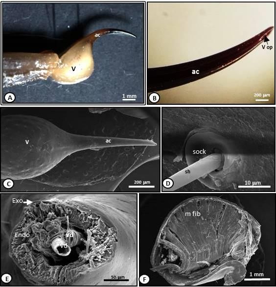
El-Atti et al.                      Venom Apparatus of the Medically Relevant Scorpion, Leiurus quinquestriatus

                                                       PLATE I

PLATE I: Morphology of venom apparatus of the scorpion: L. quinquestriatus. A: Photograph showing
lateral view of telson. X, 10. B: Photograph of sting (aculeus) showing lateral venom opening. X, 50. C: Scanning
electron micrographs of telson showing vesicle and aculeus. X, 25. D: High mag. of sensory hair projecting from
distinct socket. X, 1500.E: The very tip of the sting snipped off with scissors, showing the two venom ducts
surrounded by a spongy cuticle. X, 25. F: Sagittal section of telson under SEM showing a sheath of muscle fibres
lined the gland from its mesal side. X, 25. ac: aculeus or stinger; Endo: endocuticle; Exo: exocuticle; m. fib: a sheath
of muscle fibres; sh: sensory hair; sock: socket; V: vesicle; v. op: venous opening; Vd: venom duct.

                             Bioscience Research, 2020 volume 17(2): 1274-1288                                   1277
El-Atti et al. Venom Apparatus of the Medically Relevant Scorpion, Leiurus quinquestriatus
PLATE II: Light micrographs of venom glands of L. quinquestriatus showing their histological
structures (Stained H&E). A: T.S. of venom glands showing their histological structure. X, 40. B: T.S. of venom
gland showing two-layered cuticle. X, 250. C: T.S. of venom gland showing structure of muscles. X, 250. D: T.S. of
venom gland showing folding of venom-producing cells. X, 250. E: T.S. of venom gland showing venom-producing
cells with different types of granules. X, 400. F: T.S. of venom gland showing mucous cells. X, 250. G&H: Different
types of venom granules. X, 1000. A: light band; Ag: A-type venom granules; Bg: B-type venom granules; C.t.:
connective tissue; C: cuticle; Cg: C-type venom granules; Endo: endocuticle; Exo: exocuticle; hv: hemolymph vessel;
I: dark band; L: lumen; M: muscle; MC: mucous cells; Mg: mucous granules; N: nucleus; SC: supporting cells; Tr:
trabecula; VC: venom-producing cells.
Bioscience Research, 2020 volume 17(2): 1274-1288 1278El-Atti et al. Venom Apparatus of the Medically Relevant Scorpion, Leiurus quinquestriatus
PLATE III: Light micrographs showing hitochemistry of the venom glands of L. quinquestriatus. A:
T.S. of venom gland showing carbohydrate histochemistry of glandular secretory epithelium. (Stained PAS) X, 500.
B: T.S. of venom gland showing carbohydrate histochemistry of venom-producing cells. (Stained PAS). X, 500. C:
T.S. of venom gland showing carbohydrate histochemistry of mucous cells. (Stained PAS). X, 150. D: T.S. of venom
gland showing protein histochemistry of venom-producing cells (Stained mercuric bromophenol-blue). X, 500. E: T.S.
of venom gland showing protein histochemistry of mucous cells (Stained mercuric bromophenol-blue). X, 400.
F,G&H: Protein histochemistry of different types of venom granules. (Stained mercuric bromophenol-blue). X, 1000.
Ag: A-type venom granules; Bg: B-type venom granules; Cg: C-type venom granules; MC: mucous cells; Mg: mucous
granules; N: nucleus; SC: supporting cells; Tr: trabecula; V: vacuole; VC: venom-producing cells.
Bioscience Research, 2020 volume 17(2): 1274-1288 1279El-Atti et al. Venom Apparatus of the Medically Relevant Scorpion, Leiurus quinquestriatus
PLATE IV: Histological structures of the venom glands of L. quinquestriatus after different times
of milking (Stained H&E). A: T.S. of venom gland showing venom-producing cells just after milking. X,
400. B: T.S. of venom gland showing mucous cells just after milking. X, 400. C: T.S. of venom gland
showing venom-producing cells after one day of milking. X, 600. D: T.S. of venom gland showing mucous
cells after one day of milking. X, 600 E: T.S. of venom gland showing venom-producing cells after three
days of milking. X, 400. F: T.S. of venom gland showing mucous cells after three days of milking. X, 250.
Ag: A-type venom granules; Bg: B-type venom granules; Cg: C-type venom granules; L: lumen; MC:
mucous cells; Mg: mucous granules; N: nucleus; Tr: trabecula; V: vacuole; Vg: venom granules.
Bioscience Research, 2020 volume 17(2): 1274-1288 1280El-Atti et al. Venom Apparatus of the Medically Relevant Scorpion, Leiurus quinquestriatus
PLATE V: Transmission electron micrographs of the venom glands of L. quinquestriatus. A: The
ultrastructure of cuticle. X, 10,000. B: The ultrastructure of muscles. X, 7,000. C: Different types of cells.
X, 2,250. D: Different types of cells. X, 5,400. E: Non-secretory cell and venom-producing cells. X, 6,000.
F: Mucous cell showing round nucleus, many mitochondria and cisternae of RER. X, 5,000. G: Different
types of cells. X, 3,900. Endo: endocuticle; Exo: exocuticle; mb: muscle bundles; MC: mucous cells; Mi:
mitochondria; N: nucleus; Nu: nucleolus; RER: rough-endoplasmic reticulum; SC: supporting cells; VC:
venom-producing cells; Vg: venom granules; Z: Z-line.
Bioscience Research, 2020 volume 17(2): 1274-1288 1281El-Atti et al. Venom Apparatus of the Medically Relevant Scorpion, Leiurus quinquestriatus
PLATE VI: Transmission electron micrographs of the venom glands of L. quinquestriatus. A:
Mucous cells with microvilli at its apical portion. X, 3,400. B: Secretory cells showing discharge of venom granules. X,
2,800. C: Venom-producing cells with different types of venom granules. X, 2,250. D: Venom-producing cells with
different types of venom granules. X, 3,800. E: Venom-producing cells with different types of venom granules. X,
5,000. F: C-type venom granules. X, 6,600. Ag: A-type venom granules; Bg: B-type venom granules; bm: bound
membrane; Cg: C-type venom granules; MC: mucous cells; Mg: mucous granules; MV: microvilli; Res: reservoir.
Histochemical preparations of the venom - producing cells revealed the presence of rounded
Bioscience Research, 2020 volume 17(2): 1274-1288 1282El-Atti et al. Venom Apparatus of the Medically Relevant Scorpion, Leiurus quinquestriatus
granules of variable colour density upon staining Ultrastructure of Venom Glands of L.
with same stains. These granules showed high quinquestriatus
activity for periodic acid in the form of dark Under TEM, the venom gland of L.
magenta-red granules scattered in the cytoplasm quinquestriatus appeared surrounded by cuticle,
(Plate III: B). Moderate activity for periodic acid which consists of two layers: an outer thin
was determined in form of homogenous magenta- homogeneous exocuticle and an inner thick
red granules in these granules (Plate III: A&B). lamellar endocuticle (Plate V: A).The muscles
Numerous light magenta-red granules were also occur in several bundles and separated by thin
observed scattered throughout these cells (Plate layer of connective tissue containing numerous
III: B).The mucous cells were found to contain vesicles. The Z lines (the borders of the
large apical elongated mucous granules that sarcomeres) show irregular structure (Plate V: B).
deeply stained with both PAS (Plate III: C) and Three types of cells can be recognized in the
bromophenol blue (Plate III: E). Similarly, the folded epithelium of the glands: venom-producing
glandular venom secretory cells gave a variable cells, mucous cells and supporting cells.
positive response to mercuric bromophenol blue
that indicate the presence of variable amount of A. Venom-producing cells
protein in these cells (Plate III: E-H). The secretory venom-producing cells are tall
Microscopical preparations revealed that some of (Plate V: C). The nuclei and other organelles are
the three types of granules appeared dark blue in found in the basal area (Plate V: C). In electron
colour and others were observed in light blue micrographs, these cells appear to be undergoing
colour by mercuric bromophenol blue stain (Plate extensive protein synthesis where RER appear
III: D, F, G&H). extensive, highly distended and filled with an
amorphous substance (Plate V: C). Many
After milking mitochondria are interspersed throughout the ER
The histological architecture of the venom (Plate V: E). Most of the cell is filled by vesicles of
gland was observed after milking process to different sizes, shapes and electron densities.
reveal the mode of venom secretions. The glands Three distinct types of membrane-bounded
were stimulated with electric shock to release secretory granules can be distinguished with
venom and then allowed to recover for three days. TEM: Type-A granules are filled with large
Just after milking, the venom- producing cells electron-dense vesicles which appeared
seemed nearly empty with small amount of venom aggregated together (Plate VI: C&E). The
granules mainly of type A (Plate IV: A). On the diameter of the vesicles exceeds 3 µm and
other hand, some mucous cells were found almost reaches up to 8 µm. Type-B granules are filled
empty while others were loaded with mucous with small (1-4µm), irregular, electron-dense
granules (Plate IV: B). vesicles which less round than those of type A
After one day of milking, many granules (Plate VI: D&E). Type-C granules are rounded,
started to appear in both mucous and venom- electron-lucent vesicles and filled with electron-
producing cells. The secretory cells appeared in lucent elongated rods (about 1μm long) (Plate VI:
an active phase of secretion. The apical surface of D, E&F).
these cells appeared ruptured and membrane-
bounded secretory granules of various shapes B. Mucous cells (Goblet-cells)
and staining properties appeared released into the They are columnar with irregular basal nuclei
lumen (Plate IV: C& D). (Plate V: C).The mucous cell has electron-dense
After three days of milking, the secretory cytoplasm containing electron-lucent, confluent
epithelium became loaded again with secretory granules of mucous. A large number of rounded
granules and the apical portion of cells became RER cisternae and mitochondria are found
intact (Plate IV: E&F). throughout the cytoplasm (Plate V: D).The apical
The microscopical slides stained by both PAS surface of mucous cells is provided with long
and bromophenol blue of the glands after milking microvilli (Plate VI: A). Sometimes, the apical
support the above finding and revealed also that surface appeared ruptured and large vacuoles are
the secretion stages of venom and mucous seemed extruded into the lumen of the gland
occurred in progressive manner. (Plate VI: A &B).
C. Supportive Cells (Regenerating-cells)
Interspersed within the secretory epithelium few
Bioscience Research, 2020 volume 17(2): 1274-1288 1283El-Atti et al. Venom Apparatus of the Medically Relevant Scorpion, Leiurus quinquestriatus
numbered, squamous cells were observed. venom epithelium of L. quinquestriatus belongs to
Supportive cell has a flat-shaped nucleus and Type II according to the classification of
devoid of cilia or secretory vesicles (Plate V: G). Pawlowsky EN, (1913) i.e. it is extensively folded
Sometimes, the cytoplasm of these cell may and consists of a mass of secretory epithelium.
contain mesh of microfilaments and tubules (Plate These results are also in line with those reported
V: G). for other members of family Buthidae (Al-Asmari
AK, et al., 2007). The complex epithelial folding
DISCUSSION may be unsurprised in this dangerous scorpion
Scorpions are characterized by possession of since the amount, complexity and effectiveness of
venom apparatus located at the end of their the venom rely highly on the folding level of
telsons. Their success relies on the secretion of venom glands. In addition, the level of venom
very potent neurotoxic venom that is used mainly epithelial folding in the present work may give
for killing their preys and repelling their further evidence that the complexity of scorpion
competitors. In addition to these functions, it was venom glands is related to the scorpion family and
reported that venom glands play an important role phylogeny (Sherwan TA, 2015) and could be
during mating. According to Jiao GB, and Zhu useful and applicable in higher level scorpion
MS, (2010) the male scorpion often stings the taxonomy.
female during courtship. This sexual sting might The general histology, histochemistry and fine
represent a stimulating impulse (Inceoglu B, et al., structure of venom glands of several scorpion
2003). species were studied by several researchers
The present study confirmed that the venom (Kanwar U, et al., 1981; Quiroga M, et al., 1998;
apparatus of L. quinquestriatus comprises paired Soliman BA, et al., 2013; Navidpour S, et al.,
venom gland located inside the vesicle and a 2018). However, the histology and fine structure
pointed stinger. Compared to other less active of Egyptian scorpions was poorly investigated.
scorpion as Scorpio maurus (Navidpour S, et al., Thus, further studies of scorpion venom gland
2018), the bulb is larger and the stinger is taller morphology and ultrastructure are warranted.
and sharper. This may be correlated with fact that Based on microscopical examinations of the
this species is a dangerous active hunter. venom glands of the studied scorpion, three types
Two-layered cuticle reported in the studied of cells were recognized in the glandular
species may give the sting more flexibility and epithelium of the studied scorpion which are:
more resistance to mechanical stress that favour mucous cells, venom -producing cells and
this dangerous scorpion. There are setae and supportive cells. Similar findings were reported in
cuticular pits scattered all over the telson of different scorpions as Euscorpius mingrelicus
studied species. The microanatomical features of (Yigit N, and Benli M, 2008), L. quinquestriatus
these setae are characteristics for (Taib NT, and Jarrar BM, 1993) and S. maurus
mechanoreceptors or chemoreceptors. Similar towsendi (Navidpour S, et al., 2018). However,
setae were observed in other scorpions as some previous studies reported only one (Junqua
Euscorpius migrelicus (Yigit N, and Bayram A, C, and Vachon M, 1968) or two (Keegan HI, and
2007., Yigit N, et al., 2007). A number of ducts Lockwood WR, 1971; Quiroga M, et al., 1998)
were observed in the cuticle which discharges the types of cells in the venom glands of other
secretion of dermal glands. Gaffin DD, and scorpions. The supportive cells arranged between
Brownell PH, (1992) stated that female cuticle venom-producing cells and seem to act as
extracts induce courtship patterns in male reserve cells responsible for regeneration of the
scorpions and suggested that these glands may damaged cells.
produce a sex pheromone in females. In the present study, three types of secretory
The paired venom gland in the present study granules were found in venom-producing cells
was provided mesaly (side away from the cuticle) according to their sizes, shapes and staining
with a band of vertical striated muscle. This proprieties. Similarly, three types of secretory
arrangement of muscles was observed in other granules were described in different scorpion
scorpions as Androctonus crassicauda (Jarrar BM, species (Halse SA, et al., 1980; Sentenská L, et
and Al-Rowaily MA, 2008) and Scorpio maurus al., 2017). However, Navidpour S, et al., (2018)
townsendi (Navipour S, et al., 2018) and may act reported that these granules are grouped into four
to squeeze the gland against the cuticle for types in Euscorpius alpha while Taib NT, and
discharging of venom. Jarrar BM, (1993) grouped them into five types in
The present work revealed that the glandular L. quinquestriatus according their contents or
Bioscience Research, 2020 volume 17(2): 1274-1288 1284El-Atti et al. Venom Apparatus of the Medically Relevant Scorpion, Leiurus quinquestriatus
degree of maturity. However, Soliman BA, et al. 2013 revealed that
The venom granules were found to give a the mode of venom secretion in Androctonus
variable positive response with both histological amoreuxi occurs by the holocrine mode.
and histochemical stains. This may reflect the
richness and variability of their constituents. Most CONCLUSION
of these granules were positively stained with The outcome results critically described the
bromophenol blue due to their protein contents. histology, and ultrastructure of L. quinquestriatus
They were also found to be PAS-positive with venom gland, as well as the histochemical nature
variable intensity. This revealed that the venom of of their constituents and the mode of venom
this species contains neutral muco-substances secretions. Based on microscopic examination in
with variable quantity. Neutral muco-substances the herein study, three types of cells were
have been found in the venom of some other recognized in the venom glands. There are three
scorpion species especially in dangerous ones types of venom granules in the venom-producing
(Goyffon M, and Kovoor J, 1978; Hasle SA, et al., cells. These granules are distinct types with
1980; Taib NT, and Jarrar BM, 1993). Neutral different constituents and there are no transitional
muco-substances may have a possible role in the stages which indicate that they are at a maturation
transfer of venom protein fragments to the victim’s stage.
tissue (Jarrar BM, and Al-Rowaily MA, 2008).
According to Sentenská L, et al., (2017), the CONFLICT OF INTEREST
different types of granules in venom-producing The authors declared that present study was
cells may represent developmental stages where performed in absence of any conflict of interest.
the content of the vesicles develops from a
homogeneous state towards a finely one, forming ACKNOWLEGEMENT
a coarse to fine substance. Based on The authors thank Dr. Ahmed Said Al-
microscopical examination in the herein study, Azzouni, Associate Professor of Cytology &
one could conclude that these granules are Histology, Zoology Department, Faculty of
distinct types with different constituents and there Science, Helwan University\ for providing lab.
are no transitional stages which reflecting that facilities and reviewing the final manuscript
they are at a maturation stage.
In order to elucidate the mode of their AUTHOR CONTRIBUTIONS
secretion, the venom gland of L. quinquestriatus Mahmoud Abdel-Atti, Ali Gamal Gadel-Rub
was stimulated electrically to discharge venom and Jihad Al-Qassas performed the practical part
and examined it after different times of milking. and participate in scientific writing. Moustafa
Based on light and electron microscopic Sarhan developed the idea and participated in
examination, the secretory cells seem to be of the scientific writing and discussion. Mahmoud
apocrine type, i.e. the apices of the secretory Desouky developed the idea, fellow up the
cells break down during the secretion process and publication process and supervised the work. All
appear to pinch off “snouts”, leading to a authors read and approved the final version.
histologic picture of “decapitation secretion” into
the glandular lumen. Venom gland of the studied Copyrights: © 2020@ author (s).
scorpion has three phase of secretory cycle. This is an open access article distributed under the
1.Elaboration phase (Just after milking): in which terms of the Creative Commons Attribution License
they contain fine uniform or empty granules. (CC BY 4.0), which permits unrestricted use,
2.Accumulation phase (from 1-3 days after
distribution, and reproduction in any medium,
milking): larger granules are seen in the epithelial
provided the original author(s) and source are
cells. In this phase, the secretory cells appear
very active. Large number of secretory granules of credited and that the original publication in this
different size and shape accumulated in the apical journal is cited, in accordance with accepted
region. 3.An expulsion phase (appeared also in academic practice. No use, distribution or
unmilked glands): the venom granules and a reproduction is permitted which does not comply
portion of cytoplasm of the cell are extruded into with these terms.
the lumen of the gland. Most scorpion venom
secreting cells were found to be of apocrine type REFERENCES
(Taib NT, and Jarrar BM, 1993; Jarrar BM, and Al- Abd El-Aziz FE, El-Shehaby DM, Elghazaly SA,
Rowaily MA, 2008; Navidpour S, et al., 2018). Hetta HF 2019. Toxicological and
Bioscience Research, 2020 volume 17(2): 1274-1288 1285El-Atti et al. Venom Apparatus of the Medically Relevant Scorpion, Leiurus quinquestriatus
epidemiological studies of scorpion sting Biophys. Acta Biomembr., 1860, 11: 2155-
cases and morphological characterization of 2165.
scorpions (Leiurus quinquestriatus and DeBin JA, Maggio JE, Strichartz GR, 1993.
Androctonus crassicuda) in Luxor, Egypt. Purification and characterization of
Toxicol. Reports, 6: 329-335. chlorotoxin, a chloride channel ligand from
Adade CM, Souto-Padrón T, 2015. Venoms as the venom of the scorpion. Am. J.
Sources of Novel Anti-parasitic Agents. In: Physiol., 264: C361-C369.
Gopalakrishnakone P. (eds) Toxins and Drug Drury RA, Wallington EA, 1980. Carlton’s
Discovery. Toxinology. Springer, Dordrecht. Histochemical Technique. 5th ed. Oxford
Alajmi R, Al-ghamdi S, Barakat I, Mahmoud A, Univ. Press, Oxford.
Abdon N, Abdel Baber IA, 2020. El-Bitar AMH, Sarhan M, Abdel-Rahman
Antimicrobial activity of two novel venoms MA, Quintero-Hernandez V, Aoki-Utsubo C,
from Saudi Arabian scorpions: L. Moustafa MA, Possani LD, Hotta H, 2019.
quinquestriatus and A. crassicauda. Int. J. Smp76, a scorpine-like peptide isolated from
Pept. Res. Ther., 26: 67-74. the venom of the scorpion Scorpio maurus
Al-Asmari AK, Al-Saif AA, Abdo NM, 2007. palmatus, with a potent antiviral activity
Morphological identification of scorpion against hepatitis C Virus and Dengue
species from Jazan and Al-Medina Al- Virus. Internat. J. Pept. Res. Therap., First
Munawwara Regions, Saudi Arabia. J. Online: 06 July 2019.
Venom. Anim. Toxins Includ. Trop. Dis., 13, El-Hennawy HK, 2014. Updated list of scorpions
4: 821-843. of Egypt. Retrieved from http://
Al-Asmari AK, Riyasdeen A, Islam M, 2018. hishamelhennawycv.blogspot.com.eg/p/blog-
Scorpion venom causes apoptosis by page_19.html
increasing reactive oxygen species and cell Elrayess, R.A., Mohallal, M.E., El-Shahat, Y.M. et
cycle arrest in MDA-MB-231 and HCT-8 al. Cytotoxic Effects of Smp24 and Smp43
cancer cell lines. J. Evidence-Based Integr. Scorpion Venom Antimicrobial Peptides on
Medic., 23: 3-8. Tumour and Non-tumour Cell Lines. Int J
Amorim-Carmo B, Daniele-Silva AMS, Parente Pept Res Ther (2019).
AA, Furtado E, Carvalho JWF, Oliveira EC, Gabe M, Saint-Girons H, 1969. Donnees
Santos MS, Silva SRB, Silva A, Silva J, histologiques sur les glands Aalivaires des
Monteiro MF, Fernandes-pedrosa MF, 2019. Lepidosauriens. Memoires du Museum
Potent and broad-spectrum antimicrobial National d'Histoire Naturelle, 58: 11-12.
activity of analogs from the scorpion peptide Gaffin DD, Brownell PH, 1992. Evidence of
stigmurin, Int. J. Mol. Sci., 20: 1-21. chemical signalling in the sand scorpion,
Badry A, Younes M, Sarhan MMH, Saleh M, Paruroctonus mesaensis (Scorpionida:
2018. On the scorpion fauna of Egypt, with Vaejovida). Ethology, 91: 59-69.
an identification key (Arachnida: Scorpiones). Goyffon M, Kovoor J, 1978. Chactoid venoms. In:
Zool. in the Middle East, 64,1 Bettini S. Ed. Arthropod Venoms. Berlin:
BenAissa R, Othman H, Villard C, Peigneur S, Springer-Verlag, 395-417.
Llayah-Bellalouna S, Abdelkafi-Koubra Z, Gurr E, 1962. Staining Animal Tissue: Practical
Marrakchi N, Essafi-Benkhadir K, Tytgat J, and Theoretical. London: Leonard Hill, 631
Luis J, Srairi-Abid N (2020). AaHIV a sodium pp.
channel scorpion toxin inhibits the Halse SA, Prideaux PL, Cockson A, Zwicky KT,
proliferation of DU145 prostate cancer cells. 1980. Observations on the morphology and
Biochem. Biophys. Res. Commun., 8: 521, histochemistry of the venom glands of a
2, 340-346. scorpion, Urodacus novaehollandiae Peters
Biswas A, Gomes A, Sengupta J, Datta P, Singha (Scorpionidae). Austr. J. Zool., 28: 185-194.
S, Anjan Kr, Dasgupta AKR, 2012. Hayat MA, 1981. Principles and Techniques of
Nanoparticle-conjugated animal venom- Electron Microscopy. London: Edward
toxins and their possible therapeutic potential Arlond.
J. Ven. Res., 3: 15-21. Hayat MA, 2000. Principles and Techniques of
Crusca EJR, Basso LGM, Altei WF, Marchetto R, Electron Microscopy: Biological Applications.
2018. Biophysical characterization and 4th ed. 543pp. Cambridge: Cambridge
antitumor activity of synthetic Pantinin University Press.
peptides from scorpion's venom. Biochem. Inceoglu B, Lango J, Jing J, Chen L, Doymaz F,
Bioscience Research, 2020 volume 17(2): 1274-1288 1286El-Atti et al. Venom Apparatus of the Medically Relevant Scorpion, Leiurus quinquestriatus
Pessah IN, Hammock BD, 2003. One (Scorpiones: Scorpionidea) and
scorpion, two venoms: prevenom of Hemiscorpius lepturus (Scorpions:
Parabuthus transvaalicus acts as an Hemiscorpidea) From Iran. J. Zool. Res., 2,4:
alternative type of venom with distinct 29-34.
mechanism of action. PNAAS, 100: 922-927. Pawlowsky EN, 1913. Skorpiontomische
Jarrar BM, Al-Rowaily MA, 2008. Histology and Mitteilungen, I., Ein Beitrag zur Morphologie
histochemistry of the venom apparatus of the der Giftdrüsen der Skorpione, Z. Wiss. Zool.,
black scorpion Androctonus crassicauda 105: 157-77.
(Olivier, 1807) (Scorpiones: Buthidae). J. Perumal SR, Stiles BG, Franco OL, Sethi G & Lim
Venom. Anim. & Toxins Include. Trop. Dis., LHK, 2017. Animal venoms as antimicrobial
14, 3: 514-526. agents. Biochem. Pharmacol., 134: 127-138.
Jiao GB, Zhu MS, 2010. Courtship and mating of Possani LD, Becerril B, Delepierre M, Tytgat J,
Scorpiops Iuridus Zhu Lourenço & Qi, 2005 1999. Scorpion toxins specific for Na
(Scorpiones: Euscorpiidae) from Xizang channels. Eur. J. Biochem., 264: 287-300.
Province. China. Venom. Anim. Toxins Incl. Quiroga M, Marval MJ, Parrilla-Alvarez P,
Trop. Dis., 15:155-165. Desousa L, 1998. Tityus caripitensis n. sp.
Juichi H, Miyashita M, Nakagawa Y, Miyagawa H, Scorpion venom gland histology. Toxicon,
2019. Isolation and characterization of the 36: 12-69.
insecticidal, two-domain toxin LaIT3 from the Salama W, Geasa N, 2014. Investigation of the
Liocheles australis scorpion venom. Biosci. antimicrobial and hemolytic activity of venom
Biotechnol. Biochem., 83, 12: 2183-2189. of some Egyptian scorpion. J. Microbiol
Junqua C, Vachon M, 1968. Les Arachnides Antimicrob. 6: 21-28.
venimeux et leurs venins. Etat actuel des Salman MMA, 2018. Antioxidant Effects of
recherches. Memoires Academie Royale des Bradykinin Potentiating Factor (BPF) Isolated
Sciences d'Outre-Mer (Cl. Sci. Nat. Med., from scorpion venom in liver injury induced
N.S.), 17, 5: 1-136. by carbon tetrachloride (CCl4) in male albino
Kaltsas D, Stathi I, Fet V, 2008. Scorpion of the rats. J. Clin. Toxicol., 8, 6: 399.
East Mediterr. Adv. Arachnol. Develop. L: Sentenská L, Graber F, Richard M, Kropf C, 2017.
Biol., 12: 209-246. Sexual dimorphism in venom gland
Kanwar U, Sharma A, Nagpal N, 1981. morphology in a sexually stinging scorpion.
Morphological and cytochemical studies on Biol. J. Linn. Soc., XX: 1-15.
the venom-secreting cells of the scorpion Sherwan TA, 2015. Morphology and histology of
Buthus tamulus. J. Anim. Morph .Physiol. 28: venom gland of Scorpio maurus kruglovi
206-209. (Birula, 1910) (Scorpionidae: Scorpiones). J.
Keegan HI, Lockwood WR, 1971. Secretory Pure Appl. Sci., 27,5: 59-62.
epithelium in venom glands of two species of Soliman BA, Shoukry NM, Mohallal ME, Fetaih
scorpion of the genus Centruroides. Am. J. HAW, Khaled HS, 2013. Fine structure of the
Trop. Med. Hyg., 20: 770-785. stinger, histology and histochemistry of the
Lyons SA, O’Neal J, Sontheimer H, 2002. venom gland in the scorpion Androctonus
Chlorotoxin, a scorpion-derived peptide, amoreuxi (Buthidae) J. Bas. & App. Zool., 66:
specifically binds to gliomas and tumors of 41- 46.
neuroectodermal origin. Glia, 39: 162-173. Taib NT, Jarrar BM, 1993. Histological and
Mazia D, Brewer PA, Alfert M, 1953. The histochemical characterization of the venom
cytochemical staining and measurement with apparatus of Palestine yellow scorpion,
mercuric bromophenol blue. Biological Leiurus quinquestriatus (Hemprich &
Bulletin, 104: 57-67. Ehrenberg 1828). Trop. Zool., 6: 143-152.
Nafie MS, Abdel Daim MM, Ali IAI, Nabil ZA, Veiga ABG, Berger M, Guimarães JA, 2009.
Tantawy MA, Abel-Rahman MA, Lonomia obliqua venom: Pharmaco-
2020. Antitumor efficacy of the Egyptian toxicological effects and biotechnological
Scorpion Venom Androctonus australis: in perspectives. In: De Lima, M. E., Pimenta, A.
vitro and in vivo study. JoBAZ, 8: 8. M. C., Martin Eauclaire, M. F., Zingali, R. B.,
Navidpour S, Gharagozloyan MM, Pousty I, 2018. Rochat, H. (Eds.), Anim. Toxin: State of the
Histological study on venom gland apparatus Art: Perspectives in Health and Biotechnol.
in Odontobuthus doriae (Scorpions: UFMG, Belo Horizonte, pp. 372-390.
Buthidae), Scorpio maurus towsendi Yaqoob RHM, Tahir M, Arshad S, Naseem A,
Bioscience Research, 2020 volume 17(2): 1274-1288 1287El-Atti et al. Venom Apparatus of the Medically Relevant Scorpion, Leiurus quinquestriatus
Ahsan MM, 2016. Optimization of the
conditions for maximum recovery of venom
from scorpions by electrical stimulation.
Pakistan J. Zool., 48: 265-269.
Yigit N, Bayram A, Danishman T, 2007.
Functional morphology of venom apparatus
of Euscorpius mingerlicus (Scorpiones:
Euscorpionidae). J. App. Biol. Sci. 1: 27-31.
Yigit N, Benli M, 2008. The venom gland of the
scorpion species Euscorpius mingrelicus
(Scorpiones: Euscorpiidae): Morphological,
ultrastructural characterization. J. Venom.
Anim Toxins Incl. Trop. Dis., 14: 466-480.
Bioscience Research, 2020 volume 17(2): 1274-1288 1288You can also read