Effect of VCP modulators on gene expression profiles of retinal ganglion cells in an acute injury mouse model - Nature
←
→
Page content transcription
If your browser does not render page correctly, please read the page content below
www.nature.com/scientificreports

            OPEN           Effect of VCP modulators on gene
                           expression profiles of retinal
                           ganglion cells in an acute injury
                           mouse model
                           Tomoko Hasegawa1, Hanako Ohashi Ikeda1*, Norimoto Gotoh1, Kei Iida2, Sachiko Iwai1,
                           Noriko Nakano1, Akira Kakizuka3 & Akitaka Tsujikawa1
                           In glaucoma, retinal ganglion cells are damaged, leading to the progressive constriction of the visual
                           field. We have previously shown that the valosin-containing protein (VCP) modulators, Kyoto University
                           Substance (KUS)121 and KUS187, prevent the death of retinal ganglion cells in animal models of
                           glaucoma, including the one generated by N-methyl-D-aspartate (NMDA)-induced neurotoxicity.
                           KUSs appeared to avert endoplasmic reticulum (ER) stress by maintaining ATP levels, resulting in the
                           protection of ganglion cells from cell death. To further elucidate the protective mechanisms of KUSs, we
                           examined gene expression profiles in affected ganglion cells. We first injected KUS-treated mice with
                           NMDA and then isolated the affected retinal ganglion cells using fluorescence-activated cell sorting.
                           Gene expression in the cells was quantified using a next-generation sequencer. Resultantly, we found
                           that KUS121 upregulated several genes involved in energy metabolism. In addition, we observed the
                           upregulation of Zfp667, which has been reported to suppress apoptosis-related genes and prevent cell
                           death. These results further support the suitability of KUS121 as a therapeutic drug in protecting retinal
                           ganglion cells in ophthalmic disorders, such as glaucoma.

                           Glaucoma is one of the leading causes of blindness around the world1–4. In this disease, retinal ganglion cells are
                           damaged followed by progressive visual field constriction5,6. The most commonly used evidenced treatment for
                           glaucoma involves reducing intraocular pressure with drugs or surgery, and it effectively slows the deterioration
                           of visual function7–9. While high intraocular pressure and age are known risk factors for glaucoma progression10,
                           the possible involvement of myopia and blood flow impairment remain controversial10–14, thus, the glaucoma
                           pathologies are not fully understood.
                               We have previously synthesised novel compounds, Kyoto University Substances (KUSs), which mitigate cellu-
                           lar ATP reduction by modulating the ATPase activity of valosin-containing protein (VCP)15, the most abundant
                           soluble ATPase in the cell. KUSs prevented ATP depletion, endoplasmic reticulum (ER) stress, and consequently
                           cell death in cultured cells. KUSs consistently suppressed retinal neuronal cell death in animal models of ocular
                           diseases, such as retinitis pigmentosa15,16, glaucoma17, and central retinal artery occlusion18.
                               Intravitreous injection of N-methyl-D-aspartate (NMDA) induces neurotoxicity mainly in retinal ganglion
                           cells19. Administration of KUSs prevented the decrease of the retinal ganglion cells and nerve fibers, in the acute
                           retinal injury model induced by NMDA17. In addition to the suppression of the decrease of ATP levels, we aimed
                           to clarify the potential involvement of cellular genes by the KUS treatment. Towards this end, isolation and collec-
                           tion of retinal ganglion cells is needed because they consist of only a small proportion of retinal cells20. A two-step
                           immunopanning and magnetic separation21–24, or combined immunopanning-magnetic separation25 have been
                           used to isolate retinal ganglion cells previously26–28. The use of fluorescence-activated cell sorting (FACS) is
                           another way to collect the retinal ganglion cells. These methods allow us to isolate fresh ganglion cells for RNA
                           analyses, which faithfully reflect the in vivo state. Thy1-CFP transgenic mice29 (referred hereafter as Thy1-CFP
                           mice) express cyan fluorescent protein (CFP) in the retinal ganglion cells30 under the Thy1 promoter31,32, which

                           1
                            Department of Ophthalmology and Visual Sciences, Kyoto University Graduate School of Medicine, Kyoto, 606-
                           8501, Japan. 2Medical Research Support Center, Kyoto University Graduate School of Medicine, Kyoto, 606-8501,
                           Japan. 3Laboratory of Functional Biology, Kyoto University Graduate School of Biostudies & Solution Oriented
                           Research for Science and Technology, Kyoto, 606-8501, Japan. *email: hanakoi@kuhp.kyoto-u.ac.jp

Scientific Reports |   (2020) 10:4251 | https://doi.org/10.1038/s41598-020-61160-6                                                              1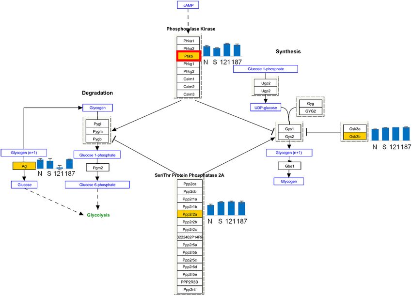
www.nature.com/scientificreports/                                                              www.nature.com/scientificreports

                           Figure 1. Relative mRNA expression at different time points after intravitreal N-methyl-D-aspartate
                           (NMDA) injection. Neural retinas were analysed 2, 4 and 6 h after intravitreal NMDA (5 nmol) injection.
                           The relative expression levels of (a) jun proto-oncogene (Jun), (b) FBJ osteosarcoma oncogene (Fos), (c) v-rel
                           reticuloendotheliosis viral oncogene homolog A (Rela), (d) capsase-3 (Casp3), (e) nuclear factor of kappa
                           light polypeptide gene enhancer in B cells inhibitor, alpha (Nfkbia), (f) tumor necrosis factor (Tnf) and (g)
                           interleukin 6 (Il6) mRNA were analysed by qRT-PCR. The ratios of mRNA expression of each gene to that of
                           actin were calculated. The ratios to actin at 2 h, 4 h and 6 h were divided by those at 0 h.

                           enabled us to purify retinal ganglion cells by FACS. Next, we used next-generation sequencing technologies33 to
                           compare gene expression profiles between with and without KUS treatments.

                           Results
                           mRNA expression of key genes was significantly altered 4 h after NMDA injection.                           To decide
                           the timing for evaluation of gene expression after intravitreous NMDA injection, the mRNA levels of 18 genes,
                           of which some were reported to be upregulated after NMDA injection and some could be influenced by admin-
                           istration of KUSs, were analysed using quantitative reverse transcription polymerase chain reaction (qRT-PCR).
                           The former were v-rel reticuloendotheliosis viral oncogene homolog A (Rela), caspase 3 (Casp3), nuclear factor
                           of kappa light polypeptide gene enhancer in B cells inhibitor - alpha (Nfkbia), interleukin 6 (Il6), FBJ osteosar-
                           coma oncogene (Fos), mitogen-activated protein kinase (Mapk)1, Mapk3, Mapk10, jun proto-oncogene (Jun),
                           tumor necrosis factor (Tnf), and high mobility group box 1 (Hmgb1) and the latter were serine/threonine-pro-
                           tein kinase (Akt)1, Akt2, mitochondrial fission 1 (Fis1), mitofusin (Mfn)1, Mfn2, dynamin 1-like (Dnm1l), optic
                           atrophy 1 (Opa1). Jun and Fos, which were reported to be immediately upregulated following external stimuli34
                           increased 2 h after the NMDA injection (Fig. 1). Expression of Rela and Casp3 (a downstream effector of apopto-
                           sis) increased prominently 6 h after the injection (Fig. 1). In contrast, expression of the other 14 genes including
                           Nfkbia, Tnf, and Il-6 increased notably 4 h after the NMDA injection (Fig. 1). Therefore, we decided to evaluate
                           the effect of KUSs on the gene expression profiles 4 h after the NMDA injection.

                           Purification of retinal ganglion cells by FACS. To study the effects of the KUSs on the affected cells,
                           we first isolated the CFP expressing retinal ganglion cells from dissociated whole neural retina of Thy1-CFP
                           mice using FACS. The CFP-positive cells were found to account for 0.04–0.12% of all retinal cells (Fig. 2a, area

Scientific Reports |   (2020) 10:4251 | https://doi.org/10.1038/s41598-020-61160-6                                                           2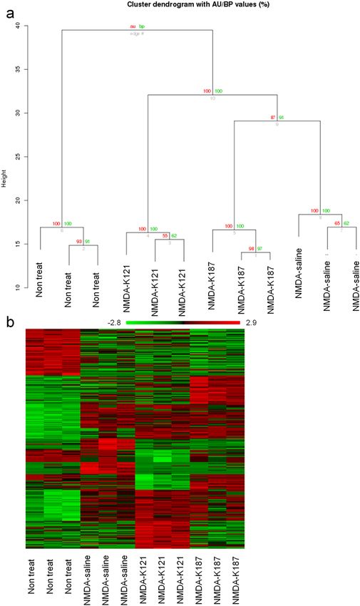
www.nature.com/scientificreports/ www.nature.com/scientificreports
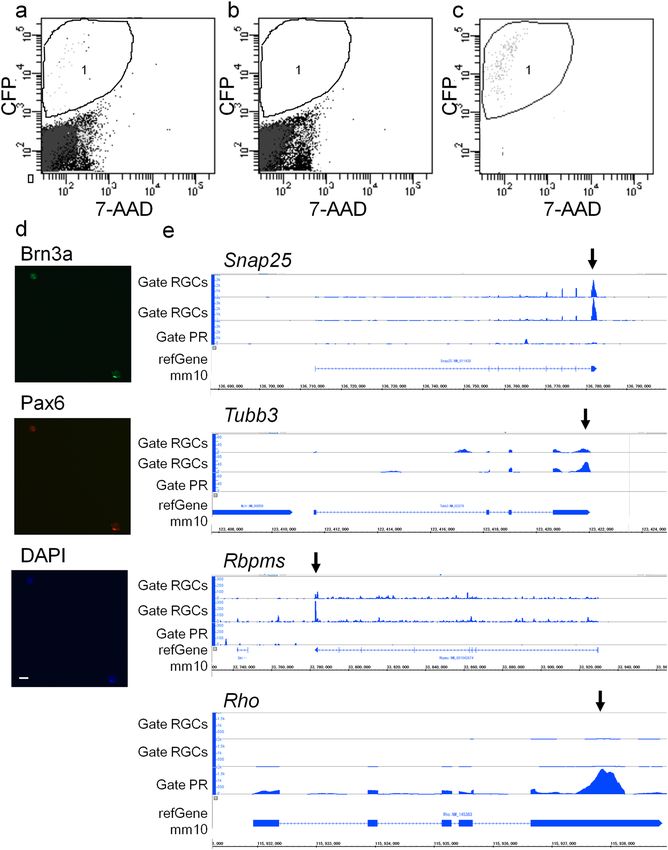
Figure 2. Analysis of dissociated retinal cells and sorted cells by flow cytometry. Retinal cells were analysed
using fluorescence-activated cell sorting. The x-axis shows fluorescent of PerCP-Cy5 to detect 7-Amino-
Actinomycin D (7-AAD) fluorescence which labels dead cells and the y-axis shows fluorescence of AmCyan-A
to detect cyan fluorescent protein (CFP) fluorescence. (a) Analysis of dissociated retinal cells of Thy1-CFP mice
which manifest CFP in retinal ganglion cells. Cells which possess relatively strong CFP fluorescent without
7-AAD fluorescence, whose CFP fluorescence was considered not to be autofluorescence, were contained in
the area 1 (0.04–0.12% of total cells). (b) Analysis of dissociated retinal cells of wild-type mice. No cells were
contained in the area 1 (CFP-positive). (c) Re-analysis of the sorted cells by gate RGCs (see Method and Fig. S1).
88.9–93.4% of the sorted cells were included in area 1 (CFP-positive). (d) The sorted cells were stained with
anti-brain-specific homeobox/POU domain protein 3A (Brn3a, green) and anti- paired box genes 6 (Pax6, red)
antibodies. Nuclei were counter stained with 4’,6-diamidino-2-phenylindole (DAPI, blue). Scale bar: 50 µm.
(e) mRNA expression of synaptosomal-associated protein 25 (Snap25), tubulin, beta 3 class III (Tubb3), RNA
binding protein with multiple splicing (Rbpms) and rhodopsin (Rho) in cells sorted by gate RGCs or gate PR
(see Methods and Supplementary, Fig. S1b) were visualised with the Integrated Genome Browser. Snap25,
Tubb3 and Rbpms were highly expressed in Gate RGCs and not in Gate PR while Rho was highly expressed in
Gate PR and not in Gate RGCs (black arrows). RGC: retinal ganglion cell, PR: photoreceptors.
1); no CFP-positive cells were observed in the retina of wild-type mice (Fig. 2b). Retinal cells expressing both
brain-specific homeobox/POU domain protein 3A (Brn3a) and paired box protein (Pax) 6 are defined as reti-
nal ganglion cells. By immunostaining the dissociated cells, we confirmed that Brn3a- and Pax6-positive retinal
Scientific Reports | (2020) 10:4251 | https://doi.org/10.1038/s41598-020-61160-6 3www.nature.com/scientificreports/ www.nature.com/scientificreports
ganglion cells accounted for 0.01–0.12% of the total retinal cells. The percentage of the retinal ganglion cells esti-
mated with the immunocytochemical analysis was almost the same as the FACS analysis.
FACS from 2 retinas of 2 Thy1-CFP mice enabled us to sort 1,494–3,550 CFP-positive cells (see method
and Fig. S1, gate RGCs) in 30–50 min, which was considered sufficiently fast to collect fresh cells to analyze
mRNA expression. Re-analysis of the sorted cells showed that 88.9–93.4% of the cells were CFP-positive, which
indicates that the FACS sorting effectively collected and concentrated the CFP-positive retinal cells (Fig. 2c).
Immunostaining of the sorted cells showed that almost all the sorted cells were Brn3a- and Pax6-positive and
were retinal ganglion cells (Fig. 2d and Fig. S2). To confirm that the collected cells were indeed retinal gan-
glion cells, their mRNAs were visualised with the Integrated Genome Browser35. We confirmed expected mRNA
expression profiles: high expression of synaptosomal-associated protein (Snap25)36, and tubulin, beta 3 class III
(Tubb3), which are expressed in neuronal cells and RNA binding protein with multiple splicing (Rbpms)37, which
is expressed in retinal ganglion cells and low expression of rhodopsin (Rho), which is expressed in rod photore-
ceptors. These results further validated our FACS sorting protocol (Fig. 2e, gate RGCs in Fig. S1). Consistent with
the above, CFP-negative cells collected by gate PR in Fig. S1, expressed high Rho levels without Snap25, Tubb3,
and Rbpms expression, indicating that they contained rod photoreceptors and not ganglion cells (Fig. 2e, gate PR).
From these data, we assumed the CFP-positive cells sorted by FACS, successfully enriched retinal ganglion cells
and were suitable for the next experiments.
mRNAs related to gene expression and energy metabolism were upregulated in KUS-treated
retinal ganglion cells. Using analysis of variance (ANOVA), 255 genes showed significant (P < 0.01) expres-
sion changes among four conditions; non-treatment (non-treat), vehicle with intravitreous injection of NMDA
(NMDA-saline), KUS121 treatment with intravitreous injection of NMDA (KUS121), and KUS187 treatment
with intravitreous injection of NMDA (KUS187) (Supplementary Table S1). Hierarchical clustering analyses of
the 255 genes distributed samples between the conditions (Fig. 3a,b). Hierarchical clustering analyses of the
ANOVA-passed genes revealed that ANOVA successfully selected genes that distinguish each condition (Fig. 3a).
Moreover, the samples of the three experimental repeats showed similar patterns of upregulated and downreg-
ulated genes on heatmap (Fig. 3b). These results showed that the experimental repeats displayed great repro-
ducibility of the gene expression profiles within each condition. While some genes showed similar expression
patterns between the NMDA-saline and KUSs-treated groups, other genes showed clearly differential expression
patterns (Fig. 3b). KUSs-treated groups were clearly separated from saline-treated groups. These data indicated
the KUSs-treated groups have characteristic gene expression profiles distinct from the non-treat or NMDA-saline
groups.
Gene ontology (GO) analysis38 was performed to annotate genes into biological ontology. After X-means
clustering, genes in hyper cluster A and B were analyzed (Supplementary Fig. S3). Genes in hyper cluster A were
found to be associated with 31 GO terms, which included RNA metabolic processes, biosynthetic processes, gene
expression and metabolic processes (Table 1, Supplementary Table S2) while genes in hyper cluster B were not
associated with enrichment of any GO terms.
Pathway analysis 39,40 showed 4 statistically significant pathways which were common in
NMDA-saline < non-treat pathways, KUS121 > NMDA-saline pathways and KUS187 > NMDA-saline pathways
(Table 2). The activated pathways included the glycogen metabolism pathway (Fig. 4). There were 7 statistically
significant pathways which were common in NMDA-saline > non-treat pathways, KUS121 < NMDA-saline path-
ways and KUS187 < NMDA-saline pathways (Table 3).
Zfp667 was upregulated in KUS121-treated retinal ganglion cells. We next performed literature
search on the functions of the 255 genes whose mRNA expression changed significantly among the four groups.
These genes included genes related to energy metabolism, cell proliferation, cell survival, and cell death, such
as zinc finger protein 667 (Zfp667), phosphorylase b kinase regulatory subunit beta (Phkb), peroxisome prolif-
erative activated receptor gamma coactivator 1 alpha (Ppargc1a), pentatricopeptide repeat domain 2 (Ptcd2),
nucleophosmin 1 (Npm1), dual specificity phosphatase 18 (Dusp18), paternally expressed gene 10 (Peg10), and
topoisomerase (DNA) 3 alpha (Top3a) (Table 4).
Of the genes studied, we focused on Zfp667, which has been reported to suppress apoptosis-related genes
and consequently prevent cell death in ischemia-reperfusion injury41,42. Western blot analysis of mouse retinal
proteins showed that expression levels of Zfp667 was not significantly different between non-treated retinas
and saline-treated NMDA-injected retinas (NMDA-saline as control) of wild-type mice (P = 0.61, Turkey HSD,
Fig. 5a,b). In contrast, expression of Zfp667 was significantly increased in KUS121-treated NMDA-injected ret-
inas compared to the NMDA-saline group (P = 0.004, Turkey HSD, Fig. 5a,b). Immunohistochemical analysis
with an anti-Zfp667 antibody showed higher expression of Zfp667 predominantly at the retinal ganglion cell layer
in KUSs-treated NMDA-injected retinas compared to NMDA-saline injected retinas (Fig. 5c).
Discussion
In this study, we successfully isolated retinal ganglion cells to a high level of purity using FACS and found that
KUSs affect the expression of a wide variety of genes. These include genes involved in the regulation of energy
metabolism and suppression of apoptosis in the retinal ganglion cells of an NMDA-induced retinal injury model
mouse.
There have been studies in which neural cells including retinal ganglion cells were isolated using FACS with
retrograde labelling43,44 or with antibodies45. In the current study, we used Thy1-CFP transgenic mice29,30, in
which CFP is expressed in retinal ganglion cells, to omit the need for antibody reaction or the retrograde labelling
step. It enabled us to collect the cells simply and quickly, which were suitable for the analysis of mRNA profiles.
Scientific Reports | (2020) 10:4251 | https://doi.org/10.1038/s41598-020-61160-6 4www.nature.com/scientificreports/ www.nature.com/scientificreports
Figure 3. mRNA expression in retinal ganglion cells in an acute injury model. (a) Tree diagram of
hierarchically clustered conditions. Red and green numbers show approximately unbiased p-values (AU) and
bootstrap probability value (BP). (b) Heat map of mRNA expression of clustered conditions. Non treat: retinal
ganglion cells of non-treated mice, NMDA-K121: retinal ganglion cells of NMDA-injected mice administered
KUS121, NMDA-K187: retinal ganglion cells of NMDA-injected mice administered KUS187, NMDA-saline:
retinal ganglion cells of NMDA-injected mice administered vehicle.
In the preliminary experiments to decide the timing for evaluating gene expression after intravitreous NMDA
injection, neural retina of wild-type mice was used. While intravitreous NMDA injection has been reported
to damage RGCs19, whole retinas of mice that received intravitreous NMDA injection were used for mRNA
evaluation by qRT-PCR46. Hence, we used neural retina in the preliminary experiments. Moreover, among the
18 genes evaluated by qRT-PCR using neural retina, Jun and Fos were included in the 255 genes evaluated by
next-generation sequencing using sorted RGCs that showed significant changes in expression among the four
conditions. The expression of Jun and Fos genes was upregulated by 1.63 and 3.59 times, respectively, in NMDA
treated mice RGCs compared to non-treated controls.
Our experiments showed that KUS121, which has already been used in a clinical trial for ischemic reti-
nal disease (UMIN000023979), upregulated gene expression and translation of Zfp667 in the retinal ganglion
cells. Zfp667 has been reported to suppress apoptosis-related genes in ischemia-reperfusion injury41,42. KUS121
also upregulated the expression of a variety of genes such as Phkb, which is involved in glycogen metabolism47;
Ppargc1a, which is a strong activator of mitochondrial function and a regulator of energy metabolism48–50;
Ptcd2, which is involved in mitochondrial gene expression51; Npm1, which promotes cell survival under stress52;
Dusp18, which may dephosphorylate and inactivate mitogen-activated protein kinase (MAPK)47; Peg10, which
is anti-apoptotic53. On the other hand, genes that were downregulated by KUS121 included Top3a, which adjusts
the DNA topological states during transcription47. Whether the translation of these genes is also altered remains
to be clarified. In addition to reducing ATP consumption, these results revealed the possibility that KUS121
Scientific Reports | (2020) 10:4251 | https://doi.org/10.1038/s41598-020-61160-6 5www.nature.com/scientificreports/ www.nature.com/scientificreports
Over-
ID p value representation Description Genes
regulation of RNA
GO:0051252 0.000903 3.1 Camta1,Cir1,Csrnp2,Fos,Fosb,Gatad2a,Nr4a1,Rfc1
metabolic process
regulation of
GO:0010556 0.001284 2.9 macromolecule Camta1,Cir1,Csrnp2,Fos,Fosb,Gatad2a,Nr4a1,Rfc1
biosynthetic process
regulation of
nucleobase-
GO:0019219 0.001308 2.9 containing Camta1,Cir1,Csrnp2,Fos,Fosb,Gatad2a,Nr4a1,Rfc1
compound
metabolic process
regulation of
GO:0051171 0.001308 2.9 nitrogen compound Camta1,Cir1,Csrnp2,Fos,Fosb,Gatad2a,Nr4a1,Rfc1
metabolic process
regulation of
GO:0031326 0.001326 2.9 cellular biosynthetic Camta1,Cir1,Csrnp2,Fos,Fosb,Gatad2a,Nr4a1,Rfc1
process
regulation of
GO:0009889 0.001332 2.9 Camta1,Cir1,Csrnp2,Fos,Fosb,Gatad2a,Nr4a1,Rfc1
biosynthetic process
regulation of gene
GO:0010468 0.00219 2.7 Camta1,Cir1,Csrnp2,Fos,Fosb,Gatad2a,Nr4a1,Rfc1
expression
regulation of
GO:0060255 0.002518 2.7 macromolecule Camta1,Cir1,Csrnp2,Fos,Fosb,Gatad2a,Nr4a1,Rfc1
metabolic process
regulation of
GO:0080090 0.003192 2.6 primary metabolic Camta1,Cir1,Csrnp2,Fos,Fosb,Gatad2a,Nr4a1,Rfc1
process
regulation of
GO:0031323 0.003291 2.6 cellular metabolic Camta1,Cir1,Csrnp2,Fos,Fosb,Gatad2a,Nr4a1,Rfc1
process
cellular
GO:0034645 0.001295 2.6 macromolecule Camta1,Cir1,Csrnp2,Eif2s3y,Extl1,Fos,Fosb,Gatad2a,Nr4a1,Rfc1
biosynthetic process
macromolecule
GO:0009059 0.001314 2.6 Camta1,Cir1,Csrnp2,Eif2s3y,Extl1,Fos,Fosb,Gatad2a,Nr4a1,Rfc1
biosynthetic process
regulation of
GO:0019222 0.003592 2.5 Camta1,Cir1,Csrnp2,Fos,Fosb,Gatad2a,Nr4a1,Rfc1
metabolic process
nucleobase-
containing
GO:0034654 0.006007 2.3 Camta1,Cir1,Csrnp2,Fos,Fosb,Gatad2a,Nr4a1,Rfc1
compound
biosynthetic process
aromatic compound
GO:0019438 0.006642 2.3 Camta1,Cir1,Csrnp2,Fos,Fosb,Gatad2a,Nr4a1,Rfc1
biosynthetic process
Table 1. Top 15 gene ontology (GO) terms for genes whose upregulation by NMDA was attenuated in KUS-
treated retinal ganglion cells.
prevents retinal ganglion cell death through several mechanisms, including activating energy production and
suppression of apoptosis. These mechanisms could be related to the modulation of VCP function or alternatively,
KUSs may have additional targets, which could be involved in transcriptional control of cell survival. These pos-
sibilities need to be elucidated by further studies.
In conclusion, KUS121 can modulate gene expression profiles in retinal ganglion cells in mice, via mech-
anisms not yet fully elucidated, which are likely to contribute to protecting the retinal ganglion cells from
NMDA-induced neurotoxicity. This study further strengthens the suitability of KUS121 as a therapeutic drug in
rescuing retinal ganglion cells in eye diseases that are currently incurable, such as glaucoma.
Methods
Experimental animals. This study was conducted in accordance with the Association for Research in
Vision and Ophthalmology (ARVO) Statement for the Use of Animals in Ophthalmic and Vision Research. All
protocols were approved by the Institutional Review Board of Kyoto University Graduate School of Medicine
(MedKyo 12245, 13221, 14213, 15531, 16501). B6.Cg-Tg(Thy 1-CFP) 23Jrs/J mice were obtained from the
Jackson Laboratory (Bar Harbor, ME, USA) and wild-type mice (C57/BL6), which share the genetic background
of Thy1-CFP mice, were purchased from Japan SLC, Inc. Mice were kept in a 14 h light/10 h dark cycle and fed
ad libitum. Male mice aged 2 to 3 months were used for the experiments. Before intravitreous NMDA injection
(5 nmol), mice were anesthetised with intraperitoneal pentobarbital (50 mg/kg) injection and pupils were dilated
with tropicamide and phenylephrine eye drops (0.5% each).
Quantitative RT-PCR of neural retinae. Changes in mRNA expression in the neural retina were exam-
ined at several time points after intravitreous NMDA injection. NMDA was intravitreally injected into wild-type
mice to induce acute damage of retinal ganglion cells17,19. Eyeballs were enucleated 2, 4, and 6 h after NMDA
injection after pentobarbital overdose. Enucleated eyeballs were immersed in cold Hanks’ balanced salt solution
Scientific Reports | (2020) 10:4251 | https://doi.org/10.1038/s41598-020-61160-6 6www.nature.com/scientificreports/ www.nature.com/scientificreports
Matched
Entities p value
Matched Entities Matched Entities (saline < non- Total p value p value (saline < non-
ID Term (K121 > saline) (K187 > saline) treat) Entities (K121 > saline) (K187 > saline) treat)
WP1251 Metapathway biotransformation 1 1 3 143 0.005723 0.004692 5.40E-09
WP317 Glycogen Metabolism 1 1 1 34 0.007623 0.006252 0.007125
PodNet-protein-protein interactions
WP2310 1 3 2 315 0.00952 3.61E-08 3.12E-05
in the podocyte
XPodNet-protein-protein
WP2309 interactions in the podocyte 1 3 2 836 0.018951 4.31E-07 1.40E-04
expanded by STRING
Table 2. Pathways which were common in NMDA-saline group < non-treat group, KUS121 group > NMDA-
saline group and KUS187 group > NMDA-saline group (fold change > 2 or fold change < 0.5, respectively).
saline: NMDA-saline group, K121: KUS121 group, K187: KUS187 group.
Figure 4. Glycogen metabolism Pathway activation in KUS-treated retinal ganglion cells. Pathway analysis of
genes which were upregulated (fold change > 2) or downregulated (fold change < 0.5) between groups were done
using an analysis software, Gene Spring14 (TOMY Digital Biology, http://genespring-support.com/). Pathways
that were common in NMDA-saline < non-treat, KUS121 > NMDA-saline and KUS187 > NMDA-saline included
glycogen metabolism pathway. Genes included in the current analysis, which passed p < 0.01 with analysis of
variance (255 genes), are shown in orange color boxes. The bar graph next to the genes in the orange color boxes
show the normalized expression values (N: non-treat, S: NMDA-saline, 121: KUS121, 187: KUS187). Phkb was
an only gene which passed the upregulated and downregulated criteria above; downregulated in NMDA-saline
compared to non-treat and was upregulated in KUS121 and KUS187 compared to NMDA-saline. The gene is
shown in red frame box. non treat: retinal ganglion cells of mice without NMDA injection nor KUS treatment,
NMDA-saline: retinal ganglion cells of NMDA-injected mice administered vehicle, KUS121: retinal ganglion
cells of NMDA-injected mice administered KUS121, KUS187: retinal ganglion cells of NMDA-injected mice
administered KUS187. In fold change analysis, >2 indicates more than 2 folds while B means statistically significant pathways of genes that
were upregulated in group A by more than 2 folds compared to group B.
immediately after enucleation. Incisions were made using pinholes in the corneas, then using the incisions, the
sclera was peeled to remove the mixture of the retinal pigment epithelium, choroid and sclera from the neural ret-
ina as previously described16. The lens and iris were removed. RNA was isolated from the neural retina using the
Scientific Reports | (2020) 10:4251 | https://doi.org/10.1038/s41598-020-61160-6 7www.nature.com/scientificreports/ www.nature.com/scientificreports
Matched Entities p value
Matched Entities Matched Entities (saline > non- Total p value p value (saline > non-
ID Term (K121 < -saline) (K187 < -saline) treat) Entities (K121 < saline) (K187 < saline) treat)
PodNet-protein-
WP2310 protein interactions in 3 1 1 315 1.14E-07 0.008276 0.017887
the podocyte
XPodNet-protein-
protein interactions in
WP2309 3 2 5 836 1.36E-06 1.21E-04 1.38E-10
the podocyte expanded
by STRING
Complement
WP200 Activation, Classical 1 1 1 17 0.002287 0.001661 0.003603
Pathway
MicroRNAs in
WP1560 Cardiomyocyte 1 1 1 104 0.004569 0.003318 0.007193
Hypertrophy
Notch Signaling
WP29 1 1 2 47 0.004569 0.003318 1.29E-05
Pathway
Prostaglandin
WP374 Synthesis and 1 1 1 31 0.006846 0.004974 0.010771
Regulation
SIDS Susceptibility
WP1266 1 1 2 61 0.006846 0.004974 3.85E-05
Pathways
Table 3. Pathways which were common in NMDA-saline group > non-treat group, KUS121 group < NMDA-
saline group and KUS187 group < NMDA-saline group (fold change > 2 or fold change < 0.5, respectively).
saline: NMDA-saline group, K121: KUS121 group, K187: KUS187 group.
Description (possible) Function
may play a role in protecting cells against
Zfp667
ischemia-reperfusion injury41,42
Phkb plays a role in glycogen metabolism47
Ppargc1a activate mitochondrial function48–50
plays a role in post-transcriptional expression of
Ptcd2
the mitochondrial genome51
Npm1 enhances cell survival under stress conditions52
Dusp18 inactivate MAP kinases47
Peg10 promote growth and suppress apoptosis53
Top3a adjust the DNA topological states47
Table 4. Focused genes which were significantly changed between KUS-treated and control retinal ganglion
cells.
RNeasy Mini Kit (QIAGEN, Venlo, Netherlands). The mRNA was reverse transcribed with the M-MLV reverse
transcriptase (Promega, WI, USA) and then complementary DNA was amplified by PCR with SYBR premix Ex
Taq polymerase (Takara Bio Inc., Shiga, Japan) and 60 °C as the annealing temperature on the 7300 Real-Time
PCR System (Applied Biosystems, CA, USA). Eyes were enucleated before and 2, 4 and 6 h after NMDA injection
and each eye was analysed separately.
The levels of the following mRNAs were analysed by qRT-PCR: Nfkbia, Il6, Rela, Casp3, Fos, Mapk1, Mapk3,
Mapk10, Jun, Tnf, Akt1, Akt2, Fis1, Mfn1, Mfn2, Dnm1l, Opa1, Hmgb1 (primers used are shown in Supplementary
Table S3). Actin was used as the internal standard.
Administration of KUSs and preparation of cell suspension for flow cytometry. Daily KUS121,
KUS187 (50 mg/kg/day each), or vehicle (5% Cremophor EL (Sigma) in phosphate buffered saline (PBS)) as
a control were given orally to Thy1-CFP mice using a feeding tube. Seven days after the start of the medica-
tion, NMDA (5 nmol) was intravitreally injected with a 33-gage needle54. Our experiment involved one-week
pre-treatment with KUSs because we previously showed that it protects against NMDA injury17. Four hours
after the NMDA injection, the retinas were collected as described in the qRT-PCR section and incubated in 0.2%
papain solution (two retinas of two mice each) including glucose (1 mg/mL), DNase 1 (Worthington, 100 U/mL),
superoxide dismutase (Worthington, 5 µg/mL) and catalase (Sigma, 5 µg/mL) at 8 °C for 30 min and at 28 °C for
9 min. The solution was centrifuged at 100 G for 5 min at 4 °C, cells were resuspended in a solution containing
antipain (Roche, 50 µg/mL), and then centrifuged again at 100G for 5 min at 4 °C55. The cell pellet was resus-
pended in 500 µL of ice-cold Ames’ medium (with L-glutamine, without sodium bicarbonate, Sigma).
Collection of retinal ganglion cells by flow cytometry. The cells in suspended Ames’ medium
described above were sorted by FACS Aria 2 (BD Biosciences) based on the size and intensity of CFP fluores-
cence. Immediately after 7-Amino-Actinomycin D (BD Biosciences, Cell Viability Solution, 20 µL) were added,
Scientific Reports | (2020) 10:4251 | https://doi.org/10.1038/s41598-020-61160-6 8www.nature.com/scientificreports/ www.nature.com/scientificreports
Figure 5. Increased expression of Zfp667 in KUS121-treated retinal ganglion cells. (a) Western blot analysis
of NMDA-injected mice retinas with KUS121 (labelled as “K”) or vehicle (NMDA-saline as control, labelled
as “C”) administration with a zing finger protein (ZFP667) antibody. Actin was used as a loading control. WT:
wild-type mouse retina. Complete scans of western blots are shown in Supplementary Fig. S4. (b) Comparison
of ZFP667 expression shown as ratio of actin. **P < 0.01, Turkey HSD. (c) Vertical retinal sections of mouse
retina were stained with anti-ZFP667 antibody (red). Fluorescence intensity of the complex of RNFL, GCL
and IPL was measured. Control: NMDA-injected mouse retina with saline administration, KUS121: NMDA-
injected mouse retina administered KUS121, KUS187: NMDA-injected mouse retina administered KUS187,
WT: wild-type mouse retina without NMDA injection or KUSs administration. RNFL: retinal nerve fiber layer;
GCL: ganglion cell layer; IPL: inner plexiform layer; INL: inner nuclear layer; OPL: outer plexiform layer; ONL:
outer nuclear layer; IS: inner segment of photoreceptors, and OS: outer segment of photoreceptors. Scale bar:
50 µm.
sorting was performed using a 85-micron nozzle into Ames’ medium at 4 °C. Forward scatter (FSC) and sideward
scatter (SSC) were used to segregate retinal ganglion cells. CFP-positive cells were contained in the high FSC
sub-population (area 2 in Supplementary Fig. S1a). To collect retinal ganglion cells with high purity, only cells
included in both gate 4 (Supplementary Fig. S1b), which is a smaller area than area 2, and gate 5 (Supplementary
Fig. S1c), which was narrower than the area 1 in Fig. 2a–c, were collected (gate RGCs). For comparison, cells
included in both gate 6 (Supplementary Fig. S1b) and gate 7 (Supplementary Fig. S1c), which were considered to
be photoreceptors, were collected in the same way (gate PR).
cDNA synthesis, amplification and next-generation sequencing. The sorted cells (1,494–7,848 cells
for each sample) were centrifuged at 500G for 5 min at 4 °C, suspended in 100 µL of buffer B (Prelude Direct Lysis
Module, NuGEN), centrifuged again at 500G for 5 min at 4 °C, and the pellet was resuspended in 1 µL of buffer A
(Prelude Direct Lysis Module, NuGEN). The lysates were then taken forward for cDNA synthesis and amplifica-
tion using Ovation RNA-Seq System V2 (NuGEN) according to the manufacturer’s instructions. The amplified
cDNA was purified using MinElute Reaction Cleanup Kit (QIAGEN, Venlo, Netherlands). The quality of the
amplified cDNAs was analyzed by a 2100 Bioanalyzer (Agilent) and was high enough for sequencing. The concen-
tration of cDNA as measured by Qubit (Invitrogen) was sufficient (84.7–185.0 ng/µL). The amplified cDNA was
then sequenced with a next-generation sequencer (Illumina HiSeq). The experiments were repeated three times.
Analysis of the RNA-sequencing results. The results of RNA-sequencing were mapped on reference
sequence (mouse mm10, USCS genome browser) using TopHat2. Reads which formed reasonable pairs (on the
same chromosome, two directionally, and distance between pair reads < 500 k b.p.) were used to calculate expres-
sion levels (61.1 ± 8.5% of reads). Reads Per Kilobase of exon model per 10 Million mapped reads (RPK10M)
Scientific Reports | (2020) 10:4251 | https://doi.org/10.1038/s41598-020-61160-6 9www.nature.com/scientificreports/ www.nature.com/scientificreports
were calculated for terminal exons with the in-house scripts as the expression values for genes, and transferred
into log2 scale. Genes whose maximum expression values were more than 3 among the conditions were consid-
ered as expressed genes and used for the following analyses. Then the expression value was normalized using
quantile normalization methods56. Using analysis of variance (ANOVA), 255 genes (including isoforms) showed
significant changes in expression among the four conditions (non-treat, NMDA-saline, KUS121 and KUS187,
p < 0.01). Hierarchical clustering of the 255 genes that passed the ANOVA test with approximately unbiased
p-values and bootstrap probability value of mRNA expression was performed with pvclust package in the sta-
tistical environment R using Euclidean distances. A heatmap was drawn with Z-value transferred expression
values. Expression profiles of ANOVA-passed genes were transferred to Z-scores and clustered with the x-means
method. X-means clustering was performed on the statistical environment R. The genes were divided into 12
clusters that were further categorized according to expression changes across conditions. The upregulation and
downregulation of gene expression were defined as the difference in the cluster centers between conditions with
more than 0.2 and less than −0.2 z-values, respectively (Supplementary Fig. S3). The clusters with upregulated
expression in NMDA-saline compared to non-treat and downregulated expression in KUS121 and KUS187
compared to NMDA-saline were defined as hyper cluster A. The clusters whose expression was upregulated in
NMDA-saline compared to non-treat and whose expression was upregulated in KUS121 and KUS187 compared
to NMDA-saline, were defined as hyper cluster B. GO analysis of the genes included in hyper cluster A (55 genes)
and B (12 genes) was performed based on hypergeometric distribution.
Pathway analysis was performed by Gene Spring14 (TOMY Digital Biology) using Wiki Pathways (http://
www.wikipathways.org/index.php/WikiPathways). Genes that were upregulated (115 genes) or downregu-
lated (57 genes) in the NMDA-saline group compared to the non-treat group, and those that were upregulated
(61 genes) or downregulated (73 genes) in the KUS121 group compared to the NMDA-saline group as well as
upregulated (50 genes) or downregulated (53 genes) in the KUS187 group compared to the NMDA-saline group
(fold change > 2 or fold change < 0.5, respectively) were analyzed. The level of statistical significance was set to
P < 0.05.
In fold change analysis, >2 indicates more than 2 folds while B means statistically significant pathways of genes that were upregulated
in group A by more than 2 folds compared to group B.
Immunocytological evaluation of cells. Dissociated retinal cells or FACS sorted cells were fixed by
adding an equal amount of 4% paraformaldehyde, centrifuged at 3000 rpm for 15 min at 4 °C. After the extra
supernatant was removed, the cell suspension (200 µL) was centrifuged at 1000 rpm for 10 min using Cytospin
(Thermo Scientific) to be pasted onto slides. The cells were stained with anti-Brn3a (CHEMICON) and anti-Pax6
(COVANCE) antibodies and imaged under an optical microscope (Axio Imager.A1, Zeiss).
Immunohistological evaluation of retinas. Non-treated eyeballs or NMDA-injected eyeballs of mice
treated with KUS121, KUS187 or vehicle (saline) were enucleated after pentobarbital overdose. A marking dye
(Davidson) was placed on the edge of the superior conjunctiva to identify the superior portion of the retina as
previously described57. The eyes were fixed in 4% paraformaldehyde for 24 h at 4 °C, embedded in O.C.T. com-
pound (Sakura Finetek Japan) and frozen. Serial 16 µm O.C.T-embedded sections were cut through the dye and
at the point of insertion of the optic nerve. Sections that included the center of the optic nerve head were stained
with an anti-ZFP667 antibody (GeneTex) and imaged under an optical microscope (BZ-9000, Keyence) at a
distance of 400 µm from the edge of the optic nerve head. The fluorescence intensity of each eye was measured in
40 µm × 300 µm squares including the retinal nerve fiber layer, the ganglion cell layer, and the inner plexiform
layer using BZ II Analyzer software (Keyence).
Western blotting of neural retinas. Neural retinas were prepared as described in the qRT-PCR sec-
tion. Neural retinas and wild-type mouse brain, which was used as a positive control, were analysed with an
anti-ZFP667 antibody (GeneTex). Actin was used as a loading control. The ratio of ZFP667 to actin was compared
between the KUS121-treated and the control retina using an unpaired t-test.
Data availability
All data generated or analyzed during this study are included in this published article and its Supplementary
Information files.
Received: 7 November 2019; Accepted: 10 February 2020;
Published: xx xx xxxx
References
1. Sommer, A. et al. Racial differences in the cause-specific prevalence of blindness in east Baltimore. N. Engl. J. Med. 325, 1412–1417,
https://doi.org/10.1056/nejm199111143252004 (1991).
2. Klaver, C. C., Wolfs, R. C., Vingerling, J. R., Hofman, A. & de Jong, P. T. Age-specific prevalence and causes of blindness and visual
impairment in an older population: the Rotterdam Study. Arch. Ophthalmol. 116, 653–658 (1998).
3. Bourne, R. R. et al. Causes of vision loss worldwide, 1990-2010: a systematic analysis. Lancet Glob. Health. 1, e339–349, https://doi.
org/10.1016/s2214-109x(13)70113-x (2013).
4. Klein, R. & Klein, B. E. The prevalence of age-related eye diseases and visual impairment in aging: current estimates. Invest.
Ophthalmol. Vis. Sci. 54, Orsf5–orsf13, https://doi.org/10.1167/iovs.13-12789 (2013).
Scientific Reports | (2020) 10:4251 | https://doi.org/10.1038/s41598-020-61160-6 10www.nature.com/scientificreports/ www.nature.com/scientificreports
5. Quigley, H. A., Addicks, E. M. & Green, W. R. Optic nerve damage in human glaucoma. III. Quantitative correlation of nerve fiber
loss and visual field defect in glaucoma, ischemic neuropathy, papilledema, and toxic neuropathy. Arch. Ophthalmol. 100, 135–146
(1982).
6. Quigley, H. A., Dunkelberger, G. R. & Green, W. R. Retinal ganglion cell atrophy correlated with automated perimetry in human
eyes with glaucoma. Am. J. Ophthalmol. 107, 453–464 (1989).
7. Leske, M. C., Heijl, A., Hyman, L., Bengtsson, B. & Komaroff, E. Factors for progression and glaucoma treatment: the Early Manifest
Glaucoma Trial. Curr. Opin. Ophthalmol. 15, 102–106 (2004).
8. Vass, C. et al. Medical interventions for primary open angle glaucoma and ocular hypertension. Cochrane Database Syst. Rev.
Cd003167 https://doi.org/10.1002/14651858.CD003167.pub3 (2007).
9. Collaborative Normal-Tension Glaucoma Study Group. The effectiveness of intraocular pressure reduction in the treatment of
normal-tension glaucoma. Am. J. Ophthalmol. 126, 498–505 (1998).
10. Ernest, P. J. et al. An evidence-based review of prognostic factors for glaucomatous visual field progression. Ophthalmology. 120,
512–519, https://doi.org/10.1016/j.ophtha.2012.09.005 (2013).
11. Flammer, J. The vascular concept of glaucoma. Surv. Ophthalmol. 38(Suppl), S3–6 (1994).
12. Marcus, M. W., de Vries, M. M., Junoy Montolio, F. G. & Jansonius, N. M. Myopia as a risk factor for open-angle glaucoma: a
systematic review and meta-analysis. Ophthalmology. 118, 1989–1994.e1982, https://doi.org/10.1016/j.ophtha.2011.03.012 (2011).
13. Mitchell, P., Hourihan, F., Sandbach, J. & Wang, J. J. The relationship between glaucoma and myopia: the Blue Mountains Eye Study.
Ophthalmology. 106, 2010–2015 (1999).
14. Lee, J. Y., Sung, K. R., Han, S. & Na, J. H. Effect of myopia on the progression of primary open-angle glaucoma. Invest. Ophthalmol.
Vis. Sci. 56, 1775–1781, https://doi.org/10.1167/iovs.14-16002 (2015).
15. Ikeda, H. O. et al. Novel VCP modulators mitigate major pathologies of rd10, a mouse model of retinitis pigmentosa. Sci. Rep. 4,
5970, https://doi.org/10.1038/srep05970 (2014).
16. Hasegawa, T. et al. Neuoroprotective efficacies by KUS121, a VCP modulator, on animal models of retinal degeneration. Sci. Rep. 6,
31184, https://doi.org/10.1038/srep31184 (2016).
17. Nakano, N. et al. Neuroprotective effects of VCP modulators in mouse models of glaucoma. Heliyon. 2, e00096 (2016).
18. Hata, M. et al. KUS121, a VCP modulator, attenuates ischemic retinal cell death via suppressing endoplasmic reticulum stress. Sci.
Rep. 7, 44873, https://doi.org/10.1038/srep44873 (2017).
19. Siliprandi, R. et al. N-methyl-D-aspartate-induced neurotoxicity in the adult rat retina. Vis. Neurosci. 8, 567–573 (1992).
20. Jeon, C. J., Strettoi, E. & Masland, R. H. The major cell populations of the mouse retina. J. Neurosci. 18, 8936–8946 (1998).
21. Shoge, K. et al. Rat retinal ganglion cells culture enriched with the magnetic cell sorter. Neurosci. Lett. 259, 111–114 (1999).
22. Tezel, G. & Wax, M. B. Increased production of tumor necrosis factor-alpha by glial cells exposed to simulated ischemia or elevated
hydrostatic pressure induces apoptosis in cocultured retinal ganglion cells. J. Neurosci. 20, 8693–8700 (2000).
23. Mukai, S. et al. Existence of ionotropic glutamate receptor subtypes in cultured rat retinal ganglion cells obtained by the magnetic
cell sorter method and inhibitory effects of 20-hydroxyecdysone, a neurosteroid, on the glutamate response. Jpn. J. Pharmacol. 89,
44–52 (2002).
24. Huang, X., Wu, D. Y., Chen, G., Manji, H. & Chen, D. F. Support of retinal ganglion cell survival and axon regeneration by lithium
through a Bcl-2-dependent mechanism. Invest. Ophthalmol. Vis. Sci. 44, 347–354 (2003).
25. Hong, S., Iizuka, Y., Kim, C. Y. & Seong, G. J. Isolation of primary mouse retinal ganglion cells using immunopanning-magnetic
separation. Mol. Vis. 18, 2922–2930 (2012).
26. Barres, B. A., Silverstein, B. E., Corey, D. P. & Chun, L. L. Immunological, morphological, and electrophysiological variation among
retinal ganglion cells purified by panning. Neuron. 1, 791–803 (1988).
27. Lindsey, J. D. & Weinreb, R. N. Survival and differentiation of purified retinal ganglion cells in a chemically defined
microenvironment. Invest. Ophthalmol. Vis. Sci. 35, 3640–3648 (1994).
28. Harada, C. et al. Role of apoptosis signal-regulating kinase 1 in stress-induced neural cell apoptosis in vivo. Am. J. Pathol. 168,
261–269, https://doi.org/10.2353/ajpath.2006.050765 (2006).
29. Feng, G. et al. Imaging neuronal subsets in transgenic mice expressing multiple spectral variants of GFP. Neuron. 28, 41–51 (2000).
30. Murata, H. et al. Imaging mouse retinal ganglion cells and their loss in vivo by a fundus camera in the normal and ischemia-
reperfusion model. Invest. Ophthalmol. Vis. Sci. 49, 5546–5552, https://doi.org/10.1167/iovs.07-1211 (2008).
31. Vidal, M., Morris, R., Grosveld, F. & Spanopoulou, E. Tissue-specific control elements of the Thy-1 gene. EMBO J. 9, 833–840 (1990).
32. Caroni, P. Overexpression of growth-associated proteins in the neurons of adult transgenic mice. J. Neurosci. Methods. 71, 3–9
(1997).
33. Mardis, E. R. DNA sequencing technologies: 2006-2016. Nat. Protoc. 12, 213–218, https://doi.org/10.1038/nprot.2016.182 (2017).
34. Healy, S., Khan, P. & Davie, J. R. Immediate early response genes and cell transformation. Pharmacol. Ther. 137, 64–77, https://doi.
org/10.1016/j.pharmthera.2012.09.001 (2013).
35. Nicol, J. W., Helt, G. A., Blanchard, S. G. Jr., Raja, A. & Loraine, A. E. The Integrated Genome Browser: free software for distribution
and exploration of genome-scale datasets. Bioinformatics. 25, 2730–2731, https://doi.org/10.1093/bioinformatics/btp472 (2009).
36. Oyler, G. A. et al. The identification of a novel synaptosomal-associated protein, SNAP-25, differentially expressed by neuronal
subpopulations. J. Cell Biol. 109, 3039–3052 (1989).
37. Rodriguez, A. R., de Sevilla Muller, L. P. & Brecha, N. C. The RNA binding protein RBPMS is a selective marker of ganglion cells in
the mammalian retina. J. Comp. Neurol. 522, 1411–1443, https://doi.org/10.1002/cne.23521 (2014).
38. Ashburner, M. et al. Gene ontology: tool for the unification of biology. The Gene Ontology Consortium. Nat. Genet. 25, 25–29,
https://doi.org/10.1038/75556 (2000).
39. Kelder, T. et al. WikiPathways: building research communities on biological pathways. Nucleic Acids Res. 40, D1301–1307, https://
doi.org/10.1093/nar/gkr1074 (2012).
40. Pico, A. R. et al. WikiPathways: pathway editing for the people. PLoS Biol. 6, e184, https://doi.org/10.1371/journal.pbio.0060184
(2008).
41. Wang, G. et al. Tissue expression and subcellular localization of Mipu1, a novel myocardial ischemia-related gene. Braz. J. Med. Biol.
Res. 43, 43–51 (2010).
42. Wang, K. et al. Mipu1, a novel direct target gene, is involved in hypoxia inducible factor 1-mediated cytoprotection. PLoS One. 8,
e82827, https://doi.org/10.1371/journal.pone.0082827 (2013).
43. Ivanov, D., Dvoriantchikova, G., Barakat, D. J., Nathanson, L. & Shestopalov, V. I. Differential gene expression profiling of large and
small retinal ganglion cells. J. Neurosci. Methods. 174, 10–17, https://doi.org/10.1016/j.jneumeth.2008.06.016 (2008).
44. Choudhury, S. et al. Novel Methodology for Creating Macaque Retinas with Sortable Photoreceptors and Ganglion Cells. Front.
Neurosci. 10, 551, https://doi.org/10.3389/fnins.2016.00551 (2016).
45. Morrison, S. J., White, P. M., Zock, C. & Anderson, D. J. Prospective identification, isolation by flow cytometry, and in vivo self-
renewal of multipotent mammalian neural crest stem cells. Cell. 96, 737–749 (1999).
46. Wada, Y. et al. PACAP attenuates NMDA-induced retinal damage in association with modulation of the microglia/macrophage
status into an acquired deactivation subtype. J. Mol. Neurosci. 51, 493–502, https://doi.org/10.1007/s12031-013-0017-5 (2013).
47. UniProt, http://www.uniprot.org/uniprot.
48. Puigserver, P. et al. A cold-inducible coactivator of nuclear receptors linked to adaptive thermogenesis. Cell. 92, 829–839 (1998).
Scientific Reports | (2020) 10:4251 | https://doi.org/10.1038/s41598-020-61160-6 11www.nature.com/scientificreports/ www.nature.com/scientificreports
49. Liang, H. & Ward, W. F. PGC-1alpha: a key regulator of energy metabolism. Adv. Physiol. Educ. 30, 145–151, https://doi.org/10.1152/
advan.00052.2006 (2006).
50. Handschin, C. & Spiegelman, B. M. Peroxisome proliferator-activated receptor gamma coactivator 1 coactivators, energy
homeostasis, and metabolism. Endocr. Rev. 27, 728–735, https://doi.org/10.1210/er.2006-0037 (2006).
51. Xu, F. et al. Disruption of a mitochondrial RNA-binding protein gene results in decreased cytochrome b expression and a marked
reduction in ubiquinol-cytochrome c reductase activity in mouse heart mitochondria. Biochem. J. 416, 15–26, https://doi.
org/10.1042/bj20080847 (2008).
52. Li, J. et al. Nucleophosmin regulates cell cycle progression and stress response in hematopoietic stem/progenitor cells. J. Biol. Chem.
281, 16536–16545, https://doi.org/10.1074/jbc.M601386200 (2006).
53. Liu, Y. et al. Suppressive effects of genomic imprinted gene PEG10 on hydrogen peroxide-induced apoptosis in L02 cells. J. Huazhong
Univ. Sci. Technol. Med. Sci. 29, 705–709, https://doi.org/10.1007/s11596-009-0606-2 (2009).
54. Nakano, N. et al. Longitudinal and simultaneous imaging of retinal ganglion cells and inner retinal layers in a mouse model of
glaucoma induced by N-methyl-D-aspartate. Invest. Ophthalmol. Vis. Sci. 52, 8754–8762, https://doi.org/10.1167/iovs.10-6654
(2011).
55. Ma, W. et al. Gene expression changes in aging retinal microglia: relationship to microglial support functions and regulation of
activation. Neurobiol. Aging. 34, 2310–2321, https://doi.org/10.1016/j.neurobiolaging.2013.03.022 (2013).
56. Bolstad, B. M., Irizarry, R. A., Astrand, M. & Speed, T. P. A comparison of normalization methods for high density oligonucleotide
array data based on variance and bias. Bioinformatics. 19, 185–193 (2003).
57. Hasegawa, T. et al. Changes in morphology and visual function over time in mouse models of retinal degeneration: an SD-OCT,
histology, and electroretinography study. Jpn. J. Ophthalmol. 60, 111–125, https://doi.org/10.1007/s10384-015-0422-0 (2016).
Acknowledgements
We thank Masami Suetsugu, Miwa Fukami, Kaori Misonou, and Eri Kawaguchi for their technical assistance.
This research was supported in part by research grants from the Astellas Foundation for Research on Metabolic
Disorders, the Japan Foundation for Applied Enzymology, the Uehara Memorial Foundation, Mochida Memorial
Foundation for Medical and Pharmaceutical Research, Yokoyama Foundation for Clinical Pharmacology
(YRY1308), Japan Intractable Diseases Research Foundation, Japan Research Foundation for Clinical
Pharmacology, Kobayashi Magobe Memorial Medical Foundation, Takeda Science Foundation, Japan National
Society for the Prevention of Blindness, a Grant-in-Aid for Young Scientists (24791850, H.O.I), grants from
SORST of JST (A.K.), the Ministry of Education, Culture, Sports, Science, and Technology of Japan (A.K. and
H.O.I), and the Ministry of Health, Labour and Welfare of Japan (A.K. and H.O.I.).
Author contributions
T.H. and H.O.I. conducted most of the experiments. T.H. and H.O.I. prepared the manuscript and figures.
N.G. and K.I. contributed to the analysis of the RNA-seqencing results. S.I. conducted western blotting. N.N.
contributed to the study design. A.K. and A.T. supervised the study and contributed to the study concept and
design.
Competing interests
In relation to this manuscript, Kyoto University applied for patents (PCT/JP2011/067320 & PCT/
JP2011/073160), and Hanako Ohashi Ikeda, Noriko Nakano, & Akira Kakizuka were inventors of the applied
patents. The other authors declare no competing interest.
Additional information
Supplementary information is available for this paper at https://doi.org/10.1038/s41598-020-61160-6.
Correspondence and requests for materials should be addressed to H.O.I.
Reprints and permissions information is available at www.nature.com/reprints.
Publisher’s note Springer Nature remains neutral with regard to jurisdictional claims in published maps and
institutional affiliations.
Open Access This article is licensed under a Creative Commons Attribution 4.0 International
License, which permits use, sharing, adaptation, distribution and reproduction in any medium or
format, as long as you give appropriate credit to the original author(s) and the source, provide a link to the Cre-
ative Commons license, and indicate if changes were made. The images or other third party material in this
article are included in the article’s Creative Commons license, unless indicated otherwise in a credit line to the
material. If material is not included in the article’s Creative Commons license and your intended use is not per-
mitted by statutory regulation or exceeds the permitted use, you will need to obtain permission directly from the
copyright holder. To view a copy of this license, visit http://creativecommons.org/licenses/by/4.0/.
© The Author(s) 2020
Scientific Reports | (2020) 10:4251 | https://doi.org/10.1038/s41598-020-61160-6 12You can also read