Morphological and immunophenotypic variations in malignant melanoma
←
→
Page content transcription
If your browser does not render page correctly, please read the page content below
Histopathology 2000, 36, 387±402

REVIEW

Morphological and immunophenotypic variations in malignant
melanoma
S S Banerjee & M Harris
Department of Histopathology, Christie Hospital, Manchester, UK

Banerjee S S & Harris M
(2000) Histopathology 36, 387±402
Morphological and immunophenotypic variations in malignant melanoma

A variety of cytomorphological features, architectural              and very rarely pseudoangiosarcomatous change, gran-
patterns and stromal changes may be observed in                     ulomatous in¯ammation or osteoclastic giant cell
malignant melanomas. Hence, melanomas may mimic                     response may be seen in the stroma. The stromal blood
carcinomas, sarcomas, benign stromal tumours, lymph-                vessels may exhibit a haemangiopericytomatous pattern,
omas, plasmacytomas and germ cell tumours. Melan-                   proliferation of glomeruloid blood vessels and perivascular
omas may be composed of large pleomorphic cells, small              hyalinization. Occasionally, differentiation to nonmelan-
cells, spindle cells and may contain clear, signet-ring,            ocytic structures (Schwannian, ®bro-/myo®broblastic,
pseudolipoblastic, rhabdoid, plasmacytoid or balloon                osteocartilaginous, smooth muscle, rhabdomyoblastic,
cells. Various inclusions and phagocytosed material                 ganglionic and ganglioneuroblastic) may be observed.
may be present in their cytoplasm. Nuclei may show bi-              Typically melanomas are S100 protein, NKIC3, HMB-45,
or multi-nucleation, lobation, inclusions, grooving and             Melan-A and tyrosinase positive but some melanomas
angulation. Architectural variations include fascicula-             may exhibit an aberrant immunophenotype and may
tion, whorling, nesting, trabeculation, pseudoglandular/            express cytokeratins, desmin, smooth muscle actin, KP1
pseudopapillary/pseudofollicular, pseudorosetting and               (CD68), CEA, EMA and VS38. Very rarely, neuro®la-
angiocentric patterns. Myxoid or desmoplastic changes               ment protein and GFAP positivity may be seen.
Keywords: immunophenotypic aberration, malignant melanoma, morphological variation

Introduction                                                        years, together with a review of the literature, we
                                                                    discuss the bewildering variations in morphology and
Most malignant melanomas, especially of the skin, are               immunophenotype which can, on occasion, mislead the
easily diagnosed but some, particularly those presenting            most experienced pathologist as well as the tyro, and we
as noncutaneous primaries or as metastatic disease may              illustrate the least well-known variants.
closely mimic other tumours (Table 1). As the late                     The variations in cytomorphology, architecture and
Arnold Levene1 remarked in a review article published               stromal components which may be observed in
20 years ago, `Among the dif®cult diagnostic ®elds in               melanocytic tumours are listed in Table 2. Sometimes
histopathology melanocytic tumours have achieved a                  a combination of these morphological changes are seen
notoriety'.                                                         in the same tumour.
   Based on personal experience of over 1200 cases of
melanoma seen in this department over the last 10
                                                                    Variation in size of cells
Correspondence: Dr S S Banerjee, Department of Histopathology,
                                                                    It is well known that some melanomas are composed
Christie Hospital, Wilmslow Road, Withington, Manchester M20 4BX,   of large pleomorphic cells and may mimic large-cell
UK.                                                                 carcinomas, anaplastic large-cell lymphomas or
q 2000 Blackwell Science Limited.
388    S S Banerjee & M Harris

Table 1. Malignant melanomas may mimic the following non-melanocytic tumours

Carcinomas
   Squamous cell carcinoma ± poorly differentiated, spindle cell and pseudo-angiosarcomatous variants
   Adenocarcinoma ± poorly differentiated and papillary variants
   Signet ring cell carcinoma
   Extramammary Paget's disease
   Sebaceous carcinoma
   Large-cell undifferentiated carcinoma
   Clear-cell carcinoma
   Small-cell carcinoma
   Metaplastic carcinoma with bone and cartilage formation

Neuroendocrine tumours ± including Merkel cell carcinoma, paraganglioma and olfactory neuroblastoma

Sarcomas
    Fibrosarcoma
    Leiomyosarcoma
    Malignant ®brous histiocytoma, atypical ®broxanthoma, myxo®brosarcoma
    Dermato®brosarcoma protuberans
    Malignant peripheral nerve sheath tumour
    Synovial sarcoma
    Rhabdomyosarcoma
    Epithelioid sarcoma
    Epithelioid angiosarcoma
    Kaposi's sarcoma
    Haemangiopericytoma
    Liposarcoma (myxoid and pleomorphic variants)
    Alveolar soft part sarcoma
    Extraskeletal Ewing's sarcoma/peripheral neuroectodermal tumour
    Osteo and chondrosarcoma
    Gastrointestinal autonomic neuronal tumour (plexosarcoma)

Malignant lymphomas (particularly the anaplastic large-cell Ki-l lymphoma) and rarely plasmacytoma and dendritic reticulum cell
sarcoma

Germ cell tumour ± particularly dysgerminoma and metastatic or extra-testicular seminoma

Benign tumours
   Benign ®brous histiocytoma
   Reticulohistiocytoma
   Xanthoma
   Cellular neurothekeoma
   Neuro®broma
   Schwannoma
   Leiomyoma
   Fibromatosis

pleomorphic sarcomas. However, the existence of a                scanty cytoplasm (Figure 1). Nucleoli may be prominent
small-cell melanoma2±14 is not widely known. These               in some tumours. Angular nuclei and nuclear moulding
tumours are composed of rather monomorphic small                 may be seen. Mitoses and fragments of nuclear debris
cells (size similar to the cells of an intradermal naevus or     are commonly observed. The cells are usually arranged
lymphoid cells) with markedly hyperchromatic round or            in sheets but vague nesting may be present. Melanin is
oval nuclei, usually small inconspicuous nucleoli and            usually scanty or absent. Perivascular concentration of
                                                                               q 2000 Blackwell Science Ltd, Histopathology, 36, 387±402.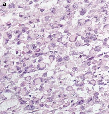
Variations in malignant melanoma     389

                                                                    the cells may give rise to pseudorosette-like structures in
                                                                    metastatic lesions (personal observation) (Figure 2).
                                                                    Monomorphism of the cells and scantiness of the
                                                                    cytoplasm are the two characteristic features of these
                                                                    tumours which separate them from conventional
                                                                    melanomas. This rare variant has been described both
                                                                    in children and adults and there is no sex predilection.
                                                                    In children they usually arise in the dermal component
                                                                    of large or giant congenital naevi.2,3,9 Some authors2
                                                                    described these neoplastic cells as lymphoblast-like
                                                                    whilst Reed9 used the term `melanoblastomas' for
                                                                    such tumours.
                                                                       Recently Barnhill et al.11,13 described ®ve cases of
                                                                    cutaneous small-cell melanoma in children, only one of
                                                                    which developed in a congenital naevus and the rest were
                                                                    de novo lesions. They were localized exclusively to the
                                                                    scalp, were thick tumours (mean Breslow thickness
                                                                    6.7 mm) and were associated with aggressive behaviour;
                                                                    all patients died of tumour. Two of these tumours
                                                                    exhibited papillomatous or verrucous epidermal surfaces
                                                                    and mimicked papillomatous naevi. In adults, primary
                                                                    de novo cutaneous melanomas of all types (nodular,
                                                                    super®cial spreading, lentigo maligna and acral lentigi-
                                                                    nous melanomas) and mucosal melanomas at various
                                                                    sites may be composed almost entirely of small cells but in
                                                                    some cases islands of small cells are present in a typical
Figure 1. A small-cell melanoma of skin. The cells contain hyper-   epithelioid or spindle cell melanoma (personal observa-
chromatic nuclei and scanty cytoplasm (H & E).                      tion). Interestingly, review of the existing literature
                                                                    suggests that small-cell melanomas are more commonly
                                                                    seen within the nasal cavity and paranasal sinuses.4,7,14
                                                                       Sometimes metastatic melanomas are composed
                                                                    entirely of small cells; Fitzgibbons et al. and Young and
                                                                    Scully described such tumours in the ovary.5,6 Attanoos
                                                                    and Grif®ths8 documented a case of metastatic small-
                                                                    cell melanoma in the stomach which mimicked a
                                                                    primary gastric lymphoma. Intraglandular melanoma
                                                                    cells in this case produced a picture reminiscent of
                                                                    lymphoepithelial lesions.
                                                                       In the skin these tumours may be mistaken either
                                                                    for benign naevocytic lesions when they are small,
                                                                    symmetrical and nested or for malignant small round
                                                                    cell tumours (such as Merkel cell carcinoma, metastatic
                                                                    small-cell carcinoma, lymphomatous/leukaemic depos-
                                                                    its, PNET, etc.) when they are diffuse, in®ltrative and
                                                                    ulcerated with an inconspicuous junctional component.
                                                                    In cases of mucosal or metastatic small-cell melanomas,
                                                                    other small-cell tumours such as rhabdomyosarcomas,
                                                                    small-cell squamous carcinomas and olfactory neuro-
                                                                    blastomas also enter the differential diagnosis.
                                                                       To differentiate a naevoid small-cell melanoma from a
                                                                    cellular compound naevus it is helpful to look for
Figure 2. Metastatic small-cell melanoma in the bone marrow show-   atypical junctional activity, pagetoid spread, poor nesting
ing perivascular pseudo-rosettes (H & E).                           of the dermal component, subtle nuclear atypia with
q 2000 Blackwell Science Ltd, Histopathology, 36, 387±402.
390 S S Banerjee & M Harris
Table 2. Morphological variations in melanomas. Some melanomas show a combination of these aberrant morphological features
Variation in size of cells
Large cells
Small cells
Variation in shape of cells
Polygonal
Spindle
Dendritic
Variation in cytoplasmic features
Clear cells
Signet-ring cells
Pseudolipoblastic cells
Rhabdoid cells
Balloon cells
Inclusions and phagocytosed material:
Hyaline
Erythrocytic
Neutrophilic
Tumour cells
Variation in nuclear features
Bi- and multi-nucleation
Lobation
Plasmacytoid features
Intranuclear cytoplasmic inclusions
Nuclear grooving and angulation
Variations in architecture
Fasciculation
Whorling
Nesting
Trabeculation
Pseudoglandular/pseudopapillary/pseudofollicular pattern
Pseudorosetting and angiocentric pattern
Myxoid change in the stroma
Stromal desmoplasia and neurotropism
Changes in stromal vascularity
Prominent vascular proliferation in the stroma
Herniation of tumour cells in vessels as seen in MPNST
Haemangiopericytomatous pattern
Proliferation of glomeruloid vascular structures (as in neuroendocrine or glial tumours)
Perivascular hyalinization
Angiomatoid/pseudoangiosarcomatous pattern
Associated granulomatous in¯ammation
Osteoclastic giant cells in the stroma
q 2000 Blackwell Science Ltd, Histopathology, 36, 387±402.Variations in malignant melanoma 391
Table 2. Continued
Differentiation towards nonmelanocytic elements
Schwannian
Fibroblastic/myo®broblastic
Smooth muscle
Rhabdomyoblastic
Osteocartilaginous
Ganglionic/ganglioneuroblastic
marked hyperchromatism and a coarse chromatin are likely to be S100 protein positive but many spindle
pattern, mitotic activity in the deeper part of the cell/desmoplastic melanomas are HMB-45 and Melan-A
lesion, apoptosis and lymphocytic in®ltration. HMB-45 negative. Ultrastructurally, melanosomes are hard to
positivity in the deeper part of the lesion is another ®nd in spindle cell desmoplastic melanomas and some of
worrying sign. Kossard and Wilkinson12 have suggested these tumours may, in fact, exhibit true Schwannian
that counting of AgNORs (nucleolar organizer regions) differentiation16 with cell processes and formation of
may help to differentiate a small-cell naevoid melanoma well developed basal lamina. The presence of an in-situ
from an ordinary naevus. The average number of melanoma in the overlying epidermis in a cutaneous
AgNORs per nucleus in 10 small-cell melanomas lesion, a previous history of excision of a melanoma,
studied in their series was 5.83 (SD 6 1.69) compared diffuse S100 protein positivity, the presence of melanin
to 2.71 (SD 6 0.50) for the 10 dermal naevi examined. pigment, prominent nucleoli and the presence of large
To differentiate a small-cell melanoma from other epithelioid or polygonal cells mingling with spindle cells
malignant small blue cell tumours one should rely on would favour a melanoma.
the demonstration of melanin pigment, positivity for Melanomas with large polygonal cells may mimic
speci®c melanocytic markers and negative staining for large-cell carcinomas, anaplastic large-cell lymphomas
cytokeratin, lymphoid markers, desmin and neuronal/ and some sarcomas such as epithelioid sarcoma,
neuroendocrine markers. epithelioid angiosarcoma, pleomorphic rhabdomyosar-
If necessary, electron microscopy should be performed coma and the epithelioid variant of MPNST.
to demonstrate melanosomes. Dendritic morphology of malignant cells is usually
seen in the in-situ component of acral lentiginous and
mucosal malignant melanomas but is uncommon in
Variation in shape of cells
invasive tumours.
Spindling of cells in malignant melanomas is a common
and well known occurrence which may lead to the
Variation in cytoplasmic features
misdiagnosis of a melanoma as a sarcoma1,15 or
sarcomatoid carcinoma. In our experience they are Both primary cutaneous/mucosal and metastatic
commonly misdiagnosed as MFH, MPNST or leiomyo- melanomas may contain glycogen-rich clear cells17±19
sarcoma. When dealing with a malignant spindle cell and may mimic clear-cell carcinomas or germ cell
tumour at any site, one should always keep the tumours. We have seen a case of glycogen-rich clear-cell
possibility of a melanoma in mind and should include metastatic amelanotic melanoma in the ovary of a
melanocyte markers in the immunopanel. Electron young woman which was initially misdiagnosed as a
microscopy may also be useful in such a situation. dysgerminoma. Rare examples of the clear-cell type of
However, differentiation of a spindle cell or desmoplastic spindle cell melanoma in the skin may mimic clear-cell
melanoma from a MPNST may in some cases be leiomyosarcoma or clear-cell dermato®broma. Melan-
extremely dif®cult if not impossible, as these two omas of soft parts (so-called clear-cell sarcomas) are well
tumours share many morphological features such as known for their glycogen-rich clear-cell component.20
fasciculation, whorling, nuclear palisading, dendritic Signet-ring cell melanoma4,21±26 is another rare
cell morphology, wavy nuclei, geographical areas of morphological variant which closely resembles a signet-
necrosis with palisading of cells around the necrotic ring cell carcinoma, a signet-ring lymphoma or a
foci, herniation of tumour cells into vascular lumina, liposarcoma. The signet-ring morphology is usually
and perivascular hyalinization. Melanoma and MPNST produced by accumulation of vimentin ®laments in the
q 2000 Blackwell Science Ltd, Histopathology, 36, 387±402.392 S S Banerjee & M Harris
cytoplasm which tends to displace the nucleus to the observation) (Figure 4a,b). The nucleolus remains
periphery and indent it to a semilunar shape (Figure prominent in these cells. Signet-ring change is usually
3a,b). Rarely, signet ring change occurs due to the seen in recurrent or metastatic lesions and is usually
formation of an intracytoplasmic vacuole (personal focal but may be diffuse. These cells do not contain
Figure 3. a, Signet-ring cell melanoma. Eosinophilic cytoplasmic
globules composed of vimentin ®laments displace nuclei to the
periphery (H & E). a, Tumour cell of signet-ring morphology with Figure 4. a, Signet-ring cell melanoma containing empty cytoplas-
crescentic nucleus indented by a mass of intermediate ®laments mic vacuoles. (H & E). b, Electron micrograph showing a mem-
containing entrapped mitochondria. Electron micrographs ´ 7000 brane-limited vacuole displacing nuclei to one side of the cell. The
(Micrograph courtesy of Brian Eyden, Christie Hospital, Manchester, vacuole bears irregular processes, which are not true glandular
UK). microvilli (´ 9000).
q 2000 Blackwell Science Ltd, Histopathology, 36, 387±402.Variations in malignant melanoma 393 mucin, fat or glycogen but may contain granular diastase-resistant PAS-positive material. In dif®cult cases the true nature of the tumour cells can be established by appropriate immuno-stains although one should be aware of the fact that a few cases of S100 protein-negative signet-ring cell melanomas have been recorded in the literature.22,23 Occasionally the presence of multiple empty vacuoles within the cytoplasm with scalloping of nucleus may impart a pseudolipoblastic appearance15 and, in a metastatic melanoma in the soft tissue with pleomorphic cells or myxoid stroma, this may lead to the erroneous diagnosis of a pleomorphic or myxoid liposarcoma. Rarely, a pseudolipoblastic appearance may be produced in a myxoid melanoma due to the presence of alcian blue positive mucinous material within the cytoplasm of the neoplastic melanocytes (personal observation). Rhabdoid change27±29 due to the presence of large glassy hyaline inclusions in the cytoplasm with eccentric round or irregular nuclei and prominent nucleoli is a rare phenomenon. The inclusions usually represent paranuclear whorls of intermediate ®laments and this variant is therefore pathogenetically related to the more common form of signet-ring melanomas. In some cases the rhabdoid appearance is caused by a collection of mitochondria and dilated rough endoplas- mic reticulum that contains microtubular arrays.28 A rhabdoid melanoma may be mistaken for a rhabdomyo- Figure 5. Metastatic balloon cell melanoma in a lymph node con- sarcoma or an extrarenal rhabdoid tumour. The taining pseudolipoblastic cells (H & E). immunohistochemical features of these tumours may be misleading. Bittesini et al.27 documented a case of tumour may be composed of these cells and the tumour rhabdoid metastatic melanoma in a lymph node in may simulate a clear-cell carcinoma, sebaceous carcin- which the cells exhibited vimentin, cytokeratin and oma or a lesion of foamy histiocytes such as a xanthoma desmin positivity but there were no ultrastructural or Rosai±Dorfman disease. Balloon cell melanomas are features of rhabdomyoblastic differentiation. Borek et S100 protein and HMB-45 positive and negative for al.29 have described three cases of primary rhabdoid cytokeratin. Ultrastructurally the cells show numerous melanoma of skin, one of which showed cytokeratin and intracytoplasmic vacuoles which probably represent focal a-SMA positivity. A diminution in the expression of enlarged and coalescent melanosomes. S100 protein and HMB-45 has been observed in some Melanoma cells may contain a variety of intracyto- cases of rhabdoid melanoma which may also lead to plasmic inclusions/phagocytosed material of which diagnostic dif®culty.28 hyaline eosinophilic PAS globules are the most In balloon cell melanomas30±35 the neoplastic cells common; similar globules are present in a variety of appear large, polygonal or round and contain abundant epithelial and mesenchymal tumours.36,37 The precise ®nely granular, reticulated or vacuolated cytoplasm nature of these globules is uncertain; they may either with delicate, focally disrupted cytoplasmic membranes. represent engulfed cellular material from necrotic or The nuclei may be central or eccentric and show a mild apoptotic cells or degenerated red cells.37 Phagocytosed to moderate degree of hyperchromatism and pleo- erythrocytes and neutrophils are rarely seen in mela- morphism with one or two eosinophilic nucleoli. Cells noma cells.38,39 One tumour cell phagocytosing with larger vacuoles and scalloped nuclei mimicking another is an extremely uncommon phenomenon.39 lipoblasts may be seen (Figure 5). The cytoplasm is devoid of glycogen and may show ®ne melanization. Rare Variations in nuclear features mitoses are seen. Usually balloon cell change occurs focally in a conventional melanoma but rarely the entire Bi- and multi-nucleation can be seen in pleomorphic q 2000 Blackwell Science Ltd, Histopathology, 36, 387±402.
394    S S Banerjee & M Harris

melanomas and Reed±Sternberg-like cells may occa-                  omas in the ovary5,6 and these tumours super®cially
sionally be present in these tumours. Melanomas with               resembled adult granulosa cell tumours. We have seen a
multiple and lobated nuclei may closely resemble                   recurrent cutaneous melanoma composed of centro-
anaplastic large-cell lymphomas, large-cell carcinomas             cyte-like cells with cleaved or grooved nuclei and
(particularly of renal, adrenocortical or pulmonary                inconspicuous nucleoli (Figure 7).
origin) or pleomorphic sarcomas. We have encountered
a few metastatic melanomas, one case of primary
                                                                   Variations in architecture
intranasal melanoma and one cutaneous melanoma,
resembling plasmacytoma. These tumours contained                   Fasciculation and whorling1,15 are seen in spindle cell
aggregates of plasmacytoid cells with eccentric nuclei             sarcomatoid melanomas. Nesting is also a common
showing coarse chromatin, inconspicuous nucleoli and               phenomenon but a trabecular pattern1 is only rarely
abundant cytoplasm with pale paranuclear zones                     seen. An amelanotic melanoma with a nested and
(Figure 6). The tumour cells in all cases were positive            trabecular pattern containing a relatively uniform
for melanocytic markers and negative for light chains.             population of epithelioid cells may be mistaken for a
In this context, one should bear in mind that the so-              neuroendocrine tumour. Melanomas are commonly
called plasma cell marker VS38 is not useful since this            Grimelius positive which may further compound the
antibody stains a large proportion of malignant                    problem, but stains for chromogranin and synaptophy-
melanomas.40                                                       sin, are usually negative in melanocytic tumours. A
   Intranuclear cytoplasmic pseudoinclusions are com-              nested melanoma with large cells may also mimic
monly seen in melanomas and, although non-speci®c,                 an alveolar soft part sarcoma. Rarely, primary and
are a useful diagnostic clue. Nuclear grooving and                 metastatic melanomas contain pseudoglandular or
angulation have been described in metastatic melan-                pseudopapillary structures mimicking adenocarcin-
                                                                   oma.1,4

Figure 6. Metastatic melanoma containing plasmacytoid cells (H &   Figure 7. Recurrent cutaneous melanoma containing centrocyte-like
E).                                                                cells (H & E).

                                                                                 q 2000 Blackwell Science Ltd, Histopathology, 36, 387±402.
Variations in malignant melanoma 395
Follicle-like structures have been described in ovarian blasts which is associated with marked collagenization
metastases of malignant melanomas.6 These structures of the intercellular matrix.
were either empty or contained thin pink ¯uid or Although the pathological features of DMM have
peripherally scalloped colloid-like material. Occasion- been well described in several excellent papers and
ally, melanomas contain pseudorosettes with malignant monographs on melanocytic lesions, these tumours are
cells radially arranged around blood vessels. Rarely, cells still frequently misdiagnosed because of their protean
of conventional melanomas and desmoplastic melan- and, at times, deceptively innocuous histological
omas concentrate around and invade walls of large appearances. Clinically, they usually present as pigmen-
blood vessels producing a striking angiocentric and ted or nonpigmented indurated areas, plaques or
angioinvasive picture. nodules in the dermis or subcutis of elderly white
individuals. Head and neck are common sites, although
they can occur at other anatomical sites including
Myxoid change
mucosae. Rarely, a conventional melanoma may recur
In 1986 Bhuta et al.41 described four cases of metastatic as a desmoplastic melanoma. There is no sex predilec-
malignant melanoma which exhibited prominent tion and the size may vary from 4 to 60 mm in
myxoid change in the stroma. Subsequently several maximum diameter. Histologically, the tumours are
additional cases of primary cutaneous, mucosal and composed of fascicles, sheets, nodules and strands of
metastatic myxoid melanomas have been documented mildly to markedly atypical spindle cells which in®ltrate
in the literature.4,42±48 The myxoid change in the the dermis and often extend to subcutis or deeper tissues
tumour may be focal or diffuse and in a primary with margins which are often ill-de®ned; satellite foci
cutaneous lesion this change may occur in any variant may be present.
of melanoma. Myxoid change is also seen in desmo- The nuclei of the neoplastic cells may be hyperchro-
plastic melanomas. Myxoid melanomas may exhibit matic or vesicular with inconspicuous or prominent
vague lobularity with rounded, pushing margins, nucleoli and variable amounts of cytoplasm. The cells
individual lobules being surrounded by delicate ®bro- may resemble ®broblasts, Schwann cells or smooth
vascular septa. Paraseptal and perivascular concentra- muscle cells. The concentration of cells may vary
tion of cells may occur in some cases. The neoplastic markedly and some tumours may be quite paucicellular.
cells in these tumours usually appear small, stellate or Multinucleated tumour cells and scattered epithelioid
spindled and are arranged singly or in cords. Rarely malignant cells may be present. Mitotic activity is
pseudoglandular structures are seen. Some tumours extremely variable but usually low. Atypical mitoses
contain large epithelioid cells. Scattered pseudolipoblas- have been noted in mitotically active tumours. Most
tic cells may be noted. Small pools of mucin may be DMM are entirely amelanotic. Fibroblastic proliferation
present producing a close resemblance to myxoid and collagenization of the stroma is prominent,
liposarcoma. Melanin pigment is usually scanty or although the degree may vary from area to area. In
absent. The stroma may be moderately vascular but a some cases there is also myxoid change. Elastosis of
chicken wire type of vascularity is never seen. Most dermal collagen is commonly observed. Curvilinear
cases are S100 protein, NKIC3 and HMB-45 positive. arrangements of fusiform cells, storiform patterns,
Myxoid melanomas should be distinguished from cellular whorls, osteogenesis and formation of small
other myxoid tumours, such as mucinous adenocarcin- pseudoglandular or pseudovascular structures are
omas, myxoid MFH/AFX, MFS, myxoid liposarcomas, additional but inconstant features. Neuroidal structures
nerve sheath myxomas, myxoid MPNST and extraske- resembling small peripheral nerves or Meissner corpus-
letal myxoid chondrosarcomas. cles may be seen. Small foci of necrosis may be present
and in®ltration by lymphocytes and formation of
lymphoid nodules is a common feature. The overlying
Stromal desmoplasia and neurotropism
epidermis is usually intact and may show an in-situ
Minor degrees of stromal desmoplasia may be observed melanoma, commonly of lentigo maligna type. Occa-
in some cases of conventional primary cutaneous, sionally DMM arise from an acral lentiginous, mucosal
mucosal or metastatic melanomas but this phenom- lentiginous or a super®cial spreading melanoma. In
enon is prominent and diffuse in so-called desmoplastic some cases no epidermal component is seen which is
malignant melanomas (DMM).16,49±64 In these probably due to complete regression of the in-situ
tumours the neoplastic cells themselves may undergo component or an origin from appendageal melanocytes.
®broblastic or myo®broblastic differentiation or may Many desmoplastic melanomas also exhibit neurotrop-
stimulate proliferation of stromal ®broblasts/myo®bro- ism with perineural and endoneural invasion of nerve
q 2000 Blackwell Science Ltd, Histopathology, 36, 387±402.396 S S Banerjee & M Harris
fascicles by malignant cells.52,56,58,62 These tumours
may grow along the involved nerves for a considerable
distance and eradication of the tumour becomes
dif®cult. In the head and neck region DMM may
extend into the brain along cranial nerves. Neurotrop-
ism may also be seen in rare cases of nondesmoplastic
epithelioid cell melanomas.
DMM with an inconspicuous or absent in-situ
component, mild atypia, low mitotic activity and
®broblastic, smooth muscle or Schwannian type features
may mimic scar tissue, benign desmoplastic and/or
neuronized naevi and benign dermal connective tissue
tumours (such as benign ®brous histiocytoma, neuro-
®broma, Schwannoma, leiomyoma or ®bromatosis).
On the other hand, a cellular, mitotically active and
cytologically atypical DMM may be mislabelled as a
MPNST, AFX, DFSP, MFS, leiomyosarcoma, spindle cell
angiosarcoma, Kaposi's sarcoma or spindle cell carcin-
oma. Careful scrutiny of the histological features and
immunohistochemical stains should help to make the
correct diagnosis. The vast majority of DMM are S100
protein positive (95±100% according to various series)
which may be diffuse or patchy.61±63 Some are NKIC3
positive but the majority are HMB-45 and Melan-A
negative. They are negative for cytokeratin, CD34 and
CD31. Smooth muscle actin (a-SMA) shows consistent
positivity indicating presence of large numbers of myo-
®broblastic cells.62,63 Ultrastructurally, premelanosomes Figure 8. Reticulin stain showing haemangiopericytomatous vascu-
are rarely found and the tumour cells may exhibit lar pattern in a metastatic melanoma.
Schwannian, perineural or transitional features.16,62
Fibroblastic and myo®broblastic cells are also present.53,55
extremely unusual vascular change which may be
observed in melanomas. Gaudin and Rosai65 provided a
Changes in stromal vascularity
detailed description of this ¯orid vascular lesion in
Mucosal and metastatic melanomas usually tend to neuronal and neuroendocrine tumours. Similar
show prominent stromal vascularity with formation of changes are also seen in high-grade astrocytomas. In
both well formed and poorly formed blood vessels. Foci a letter subsequently published in response to Gaudin
of stromal haemorrhage in these tumours are not and Rosai's article, it was pointed out that such vascular
uncommon. Occasionally, the neoplastic melanocytes proliferations are not entirely speci®c for neuronal/
invade walls of blood vessels and herniate into the neuroendocrine tumours and a case of metastatic
lumen a feature which has been described in MPNST. melanoma in the pleura containing glomeruloid vessels
Prominent haemangiopericytomatous vascular prolif- was illustrated.66 We have also encountered glomer-
eration4,15 has been seen in rare cases and we have uloid stromal vascular proliferation (Figure 9a,b) in two
noted this phenomenon (Figure 8) in a uterine and cases of melanoma, one of which was a primary palatal
mesenteric metastasis of a malignant melanoma from mucosal melanoma and the other was a metastatic
an unknown primary site. The tumour was initially lesion in the subcutis from a primary cutaneous
misdiagnosed as a haemangiopericytomatous leiomyo- melanoma. The changes were focal in both cases. It is
sarcoma but subsequent investigations showed lack of possible that some melanomas produce potent angio-
staining for a-SMA and muscle speci®c actin, occasional genic factors which may lead to vascular proliferation of
desmin positive cells, diffuse S100 protein and patchy different morphological types. Perivascular hyaliniza-
HMB-45 positivity. Electron microscopy revealed pre- tion has been described in cases of metastatic spindle
melanosomes within the tumour cells. Proliferation of cell melanoma in the lung mimicking Schwannian
glomeruloid blood vessels is another interesting but tumours.67
q 2000 Blackwell Science Ltd, Histopathology, 36, 387±402.Variations in malignant melanoma 397
Figure 9. a,b, Glomeruloid vascular proliferation in the stroma of a mucosal melanoma. a, H & E; b, immunostain for CD34 (streptavidin per-
oxidase complex technique).
Angiomatoid/pseudoangiosarcomatous tumours mimicked pseudovascular/pseudoangiosarco-
pattern matous squamous cell carcinoma69,70 and angiosarc-
oma, respectively.
Jain and Allen58 described endothelioid cells and small
pseudovascular lumina in DMM of skin. Adler et al.68
Associated granulomatous in¯ammation
documented a case of metastatic angiomatoid melan-
oma in the skin which contained large cavernous Two cases of sarcoid-like epithelioid granulomatous
pseudovascular spaces ®lled with erythrocytes which response to metastatic melanomas in the lymph nodes
initially raised the suspicion of angiosarcoma. The have been documented.71 In one case small granul-
neoplastic cells were, however, S100 protein and HMB- omata containing epithelioid histiocytes and Langhans-
45 positive and yielded negative results with endothelial type giant cells intermingled with melanoma cells whilst
cell markers. We have observed complex anastomosing in the other well formed sarcoid-like granulomas were
pseudo-vascular channels (Figure 10) lined by malig- seen adjacent to the metastatic tumour deposit. A few
nant melanocytic cells in two cases one in an oral cases of tuberculoid granulomas in lymph nodes
melanoma and the other in a metastatic melanoma in draining cutaneous melanomas have also been
the small intestine. Occasional cellular tufts were also recorded.72 Rarely, Langerhans cell granulomatosis is
seen projecting within the pseudovascular spaces. These seen in such lymph nodes.73,74
q 2000 Blackwell Science Ltd, Histopathology, 36, 387±402.398 S S Banerjee & M Harris
Differentiation towards non-melanocytic
elements
As mentioned earlier Schwannian, perineural, ®bro-
blastic and myo®broblastic differentiation is not uncom-
mon in DMM. Rarely melanomas may undergo smooth
muscle77 or rhabdomyoblastic differentiation.58,78
A small number of cases of osteocartilaginous
differentiation in cutaneous54,79±84 (particularly sub-
ungual) and mucosal melanomas85,86 have been
documented. These tumours may mimic osteo/chon-
drosarcomas, metaplastic carcinoma or carcinosarc-
oma. Ganglionic differentiation is an extremely
uncommon occurrence78,79 and recently a case of
Figure 10. Pseudoangiosarcomatous change in a malignant melan- metastatic melanoma showing ganglioneuroblastic
oma (H & E).
differention87 has been published. Within the nodal
deposit of a metastatic melanoma there were distinct
islands of ganglion cells and neuroblasts embedded in a
®brillar stroma. Occasional Homer±Wright rosettes
Osteoclastic giant cell reaction
were also present. Interestingly, subsequent metastatic
Osteoclast-like giant cells are sometimes seen in the deposits contained only neuroblastic elements and no
stroma of poorly differentiated carcinomas particu- ganglion cells were seen.
larly of breast, pancreas and thyroid. Similar giant cell
reaction has been recorded in metastatic melanomas in
Immunophenotype of melanomas
lymph nodes,75 bone,76 soft tissue15 and lung.67 In the
bone, a melanoma with osteoclast-like giant cells may Malignant melanomas are typically vimentin, NSE,
simulate a primary giant cell tumour. Elsewhere a S100 protein,88±90 NKIC391±93 and HMB-45 posi-
spindle cell melanoma with osteoclastic giant cells may tive.90,94±96 Recently anti-Melan-A97±102and anti-
mimic a giant cell variant of malignant ®brous tyrosinase103 antibodies have been introduced as two
histiocytoma or a giant cell rich anaplastic carcinoma. other melanocytic markers (Table 3). Of these, vimentin
Table 3.
Typical Aberrant Immunophenotypes of
melanomas
S100 protein Cytokeratin
HMB-45 Desmin
NK1C3 Neuro®lament protein (NFP)
Melan-A Glial ®brillary acidic protein (GFAP)
Anti-tyrosinase a-smooth muscle actin (SMA)
Vimentin Epithelial membrane antigen (EMA)
Neurone speci®c enolase Carcinoembryonic antigen (CEA)
KP1
VS38
Factor XIIIA
q 2000 Blackwell Science Ltd, Histopathology, 36, 387±402.Variations in malignant melanoma 399
and NSE are least useful as they stain a wide variety of processed paraf®n-embedded material. The positivity is
tumours. S100 protein and NKIC3 are very useful for more frequent with CAM5.2 and it is more commonly
screening purposes as both are highly sensitive markers seen in metastatic tumours. Usually only scattered cells
for melanocytic tumours but both lack speci®city. Of are positive in contrast to the diffuse staining pattern of
these two, the S100 protein is more widely used by epithelial tumours. The other epithelial markers which
practising histopathologists. After careful analysis of the may label melanoma cells are anti-CEA and EMA
published results, Bishop et al.104 found 94% and 95% antibodies.112±115 The CEA staining is usually seen
overall S100 protein positivity for primary and meta- with polyclonal antibodies and it has been demon-
static melanomas, respectively. Usually the morpholo- strated in conventional as well as balloon cell melan-
gically atypical tumours, such as signet-ring cell and omas.34 EMA positivity has been noted rather
rhabdoid melanomas, fail to express this antigen. infrequently and usually focally in conventional melan-
Fortunately, DMM, a diagnostically dif®cult group, omas but in one series62 43% of DMM exhibited EMA
express this antigen focally or diffusely in almost all positivity presumably due to perineurial differentiation.
cases. NKIC3 is slightly more speci®c than S100 protein In addition to vimentin and cytokeratin, melanoma cells
but it also stains many nonmelanocytic tumours such occasionally contain other intermediate ®laments.
as some carcinomas,92 neuroendocrine tumours,92 Desmin positivity occurs only rarely in melan-
neurothekeomas,105 gastrointestinal autonomic neuro- omas,77,116 although Reiman et al.55 found a high
nal tumours106 and any tumour in which the cells are rate of desmin positivity (seven of the nine DMM and all
rich in lysosomes such as granular cell tumours.107 10 conventional melanomas studied in their series were
HMB-45 is a fairly speci®c and reliable marker for positive), a ®nding which has not been substantiated by
melanomas and the rate of positivity for conventional others. NFP positivity has been noted in rare cases using
primary melanomas varied between 90 and 100% in fresh frozen tissue110 and we have observed occasional
different studies. The positivity rate drops to 80% in NFP-positive cells in a few nasal melanomas and in one
recurrent or metastatic melanomas and spindle cell case of primary cutaneous small-cell melanoma.
melanomas are less often positive. The staining in DMM We have also noted GFAP positivity in a few cases of
is disappointing as only a few of these tumours express DMM which was probably related to the Schwannian
this antigen. For detailed information on HMB-45 the differentiation in these tumours. a-SMA positivity is
readers are referred to the excellent review article of extremely uncommon in conventional cutaneous or
Bacchi et al.96 Anti-Melan-A (MART-I) monoclonal metastatic melanomas104 but many DMM show diffuse
antibody is a recently discovered melanocytic marker. It positivity for this marker.62 In our experience, spindle
stains both benign and malignant melanocytic tumours cell nondesmoplastic melanomas usually do not show
but, like HMB-45, it fails to label most cases of DMM. a-SMA positivity a feature which helps to differentiate
The anti-Melan-A antibody, A103, also stains adreno- these tumours from leiomyosarcomas. Melanomas may
cortical tumours, Leydig cell tumours, Sertoli cell also stain with the so-called plasma cell marker VS3840
tumours and granulosa cell tumours.102,108 Tumours and macrophage marker anti-CD68 (KPI).45,46,117,118
of `perivascular epithelioid cells' (angiomyolipomas, DMM may stain with the dermal dendrocytic marker
clear-cell sugar tumours and lymphangioleiomyomato- Factor XIIIa.62
sis) which stain with HMB-45, also exhibit positivity for
A103 and the other anti-Melan-A antibody M2±
Conclusion
7C10.109 Anti-tyrosinase antibody103 has also been
used as a marker for melanocytic lesions but as far as we In the review cited in our introduction Arnold
are aware, its sensitivity and speci®city have not been Levene1 concluded with some notes on diagnostic
adequately assessed. methods. At that time haematoxylin and eosin plus
pigment stains were virtually all the histological
approaches that were available. Today they remain
Immunophenotypic aberration
the bedrock of diagnosis but the advent of electron
In addition to their protean morphology, melanomas are microscopy and, more importantly, immunohisto-
also known to exhibit immunophenotypic aberrations chemistry have greatly expanded the diagnostic
which may create further diagnostic problems (Table 3). armamentarium. They have clari®ed some issues and
The most frequent of these is the cytokeratin expres- removed some diagnostic uncertainties but at the
sion.104,110±113 With the commonly used anticyto- same time have sewn other seeds of potential
keratin antibodies (CAM5.2, AE1/3, etc.) up to 10% confusion. The notoriety of melanoma referred to by
of melanomas show positive staining in routinely Levene persists.
q 2000 Blackwell Science Ltd, Histopathology, 36, 387±402.400 S S Banerjee & M Harris
Acknowledgements 20. Chung EB, Enzinger FM. Malignant melanoma of soft parts: a
reassessment of clear cell sarcoma. Am. J. Surg. Pathol. 1983; 7;
We are extremely grateful to Mrs E. Ryan for typing the 405±413.
manuscript and to Mr D. Edmondson for photographic 21. Sheibani K, Battifora H. Signet ring cell melanoma: a rare
morphologic variant of malignant melanoma. Am. J. Surg.
assistance. Pathol. 1988; 12; 28±34.
22. Bonetti F, Colombari R, Zamboni G, Chilosi M, Legnami F. Signet
References ring melanoma, S100 negative (letter). Am. J. Surg. Pathol.
1989; 13; 522±523.
1. Levene A. On the histological diagnosis and prognosis of 23. Al-Talib RK, Theaker JM. Signet-ring cell melanoma: light
malignant melanoma. J. Clin. Pathol. 1980; 33; 101±124. microscopic, immunohistochemical and ultrastructural fea-
2. Reed WB, Becker SW Sr, Becker SW Jr, Nickel WR. Giant tures. Histopathology 1991; 18; 572±575.
pigmented nevi, melanoma and leptomeningeal melanocytosis. 24. Eckert F, Baricevic B, Landthaler M, Schmid U. Metastatic signet-
Arch. Dermatol. 1965; 91; 100±119. ring cell melanoma in a patient with an unknown primary
3. Hendrickson MR, Ross JC. Neoplasms arising in congenital giant tumor. J. Am. Acad. Dermatol. 1992; 26; 870±875.
nevi: Morphologic study of seven cases and a review of the 25. Livolsi VA, Brooks JJ, Soslow R, Johnson BL, Elder DE. Signet cell
literature. Am. J. Surg. Pathol. 1981; 5; 109±135. melanocytic lesions. Mod. Pathol. 1992; 5; 515±520.
4. Nakhleh RE, Wick MR, Rocamora A, Swanson PE, Dehner LP. 26. Tsang WYW, Chan JKC, Chow LTC. Signet-ring cell melanoma
Morphologic diversity in malignant melanomas. Am. J. Clin. mimicking adenocarcinoma: a case report. Acta Cytol. 1993; 37;
Pathol. 1990; 93; 731±740. 559±562.
5. Fitzgibbons PL, Martin SE, Simmons TJ. Malignant melanoma 27. Bittesini L, Dei Tos AP, Fletcher CDM. Metastatic malignant
metastatic to the ovary. Am. J. Surg. Pathol. 1987; 11; 959±964. melanoma showing a rhabdoid phenotype: further evidence of a
6. Young RH, Scully RE. Malignant melanoma metastatic to the non-speci®c histological pattern. Histopathology 1992; 20; 167±
ovary: a clinico-pathologic analysis of 20 cases. Am. J. Surg. 170.
Pathol. 1991; 15; 849±860. 28. Chang ES, Wick MR, Swanson PE, Dehner LP. Metastatic
7. Franquemont DW, Mills SE. Sinonasal malignant melanoma. A malignant melanoma with `rhabdoid' features. Am. J. Clin.
clinicopathologic and immunohistochemical study of 14 cases. Pathol. 1994; 102; 426±431.
Am. J. Clin. Pathol. 1991; 96; 689±697. 29. Borek BT, McKee PH, Freeman JA, Maguire B, Brander WL,
8. Attanoos R, Grif®ths DFR. Metastatic small cell melanoma to the Calonje E. Primary malignant melanoma with rhabdoid
stomach mimicking primary gastric lymphoma. Histopathology features: a histologic and immunocytochemical study of three
1992; 21; 173±175. cases. Am. J. Dermatopathol. 1998; 20; 123±127.
9. Reed RJ. Giant congenital nevi: a conceptualization of patterns. 30. Gardner WA, Vazquez M. Balloon cell melanoma. Arch. Pathol.
J. Invest. Dermatol. 1993; 10 (Suppl.); 300±312. Lab. Med. 1970; 89; 470±472.
10. House NS, Fedok F, Maloney ME, Helm KF. Malignant melanoma 31. Ranchod M. Metastatic melanoma with balloon cell changes.
with clinical and histologic features of Merkel cell carcinoma. J. Cancer 1972; 30; 1006±1013.
Am. Acad. Dermatol. 1994; 31; 839±842. 32. Sondergaard K, Henschel A, Hou-Jensen K. Metastatic mela-
11. Barnhill RL, Flotte TJ, Fleischli M, Perez-Atayde A. Cutaneous noma with balloon cell changes: an electron microscopic study.
melanoma and atypical Spitz tumors in childhood. Cancer 1995; Ultrastruct. Pathol. 1980; 1; 357±360.
76; 1833±1845. 33. Peters MS, Su DWP. Balloon cell malignant melanoma. J. Am.
12. Kossard S, Wilkinson B. Nucleolar organizer regions and image Acad. Dermatol. 1985; 13; 351±354.
analysis nuclear morphometry of small cell (nevoid) melanoma. 34. Kao GF, Helwig EB, Graham JH. Balloon cell malignant
J. Cutan. Pathol. 1995; 22; 132±136. melanoma of the skin: a clinicopathologic study of 34 cases
13. Barnhill RL. Childhood melanoma. Sem. Diag. Pathol. 1998; 15; with histochemical, immunohistochemical and ultrastructural
189±194. observations. Cancer 1992; 69; 2942±2952.
14. Regauer S, Anderhuber W, Richtig E, Schachenreiter J, Ott A, 35. Mowat A, Reid R, MacKie R. Balloon cell metastatic melanoma:
Beham A. Primary mucosal melanomas of the nasal cavity and an important differential diagnosis of clear cell tumours.
paranasal sinuses. A clinicopathological analysis of 14 cases. Histopathology 1994; 24; 469±472.
APMIS 1998; 106; 403±410. 36. Al-Nafussi AI, Hughes DE, Williams AR. Hyaline globules in
15. Lodding P, Kindblom L, Angervall L. Metastases of malignant ovarian tumours. Histopathology 1993; 23; 563±566.
melanoma simulating soft tissue sarcoma. Virch. Archiv. A. 37. Severi B, Pasquinelli G, Martinelli GN, Zanetti GF, Santini D.
Pathol. Anat. 1990; 417; 377±388. Hyaline globules in ovarian tumours. Histopathology 1994; 25;
16. Dimaio SM, MacKay B, Smith JL Jr, Dickersin GR. Neurosarco- 299.
matous transformation in malignant melanoma: an ultrastruc- 38. Monteagudo C, Jorda E, Carda C, Illueca C, Peydro A, Llombart-
tural study. Cancer 1982; 50; 2345±2354. Bosch A. Erythrophagocytic tumour cells in melanoma and
17. Macak J, Krc I, Elleder M, Lukas Z. Clear cell melanoma of the squamous cell carcinoma of the skin. Histopathology 1997; 31;
skin with regressive changes. Histopathology 1991; 18; 276± 367±373.
277. 39. Mooi WJ, Krausz T. Cutaneous melanoma. In: Biopsy Pathology of
18. Mourad WA, MacKay B, Ordonez NG, Ro JY, Swanson DA. Clear Melanocytic Disorders. London: Chapman & Hall, 1992: 283±
cell melanoma of the bladder. Ultrastruct. Pathol. 1993; 17; 287.
463±468. 40. Shanks JH, Banerjee SS. VS38 immunostaining in melanocytic
19. Ehara H, Takahashi Y, Saitoh A, Kawada Y, Shimokawa K, lesions. J. Clin. Pathol. 1996; 49; 205±207.
Kanemura T. Clear cell melanoma of the renal pelvis presenting 41. Bhuta S, Mirra JM, Cochran AJ. Myxoid malignant melanoma: a
as a primary tumour. J. Urol. 1997; 157; 634. previously undescribed histologic pattern noted in metastatic
q 2000 Blackwell Science Ltd, Histopathology, 36, 387±402.Variations in malignant melanoma 401
lesions and a report of four cases. Am. J. Surg. Pathol. 1986; 10; neural and neuroendocrine neoplasms. Am. J. Surg. Pathol.
203±211. 1995; 19; 642±652.
42. Nottingham JF, Slater DN. Malignant melanoma: a new mimic of 66. Kepes JJ. Vascular proliferation (letter). Am. J. Surg. Pathol. 1996;
colloid adenocarcinoma. Histopathology 1988; 13; 576±578. 20; 384±386.
43. Garcia-Caballero T, Fraga M, Antunez JR, Perez-Becerra E, 67. Suster S, Moran CA. Unusual manifestations of metastatic
Beiras A, Forteza J. Myxoid metastatic melanoma. Histopathology tumors to the lungs. Sem. Diagn. Pathol. 1995; 12; 193±206.
1991; 18; 371±373. 68. Adler MJ, Beckstead J, White CR. Angiomatoid melanoma: a case
44. Sarode VR, Joshi K, Ravichandran P, Das R. Myxoid variant of of metastatic melanoma mimicking a vascular malignancy. Am.
primary cutaneous malignant melanoma. Histopathology 1992; J. Dermatopathol. 1997; 19; 606±609.
20; 186±187. 69. Banerjee SS, Eyden BP, Wells S, McWilliam LJ, Harris M.
45. Chetty R, Slavin JL, Pitson GA, Dowling JP. Melanoma Pseudoangiosarcomatous carcinoma: a clinicopathological
botryoides: a distinctive myxoid pattern of sino-nasal malignant study of seven cases. Histopathology 1992; 21; 13±23.
melanoma. Histopathology 1994; 24; 377±379. 70. Nappi O, Wick MR, Pettinato G, Ghiselli RW, Swanson PE.
46. McCluggage WG, Shah V, Toner PG. Primary cutaneous myxoid Pseudovascular adenoid squamous cell carcinoma of the skin: a
malignant melanoma. Histopathology 1996; 28; 179±182. neoplasm that may be mistaken for angiosarcoma. Am. J. Surg.
47. Collina G, Losi L, Taccagni GL, Mairona A. Myxoid metastases of Pathol. 1992; 16; 429±438.
melanoma: Report of three cases and review of the literature. 71. Coyne JD, Banerjee SS, Menasce LP, Mene A. Granulomatous
Am. J. Dermatopathol. 1997; 19; 52±57. lymphadenitis associated with metastatic malignant melanoma.
48. Hitchcock MG, White WL. Malicious masquerade: Myxoid Histopathology 1996; 28; 470±472.
melanoma. Sem. Diagn. Pathol. 1998; 15; 195±202. 72. de Dulants F, Armijo-Moreno M, Camacho-Martinez F. Tuber-
49. Conley J, Lattes R, Orr W. Desmoplastic melanoma (a rare culoid granulomata in regional lymph nodes of malignant
variant of spindle cell melanoma). Cancer 1971; 28; 914±936. cutaneous melanoma. Dermatologica 1974; 148; 228±232.
50. Frolow GR, Shapiro L, Brownstein MH. Desmoplastic malignant 73. Richmond I, Eyden BP, Banerjee SS. Intranodal Langerhans' cell
melanoma. Arch. Dermatol. 1975; 111; 753±754. histiocytosis associated with malignant melanoma. Histopathol-
51. Labreque PG, Hu C, Winkelman RK. On the nature of ogy 1995; 26; 380±382.
desmoplastic melanoma. Cancer 1976; 38; 1205±1213. 74. Roufosse C, Lespagnard L, Sales F, Bron D, Dargent J.
52. Reed RJ, Leonard DD. Neurotropic melanoma (a variant of Langerhans' cell histiocytosis associated with simultaneous
desmoplastic melanoma). Am. J. Surg. Pathol. 1979; 3; 301± lymphocytic predominance Hodgkin's disease and malignant
311. melanoma (letter). Hum. Pathol. 1998; 29; 200±201.
53. From L, Hanna W, Kahn HJ, Gross J, Marks A, Baumal R. Origin 75. Denton KJ, Stretch J, Athanasou N. Osteoclast-like giant cells in
of desmoplasia in desmoplastic malignant melanoma. Hum. malignant melanoma. Histopathology 1992; 20; 179±181.
Pathol. 1983; 4; 1072±1080. 76. Daroca PJ, Reed RJ, Martin PC. Metastatic amelanotic mela-
54. Moreno A, Lamarca J, Martinez R, Guix M. Osteoid and bone noma simulating giant cell tumor of bone. Hum. Pathol. 1990;
formation in desmoplastic malignant melanoma. J. Cut. Pathol. 21; 978±980.
1986; 13; 128±134. 77. Banerjee SS, Bishop PW, Nicholson CM, Eyden BP. Malignant
55. Reiman H, Goellner JR, Woods JE, Mixter RC. Desmoplastic melanoma showing smooth muscle differentiation. J. Clin.
melanoma of the head and neck. Cancer 1987; 60; 2269±2274. Pathol. 1996; 49; 950±951.
56. Kossard S, Doherty E, Murray E. Neurotropic melanoma: a 78. Slaughter JC, Hardman JM, Kempe LG, Earle KM. Neurocuta-
variant of desmoplastic melanoma. Arch. Dermatol. 1987; 123; neous melanosis and leptomeningeal melanomatosis in chil-
907±912. dren. Arch. Pathol. 1969; 88; 298±304.
57. Egbert B, Kempson R, Sagebiel R. Desmoplastic malignant 79. Wahlstrom T, Saxen L. Malignant skin tumors of neural crest
melanoma: a clinicohistopathologic study of 25 cases. Cancer origin. Cancer 1976; 38; 2022±2026.
1988; 62; 2033±2041. 80. Grunwald MH, Rothem A, Feuerman EJ. Metastatic malignant
58. Jain S, Allen PW. Desmoplastic malignant melanoma and its melanoma with cartilaginous metaplasia. Dermatologica 1985;
variants: a study of 45 cases. Am. J. Surg. Pathol. 1989; 13; 358± 170; 249±252.
373. 81. Brisigotti M, Moreno A, Llistosella E, Prat J. Malignant
59. Bruijn JA, Mihm MC Jr, Barnhill RL. Desmoplastic melanoma. melanoma with osteocartilaginous differentiation. Surg. Pathol.
Histopathology 1992; 20; 197±205. 1989; 2; 73±78.
60. Anstey A, McKee P, Wilson-Jones E. Desmoplastic malignant 82. Pellegrini AE, Scalamogna PA. Malignant melanoma with
melanoma: a clinicopathologic study of 25 cases. Br. J. Dermatol. osteoid formation. Am. J. Dermatopathol. 1990; 12; 607±611.
1993; 129; 359±371. 83. Nakagawa H, Imakado S, Nogita T, Ishibahi Y. Osteosarcoma-
61. Anstey A, Cerio R, Ramnarain N. Desmoplastic malignant tous changes in malignant melanoma: immunohistochemical
melanoma, an immunocytochemical study of 25 cases. Am. J. and ultrastructural studies of a case. Am. J. Dermatopathol. 1990;
Dermatopathol. 1994; 16; 14±22. 12; 162±168.
62. Carlson JA, Dickersin GR, Sober AJ, Barnhill RL. Desmoplastic 84. Lucas DR, Tazelaar HD, Unni KK et al. Osteogenic melanoma: a
neurotropic melanoma. Cancer 1995; 75; 478±494. rare variant of malignant melanoma. Am. J. Surg. Pathol. 1993;
63. Longacre TA, Egbert BM, Rouse RV. Desmoplastic and spindle 17; 400±409.
cell malignant melanoma. An immunohistochemical study. Am. 85. Hoorweg JJ, Loftus BM, Hilgers FJM. Osteoid and bone formation
J. Surg. Pathol. 1996; 20; 1489±1500. in a nasal mucosal melanoma and its metastasis. Histopathology
64. Wharton JM, Carlson JA, Mihm MC (Jr). Desmoplastic malignant 1997; 31; 465±468.
melanoma: Diagnosis of early clinical lesions. Hum. Pathol. 86. Banerjee SS, Coyne JD, Menasce LP, Lobo CJ, Hirsch PJ.
1999; 33; 537±542. Diagnostic lessons of mucosal melanoma with osteocartilaginous
65. Gaudin PB, Rosai J. Florid vascular proliferation associated with differentiation. Histopathology 1998; 33; 255±260.
q 2000 Blackwell Science Ltd, Histopathology, 36, 387±402.402 S S Banerjee & M Harris
87. Banerjee SS, Menasce LP, Eyden BP, Brain AN. Malignant 103. Takimoto H, Suzuki S, Masui S et al. MAT-1, a monoclonal
melanoma showing ganglioneuroblastic differentiation: Report antibody that speci®cally recognizes human tyrosinase. J. Invest.
of a unique case. Am. J. Surg. Pathol. 1999; 23; 582±588. Dermatol. 1995; 105; 764±768.
88. Cochran AJ, Wen D-R, Herschman HR, Gaynor RB. Detection of 104. Bishop PW, Menasce LP, Yates AJ, Win NA, Banerjee SS. An
S100 protein as an aid to the identi®cation of melanocytic immunophenotypic survey of malignant melanomas. Histo-
tumors. Int. J. Cancer 1982; 30; 295±297. pathology 1993; 23; 159±166.
89. Wick MR, Swanson PE, De Manivel JC. Immunohistochemical 105. Calonje E, Wilson-Jones E, Smith NP, Fletcher CDM. Cellular
®ndings in tumors of the skin. In: De Lellis RA, eds. Advances in `neurothekeoma': an epithelioid variant of pilar leiomyoma?
Immunohistochemistry. New York: Raven, 1988: 395±429. Morphological and immunohistochemical analysis of a series.
90. Ordonez NG, Xiaolong JI, Hickey RC. Comparison of HMB-45 Histopathology 1992; 20; 397±404.
monoclonal antibody and S100 protein in the immunohisto- 106. Shanks JH, Harris M, Banerjee SS, Eyden BP. Gastrointestinal
chemical diagnosis of melanoma. Am. J. Clin. Pathol. 1988; 90; autonomic nerve tumours: a report of nine cases. Histopathology
385±390. 1996; 29; 111±121.
91. MacKie RM, Campbell I, Turbitt ML. Use of NKIC3 monoclonal 107. Buley ID, Gatter KC, Kelly PMA, Heryet A, Millard PR. Granular
antibody in the assessment of benign and malignant melano- cell tumours revisited. An immunohistological and ultrastruc-
cytic lesions. J. Clin. Pathol. 1984; 37; 367±372. tural study. Histopathology 1988; 12; 263±274.
92. Vennegoor C, Calafat J, Hageman Ph et al. Biochemical 108. Busam KJ, Iversen K, Coplan KA et al. Immunoreactivity for
characterization and cellular localization of a formalin A103, an antibody to Melan-A (MART-1), in adrenocortical
resistant melanoma-associated antigen reacting with mono- and other steroid tumors. Am. J. Surg. Pathol. 1998; 22; 57±
clonal antibody NKI/C-3. Int. J. Cancer 1985; 35; 287±295. 63.
93. Thomson W, MacKie RM. Comparison of ®ve antimelanoma 109. Fetsch PA, Fetsch JF, Marincola FM, Travis W, Batts KP, Abati A.
antibodies for identi®cation of melanocytic cells on tissue Comparison of melanoma antigen recognized by T cells (MART-1)
sections in rountine dermatopathology. J. Am. Acad. Dermatol. to HMB-45: additional evidence to support a common lineage
1989; 21; 1280±1284. for angiomyolipoma, lymphangiomyomatosis, and clear cell
94. Gown AM, Vogel AM, Hoak D, Gough F, McNutt MA. sugar tumor. Mod. Pathol. 1998; 11; 699±703.
Monoclonal antibodies speci®c for melanocytic tumors distin- 110. Miettinen M, Franssila K. Immunohistochemical spectrum of
guish subpopulations of melanocytes. Am. J. Pathol. 1986; 123; malignant melanoma: the common presence of keratins. Lab.
195±203. Invest. 1989; 61; 623±628.
95. Wick MR, Swanson PE, Rocamora A. Rocognition of malignant 111. Zarbo RJ, Gown AM, Nagle RB, Visscher DW, Crissman JD.
melanoma by monoclonal antibody HMB-45. An immunohis- Anomalous cytokeratin expression in malignant melanoma:
tochemical study of 200 paraf®n-embedded cutaneous tumors. one-and two-dimensional western blot analysis and
J. Cutan. Pathol. 1988; 15; 201±207. immunohistochemical survey of 100 melanomas. Mod. Pathol.
96. Bacchi CE, Bonetti F, Pea M, Martignoni G, Gown AM. HMB-45: 1990; 3; 494±501.
a review. Appl. Immunohistochem. 1996; 4; 73±85. 112. Ben-Izhak O, Stark P, Levy R, Bergman R, Lichtig C. Epithelial
97. Chen YT, Stockert E, Jungbluth A et al. Serologic analysis of markers in malignant melanoma. A study of primary lesions and
Melan-A (MART-1), a melanocyte speci®c protein homoge- their metastases. Am. J. Dermatopathol. 1994; 16; 241±246.
neously expressed in human melanomas. Proc. Natl. Acad. Sci. 113. Ben-Izhak O, Levy R, Weill S et al. Anorectal malignant
1996; 93; 5915±5919. melanoma. A clinicopathologic study, including immunohisto-
98. Fetsch PA, Cormier J, Hijazi YM. Immunocytochemical detection chemistry and DNA ¯ow cytometry. Cancer 1997; 79; 18±25.
of MART-1 in fresh and paraf®n embedded malignant melan- 114. Selby WL, Nance KV, Park HK. CEA immunoreactivity in
omas. J. Immunother. 1997; 20; 60±64. metastatic malignant melanoma. Mod. Pathol. 1992; 5; 415±
99. Jungbluth AA, Busam KJ, Gerald WL et al. A103: an anti-melan A 419.
monoclonal antibody for the detection of malignant melanoma 115. Sanders DS, Evans AT, Allen CA et al. Classi®cation of CEA-
in paraf®n-embedded tissues. Am. J. Surg. Pathol. 1998; 22; related positivity in primary and metastatic malignant mela-
595±602. noma. J. Pathol. 1994; 172; 343±348.
100. Busam KJ, Chen Y-T, Old LJ et al. Expression of Melan-A (MART-1) 116. Truong LD, Rangdaeng S, Cagle P, Ro JY, Hawkins H, Font
in benign melanocytic naevi and primary cutaneous malignant RL. The diagnostic utility of desmin: a study of 584 cases
melanoma. Am. J. Surg. Pathol. 1998; 22; 976±982. and review of the literature. Am. J. Clin. Pathol. 1990; 93;
101. Blessing K, Sanders DSA, Grant JJH. Comparison of immuno- 305±314.
histochemical staining of the novel antibody Melan-A with S100 117. Facchetti F, Bertalot G, Grigolato PG. KP1 (CD68) staining of
protein and HMB-45 in malignant melanoma and melanoma malignant melanomas. Histopathology 1991; 19; 141±145.
variants. Histopathology 1998; 32; 139±146. 118. Gloghini A, Rizzo A, Zanette I et al. KP1/CD68 expression in
102. Busam KJ, Jungbluth AA. Melan-A, a new melanocytic malignant neoplasms including lymphomas, sarcomas and
differentiation marker. Adv. Anat. Pathol. 1999; 6; 12±18. carcinomas. Am. J. Clin. Pathol. 1995; 103; 425±431.
q 2000 Blackwell Science Ltd, Histopathology, 36, 387±402.You can also read