Radioprotective effects of centipedegrass extract on NIH 3T3 fibroblasts via anti oxidative activity
←
→
Page content transcription
If your browser does not render page correctly, please read the page content below
EXPERIMENTAL AND THERAPEUTIC MEDICINE 21: 419, 2021

                  Radioprotective effects of centipedegrass extract on
                    NIH‑3T3 fibroblasts via anti‑oxidative activity
                   SEONG HEE KANG1,2*, DONG‑HO BAK1*, SEUNG SIK LEE1,3, HYOUNG‑WOO BAI1,3,
                                 BYUNG YEOUP CHUNG1 and BO SUN KANG2

                          1
                     Research Division for Biotechnology, Advanced Radiation Technology Institute (ARTI),
        Korea Atomic Energy Research Institute (KAERI), Jeongeup, Jeollabuk 56212; 2Department of Radiological Science,
                Konyang University, Daejeon 35365; 3Radiation Biotechnology and Applied Radioisotope Science,
                        University of Science and Technology (UST), Daejeon 34113, Republic of Korea

                                       Received January 14, 2020; Accepted February 10, 2021

                                                       DOI: 10.3892/etm.2021.9863

Abstract. Centipedegrass originates from China and South                 and JNK) were measured in vitro. The results demonstrated
America, and has been reported to contain several C‑glycosyl             that when 3T3 fibroblasts pretreated with CGE were subjected
f lavones and phenolic compounds, including maysin                       to H2O2‑induced cell damage, their viability was significantly
and luteolin. The present study aimed to investigate the                 decreased. Additionally, CGE pretreatment decreased ROS
radioprotective activity of centipedegrass extract (CGE) in radi‑        levels and the protein expression levels of cleaved PARP upon
ation exposed‑fibroblasts and to assess the affected molecular           H2O2 treatment, indicating that CGE induced cytoprotective
pathway. The radioprotective effects of CGE were determined              effects against H 2O2 ‑induced oxidative stress. Moreover,
in NIH‑3T3 cells using Cell Counting Kit‑8 and morphological             significant protective effects of CGE against intracellular
changes were observed. Reactive oxygen species (ROS) levels              ROS, induced upon exposure to ionizing radiation (IR), were
and the apoptotic profile of NIH‑3T3 cells were also measured.           observed. The protective effects of CGE pretreatment were
The expression levels of B‑cell lymphoma‑2 (Bcl‑2) family                also determined by morphological observation of NIH‑3T3
proteins [Bcl‑2, Bcl‑2 like protein 4 (Bax), Bcl‑2‑associated            cells following exposure to IR. CGE pretreatment increased
death promoter (Bad), caspase‑3, poly(ADP‑ribose) poly‑                  the expression levels of anti‑apoptotic signals (Bcl‑2, p‑BAD)
merase (PARP)], AKT and MAPK family proteins (ERK, p38                   and decreased the levels of pro‑apoptotic signals (Bax,
                                                                         Bad), and led to cleavage of PARP and caspase‑3 proteins.
                                                                         Additionally, in cells pretreated with CGE, the phosphoryla‑
                                                                         tion of AKT and ERK was increased and that of p38 and JNK
                                                                         was decreased compared with in cells subjected only to IR.
Correspondence to: Dr Bo Sun Kang, Department of Radiological
Science, Konyang University, 158 Gwanjeodong‑ro, Daejeon 35365,
                                                                         These results indicated that CGE may act as a radioprotector
Republic of Korea                                                        due to its anti‑oxidative activity, restoring cell homeostasis and
E‑mail: bskang@konyang.ac.kr                                             redox balance in radiation‑exposed fibroblast cells. Therefore,
                                                                         it could be suggested that CGE may be an effective candidate
Dr Byung Yeoup Chung, Research Division for Biotechnology,
                                                                         in the treatment of oxidative stress‑related diseases and in
Advanced Radiation Technology Institute (ARTI), Korea Atomic
Energy Research Institute (KAERI), 29 Geumgu‑gil, Jeongeup,
                                                                         radioprotection.
Jeollabuk 56212, Republic of Korea
E‑mail: bychung@kaeri.re.kr                                              Introduction

*
    Contributed equally                                                  Centipedegrass belongs to the genus Eremochloa (Poaceae),
                                                                         which includes eight species that inhabit China and Southeast
Abbreviations: Bcl‑2, B‑cell lymphoma‑2; Bax, Bcl‑2 like protein 4;      Asia, and is one of the most popular grasses in South
Bad, Bcl‑2‑associated death promoter; CGE, centipedegrass                America (1). It contains C‑glycosyl flavones and phenolic
extract;    H2DCFDA,         2,7‑dichlorodihydrofluorescein‑diacetate;   constituents as its common structural skeleton, which is
DHE, dihydroethidium; DMSO, dimethyl sulfoxide; DMEM,                    biologically active (2). In a previous study, we isolated and
Dulbecco's modified Eagle's medium; EA, ethyl acetate; FBS,
                                                                         analyzed the extract from centipedegrass, and found that these
fetal bovine serum; HRP, horseradish peroxidase; IR, ionizing
radiation; ROS, reactive oxygen species; MeOH, methanol; PARP,
                                                                         components included maysin, and maysin derivatives such
poly(ADP‑ribose) polymerase; RISR, radiation‑induced skin                as luteoin‑6‑C‑boivinopyranose, luteolin, Isoorientin, rham‑
reaction; RT, radiation therapy                                          nosylisoorientin, and derhamnosylmaysin (3). Particularly,
                                                                         centipedegrass extract (CGE), a flavonoid‑rich chemical
Key words: centipedegrass, fibroblasts, radioprotection, ROS             compound extracted from centipedegrass, has been reported to
scavenging, apoptosis                                                    demonstrate free radical‑scavenging activity in vitro biochem‑
                                                                         ical assays using DPPH‑radical scavenging activity (4). It was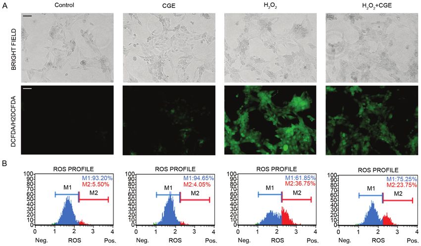
2                                         KANG et al: THE RADIOPROTECTIVE ROLE OF CGE

also reported that maysin (a C‑glycoside flavone from centipe‑       Additionally, persistent oxidative stress during RT can lead
degrass) and its precursor chemical components could exhibit         to severe cellular damage and irreversible tissue conditions,
anti‑oxidative activity via DPPH radical scavenging in vitro         which makes tissue regeneration impossible (17). This consid‑
antioxidant model (5), and protect SK‑N‑MC cell lines against        eration marked the beginning of the active development of a
inhibition of H2O2‑induced apoptotic cell death (6). Despite         radioprotector for the prevention and treatment of IR‑induced
these health benefits of maysin and flavonoid derivatives, its       free radical pathology with the use of antioxidant agents (18).
cytoprotective effects against ionizing radiation (IR)‑induced       Studies involving the use of vitamins, amino acids, animal‑
cell death in fibroblasts and the underlying mechanisms have         and plant‑based agents containing antioxidant enzymes are
not been elucidated yet.                                             considered to be highly significant but less developed in the
    Cancer remains a big challenge in global healthcare              field of radiobiology. Considering the lack of efficient radio‑
because it is one of the most rampant causes of death. Radiation     protective agents made of natural raw materials, many studies
therapy (RT) is one of the most important treatment strategies       have been undertaken with the purpose of finding radiopro‑
used in the treatment of more than two thirds of cancer cases        tective agents (19,20).
worldwide (7). Advances in technology over the last decades              In this study, CGE, which contains flavonoid derivatives,
have prompted the development of 3‑dimensional conformal             was evaluated for its radioprotective effects against IR‑induced
RT techniques, including intensity‑modulated radiation               cell damage in NIH‑3T3 fibroblast cells.
therapy and stereotactic body radiation therapy (8). In addition
to these advanced medical technologies, therapeutic strategies       Materials and methods
in radiation oncology have grown significantly with advances
in physiology, immunology, and molecular biology, allowing           Preparation of CGE. CGE was prepared using the method
us to explore the treatment outcome from a better, all‑inclusive     described in our previous report (21). Briefly, seeds of centipe‑
perspective, and perform radiotherapy more efficiently.              degrass imported from the Fukukaen Nursery (Blu Co. Ltd.)
    However, long‑term stimulation of IR in human tissues            were cultivated at the Korea Atomic Energy Research
unavoidably leads to the development of free radical                 Institute in 2016 (KAERI). The leaves of the centipedegrass
pathology. Generally, IR affects the body of patients indi‑          were harvested and stored at ‑80˚C until use. Dry leaves of
rectly; for example, upon interaction with water molecules           centipede (5 kg) were crushed in a Wiley mill (Indian Weiber)
in cells exposed to radiation, large amounts of free radicals        and filtered through a sieve of pore size 420 µm. The final
and reactive oxygen species (ROS) are produced, which                sample of ground leaves (1 kg) was extracted thrice using 80%
oxidize cellular components, causing cellular damage (9). The        methanol (MeOH, 100 liters; Merck) for 24 h with constant
cells that are exposed to radiation respond via generation of        shaking at ambient temperature in the dark. The extracts
antioxidant enzymes depending on the degree of exposure,             were filtered using a No. 2 filter paper (Advantech) and
followed which free radical‑induced damage to the cellular           concentrated in vacuum. MeOH extracts were fractionated
structure is minimized or eliminated (10). However, when             successively using n‑hexane and ethyl acetate (EA). The layer
ROS are produced due to exposure of the body to radiation and        separated using EA was concentrated in vacuum and the
excessive amounts of free radicals, they cannot be completely        dried compound was dissolved in MeOH. The active MeOH
removed by intrinsic antioxidative enzymes.                          extracts were diluted with 20% MeOH and chromatographed
    Radiation‑induced skin reaction (RISR) is one of the main        on a Toyopearl HW‑40C resin (Tosoh Corp.) column using
side effects of radiotherapy, and more than 95% of patients          70% MeOH (elution volume, 700 ml). Fractions were evapo‑
undergoing RT experience tissue damage (11). Moreover, acute         rated and lyophilized. The dried extract was diluted in dimethyl
RISR often has a big impact on the progress of RT due to limita‑     sulfoxide (DMSO) to carry out further experiments. Lastly, to
tion of the total therapeutic dose or breaks in radiotherapy (12).   confirm the active substances content, maysin, and maysin
Radiation‑induced oxidative stress is known to be the main           derivatives (the active ingredient of CGE) were confirmed by
cause of RISR (13). Oxidative stress induced by IR results in        high‑performance liquid chromatography/mass (HPLC‑MS)
damages of DNA, lipids, and proteins. The harmful effects            analysis as previously described method (3).
stimulate early transcription factors and molecular signals
leading to cellular damage. As a result, it causes damage to the     Chemicals and reagents. All the chemicals and reagents were
skin tissue (14). Thus, in radiotherapy, IR‑induced oxidative        used without further purification. A cell counting kit‑8 was
stress is an important variable before and after IR, and it is       purchased from Sigma‑Aldrich; Merck KGaA. Anti‑AKT
essential to scavenge the IR‑induced free radicals or ROS to         (#4691), anti‑p‑AKT (#4060), anti‑ERK (#4695), anti‑p‑ERK
reduce/mitigate the damage caused by radiation.                      (#4377), anti‑p38 (#9212), anti‑p‑p38 (#9215), anti‑JNK (#9252),
    Fibroblasts, the main type of cells constituting the dermal      anti‑p‑JNK (#9251), anti‑caspase‑3 (#9662), anti‑cleaved
skin, play important roles in the development of healthy skin        caspase‑3 (#9664), anti‑Bcl‑2 (#2870), anti‑Bax (#5023),
by producing the extracellular matrix and collagen. The              anti‑Bad (#9292), anti‑p‑Bad (#9291), anti‑poly(ADP‑ribose)
occurrence of damaged fibroblasts, resulting in the presence         polymerase (PARP) (#9532), anti‑cleaved PARP (#9541),
of low levels of extracellular matrix proteins, is the result of     anti‑GAPDH (#2118), and anti‑ α ‑tubulin (#2144) were
skin aging, and is consequently responsible for the forma‑           purchased from Cell Signaling Technology, Inc. Dulbecco's
tion of wrinkles (15). IR‑induced oxidative stress has been          modified Eagle's medium (DMEM), penicillin/strepto‑
reported to cause damage with ROS inducing apoptosis in              mycin (P/S), and fetal bovine serum (FBS) were purchased
a variety of cells, including fibroblasts and keratinocytes,         from Lonza. Annexin V and oxidative stress kits were
thereby reducing cell numbers and regenerative capacity (16).        purchased from Millipore.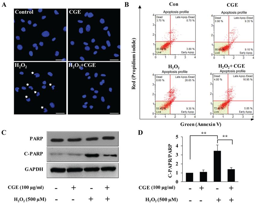
EXPERIMENTAL AND THERAPEUTIC MEDICINE 21: 419, 2021 3
Cell culture. The NIH‑3T3 cell line (mouse, fibroblast) was expressed as the percentage of the live, early/late apoptotic,
obtained from the Korean Cell Line Bank (Seoul, Korea). and dead cells, which was determined by the Muse analysis
The cells were cultured under sterile conditions at 37˚C in a software (Muse 1.1.2; EMD Millipore).
humid environment containing 5% CO2. The culture medium
consisted of DMEM supplemented with FBS (10%), glutamine gamma‑ray irradiation. gamma‑ray irradiation was performed
(4 mM), and penicillin/streptomycin (1%). at room temperature at the dose rate of 1 Gy/min using a Cs137
gamma cell (MDS Nordion). The cells were directly irradiated
Morphological analysis and cell viability. The morphology in the flasks. After irradiation, they were incubated at 37˚C
of NIH‑3T3 cells was monitored using an Olympus IX71 and 5% CO2 until they could be harvested for direct experi‑
fluorescence microscope (Olympus Corp.). In order to deter‑ mentation or were stored at appropriate temperatures until
mine the half‑maximal inhibitory concentration (IC50 value) further use.
of H2O2 or the effects of CGE on NIH‑3T3 cells, 1x10⁴ cells
(from a single‑cell suspension) were seeded into individual Clonogenic assay. The NIH‑3T3 cells (1,000 cells/well)
wells of 96‑well plates and incubated for 24 h at 37˚C before grown on coverslips in 6‑well plates were incubated with CGE
H2O2 treatment (125‑500 µM). For H2O2 treatment, each well (100 µg/ml) for 24 h at 37˚C prior to IR exposure (4 Gy). After
received a 1:2 sequential dilution of H 2O2 from 500 µM to exposure to various doses (1‑12 Gy) of gamma‑rays, the cells
125 µM. Alternatively, CGE was also added sequentially to the were incubated with fresh media for 7 days, fixed using pure
wells (25‑100 µg/ml), after which, the cells were incubated for MeOH for 20 min at room temperature and stained using
24 h. 0.1% DMSO was used as vehicle control. CCK‑8 solution Wright stain (Thermos Fisher Scientific, Inc.) for 20 min. The
was added to each well and the plates were incubated for 1 h at number of colonies containing more than 50 cells was counted.
37˚C to allow the reaction to take place before removal of the
culture medium. Cell viability was determined using a spec‑ Western blot analysis. NIH‑3T3 cells were washed with
trophotometer and the absorbance was measured at 450 nm PBS and lysed with radioimmunoprecipitation assay buffer.
(Tecan). The cell viability for each group was calculated as a The proteins (30‑50 µg) were separated via 10% sodium
percentage of that of the control group. dodecyl sulfate‑polyacrylamide gel electrophoresis and were
then transferred onto polyvinylidene difluoride membranes.
Measurement of intracellular ROS levels. NIH‑3T3 cells The membranes were blocked with 5% non‑fat dry milk for
(1x10⁵ cells/well for fluorescence and oxidative stress assays, 1 h at room temperature and then incubated overnight with
1x10 4 cells for the DCF‑DA assay) grown on coverslips in 1:1,000‑diluted primary antibodies at 4˚C. The membranes
6‑well/96‑well plates were incubated with CGE (100 µg/ml) for were washed with tris‑buffered saline and incubated with
24 h at 37˚C before H2O2 treatment (500 µM) or IR exposure horseradish peroxidase (HRP)‑conjugated secondary anti‑
(4 Gy). The following procedures were conducted: i) DCF‑DA bodies (anti‑rabbit IgG, HRP‑linked antibody, #7074; Cell
staining: the cells were treated with DCF‑DA according to the Signaling Technology, Inc.) for 2 h at room temperature. The
manufacturer's instructions and examined using a fluorescence proteins were then visualized using an enhanced chemilu‑
microscope or microplate reader (excitation: 485 nm, emission: minescence reagent (Millipore Corp.) and exposure to an
535 nm). ii) Oxidative stress assay: Cellular populations under‑ X‑ray film.
going oxidative stress were measured quantitatively using
the Muse™ Cell Analyzer and Muse™ Oxidative Stress kit Statistical analysis. Each experiment was performed at least
(EMD Millipore). According to the manufacturer's protocol, three times, and the results were expressed as the mean ± stan‑
the cells were detached, resuspended to obtain 1x10⁶ cells/ml dard deviation. For multiple comparisons, one‑way analysis of
and incubated at 37˚C for 30 min with the Muse™ Oxidative variance was used followed by Tukey's multiple comparisons
Stress working solution. The number of oxidized cells was test. P≤0.05 was considered to indicate a statistically signifi‑
counted using the Muse™ Cell Analyzer based on the inten‑ cant difference.
sity of red fluorescence. The results were obtained from four
independent experiments. Results
Apoptotic assay. NIH‑3T3 cells (3x10⁵ cells/well) grown in a CGE protects NIH‑3T3 cells from H 2O2‑induced damage.
65‑mm culture dish were incubated with CGE (100 µg/ml) for The cytotoxic effect of CGE was determined by measuring
24 h at 37˚C before H2O2 treatment (500 µM). The following the viability of cells treated with increasing concentrations of
procedures were conducted to determine apoptosis in the cells: CGE using a colorimetric CCK‑8 assay. Stimulation with CGE
i) Fluorescence imaging: Cells were incubated with DAPI did not have a significant effect on cell viability at any tested
according to the manufacturer's instructions and examined concentration (25‑100 µg/ml) over a period of 24 h (Fig. 1A).
using a confocal microscope (Zeiss AG). ii) Annexin V assay: Thus, all the subsequent experiments were conducted at the
To quantitatively analyze the apoptotic and necrotic dead cells, concentration of 100 µg/ml. Next, the effect of H2O2 (potent
Muse Annexin V and Dead Cell Assay kits (MCH100105; pro‑oxidant)‑induced damage in NIH‑3T3 cells was examined
EMD Millipore) were used. The cells were harvested to determine the protective effect of CGE against oxidative
and washed with DPBS. The cells were then stained with stress. It was observed that the viability of H 2O2‑stimulated
Annexin V and the Dead Cell reagent for 20 min, following NIH‑3T3 cells had reduced to 65 and 43% at the concentra‑
which flow cytometric assessment was performed using the tions of 250 and 500 µM, respectively (Fig. 1B). Therefore, the
Muse™ Cell Analyzer. The number of apoptotic cells were cell viability was evaluated next, and the cellular morphologies4 KANG et al: THE RADIOPROTECTIVE ROLE OF CGE
Figure 1. CGE protects NIH‑3T3 fibroblast cells from H2O2‑induced damage. (A) NIH‑3T3 cells treated with increasing concentrations of CGE or a vehicle
control for 24 h. (B) NIH‑3T3 cells stimulated with H2O2 (125‑500 µM) for 24 h. Cell viability was assessed by the CCK‑8 assay. (C and D) NIH‑3T3 cells
were treated with CGE (100 µg/ml) for 24 h before H2O2 stimulation (500 µM). After 24 h, the effects of CGE on (C) cell viability and (D) protection of the
NIH‑3T3 cells were assessed with an inverted phase‑contrast microscope. Scale bar, 200 µM. Data are presented as mean ± SD. *P≤0.05, **P≤0.01 vs. Co;
#
P≤0.05 vs. H2O2 only. CGE, centipedegrass extract; H2O2, hydrogen peroxide; Con, control; SD, standard deviation.
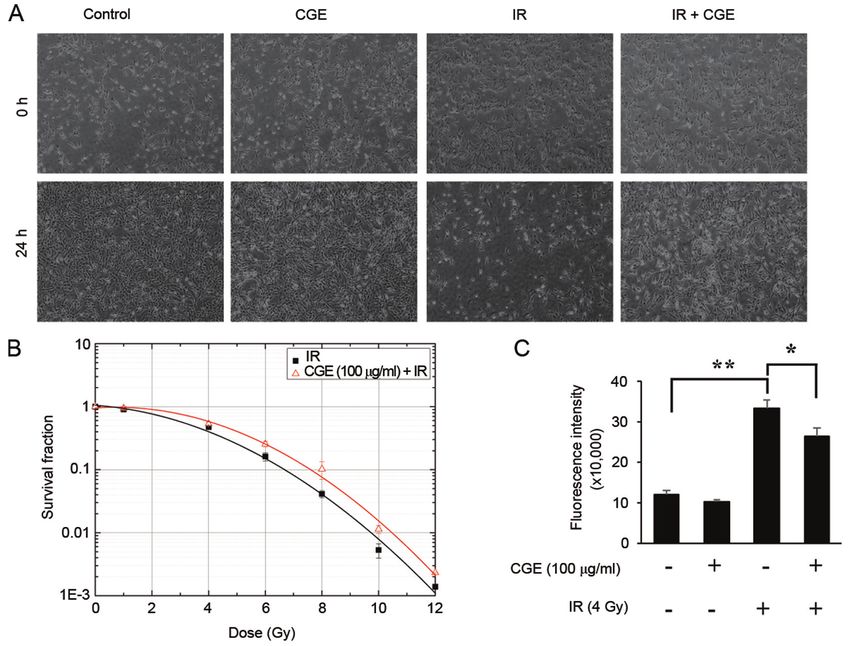
were observed to examine whether CGE exhibits any protec‑ alone had significantly increased the number of cells with
tive effect in NIH‑3T3 cells stimulated with 500 µM of H2O2. condensed or blebbing nuclei. Contrarily, when cells were
As shown in Fig. 1C, CGE exhibited a protective effect on pretreated with CGE, the nuclear damages were markedly
H 2O 2 ‑induced cytotoxicity; additionally, this effect was reduced (Fig. 3A). The results of the flow cytometry consistently
confirmed by a decrease in the irregular morphology observed indicated that H2O2 treatment had increased the population of
due to H2O2‑induced cellular damage (Fig. 1D). These data Annexin V+/PI‑apoptotic cells. However, as shown in Fig. 3B,
indicate that CGE could inhibit H2O2‑induced cytotoxicity in pretreatment of the cells with CGE prior to their exposure to
NIH‑3T3 cells. H2O2 led to effective protection of the cells against apoptosis.
Likewise, results of western blotting indicated a significant
CGE regulates H2O2‑mediated intracellular ROS levels. To increase in the expression of cleaved PARP, a well‑known
investigate the effect of CGE on ROS levels, we determined substrate of caspase‑3 in the apoptotic cell death, compared
whether CGE treatment could attenuate H2O2‑mediated oxida‑ to that of the control. However, in alignment with previous
tive stress. The NIH‑3T3 cells were stimulated with H2O2 in results, Fig. 3C showed that the H2O2‑induced PARP degrada‑
the presence or absence of CGE, after which, the production tion was reduced by pretreatment of the cells with CGE. Taken
of ROS was observed using the fluorescent probe 2,7‑dichloro‑ together, these results indicated that CGE pretreatment could
dihydrofluorescein‑diacetate (H2DCFDA) of the cellular ROS inhibit H2O2‑induced apoptotic cell death in NIH‑3T3 cells.
detection kit. Treatment with H2O2 alone resulted in robust
intracellular generation of ROS, whereas H2O2‑induced ROS CGE increases the viability of IR‑exposed NIH‑3T3 cells.
generation in NIH‑3T3 cells was significantly attenuated by To investigate whether the IR‑induced cell damage could
CGE treatment (Fig. 2A). Moreover, decrease in the oxidative be prevented by CGE pretreatment, first, the morpho‑
stress was quantified via the dihydroethidium (DHE) reac‑ logical changes in NIH‑3T3 cells were evaluated. As shown
tion. DHE is cell permeable and is considered to react with in Fig. 4A, it was confirmed that pretreatment with CGE could
superoxide anions, thus undergoing oxidation upon binding decrease the irregular morphology observed in IR‑exposed
to DNA (22). As shown in Fig. 2B, CGE treatment led to a NIH‑3T3 cells. In addition, to further investigate the relation‑
decrease in the number of cells demonstrating high ROS ship between cell fractions retaining reproductive integrity
production. A significant decrease in the oxidative stress was and absorbed radiation dose, the clonogenic survival curves
observed in the NIH‑3T3 cells. These results implied that were determined. The cell survival fraction in the IR‑exposed
pretreatment with CGE reduces the accumulation of intracel‑ NIH3/T3 cells was evaluated by the cell survival curve. As
lular ROS in NIH‑3T3 cells. shown in Fig. 4B, IR caused cell death in proportion to the
amount of exposure. However, pretreatment with CGE signifi‑
CGE inhibits H 2 O 2 ‑induced cell death in NIH‑3T3 cantly increased cell viability 24 h after irradiation over the
cells. To elucidate the protective effects of CGE against full range of doses. Particularly, the 50% lethal dose (LD 50%)
H2O2‑induced damage in NIH‑3T3 cells, the effects of CGE of cells pretreated with CGE reduced from 4.51 to 3.41 Gy.
on H 2O2‑mediated apoptosis were investigated. The results Previous experiments have confirmed that CGE is capable of
of the cell nucleus staining showed that treatment with H2O2 inhibiting H2O2‑induced cell damage. As IR has been shownEXPERIMENTAL AND THERAPEUTIC MEDICINE 21: 419, 2021 5
Figure 2. CGE regulates H2O2‑mediated intracellular ROS generation. NIH‑3T3 cells were treated with CGE (100 µg/ml) for 24 h and stimulated with H2O2
(500 µM) for 4 h. (A) Cells were stained with H2DCFDA for 30 min to measure intracellular hydrogen peroxide (H2O2) levels using fluorescence microscopy.
Scale bar, 25 µM. (B) Cells were treated with DHE for 30 min to measure intracellular superoxide (O2‑) levels and were analyzed using a Muse™ Cell
analyzer. Representative oxidative stress plots for the NIH‑3T3 cells. CGE, centipedegrass extract; H2O2, hydrogen peroxide; ROS, reactive oxygen species;
H2DCFDA, 2,7‑dichlorodihydrofluorescein‑diacetate; DHE, dihydroethidium; Con, vehicle control (0.1% DMSO).
to adversely affect cells via ROS generation, we investigated PARP, and cleaved caspase‑3, whereas it inhibits the expres‑
whether one possible mechanism for the cytoprotective effect sion of the anti‑apoptotic protein Bcl‑2. However, when cells
of CGE against damages induced by IR is based on its ROS were pretreated with CGE, the expression of anti‑apoptotic
scavenging capacity. To investigate whether CGE could reduce proteins was significantly decreased. Additionally, pretreat‑
IR‑induced ROS generation, H2DCFDA was used for a fluo‑ ment with CGE has been shown to lead to phosphorylation of
rescence assay. Fig. 4C represents the quantitative analysis of BAD by inducting AKT phosphorylation, thereby rendering
the cellular ROS levels. When the cells were pretreated with these signal proteins inactive in apoptotic processes.
CGE, the fluorescence intensity of H2DCFDA was found to Previous studies have reported that exposing cells to IR
be significantly reduced. These results demonstrated that IR and various other toxic stresses can induce simultaneous
caused cell damage by inducing overproduction and accumu‑ target activation of multiple MAPK pathways (25). This has
lation of intracellular ROS, which was effectively prevented been related with the factor‑mediated regulation of various
by CGE. cell longevity factors such as proliferation, differentiation,
aging, and apoptosis. Thus, the expression of MAPK family
CGE inhibits apoptosis in IR‑exposed NIH‑3T3 cells via regu‑ (ERK1/2, p38 and JNK), which plays a role in the extrinsic
lation of the ERK‑, p38‑, JNK‑MAPKs signaling. Radiation apoptotic pathway, in IR‑exposed cells was explored using
is known to mainly induce intrinsic apoptotic cascades such immunoblot analysis. Fig. 6A and B shows that the expression
as the mitochondrial release of cytochrome c and subsequent levels of p‑ERK in IR‑exposed NIH‑3T3 cells were downregu‑
formation of the apoptosome. However, depending on dosage lated whereas those of p‑JNK and p‑p38 were upregulated.
and cell type, the extrinsic apoptotic pathway might also Conversely, these changes in the expression levels were largely
induce cell death (23). In this study, to explore the possible alleviated by pretreatment with CGE. Collectively, these results
molecular mechanism underlying the radioprotective effects supported the hypothesis that pretreatment with CGE protects
of CGE in NIH‑3T3 cells exposed to radiation, first, the the NIH‑3T3 cells against IR‑induced cell death via inhibition
protein expression related to the intrinsic apoptotic pathway of the intrinsic apoptotic pathway as well as the regulation of
was examined. The intrinsic apoptotic pathway is controlled the ERK‑, p38‑, JNK‑MAPKs signaling.
and regulated by the activities of the members of the Bcl‑2
protein family involved in mitochondrial membrane perme‑ Discussion
ability and which may be pro‑apoptotic or anti‑apoptotic (24).
As shown in Fig. 5, radiation activates the expression of the ROS plays diverse roles depending on the concentration
pro‑apoptotic Bcl‑2 proteins Bax (Bcl‑2 like protein 4), Bad, present in cells. ROS participate in the molecular signaling6 KANG et al: THE RADIOPROTECTIVE ROLE OF CGE Figure 3. CGE suppresses H2O2‑induced apoptosis. NIH‑3T3 cells were treated with or without CGE for 24 h and incubated with H 2O2 (500 µM) for 24 h. (A) Changes in nuclear morphology examined by Hoechst 33342 nuclear staining. Arrows indicate apoptotic nuclei. Scale bar: 50 µm. (B) Cells stained with Annexin V‑FITC and analyzed using a cell sorting system with Muse™. (C) Whole cell lysates prepared and immunoblotted with antibodies against cleaved PARP. (D) Western blots were analyzed quantitatively. The band intensities were normalized to GAPDH. Data are presented as mean ± SD. **P≤0.01 vs. H2O2 only. CGE, centipedegrass extract; H2O2, hydrogen peroxide; ROS, reactive oxygen species; PARP, poly(ADP‑ribose) polymerase; GAPDH, glyceraldehyde 3‑phosphate dehydrogenase; Con, vehicle control (0.1% DMSO); SD, standard deviation. Figure 4. CGE increases the viability of IR‑exposed NIH‑3T3 cells. (A) The protection of NIH‑3T3 cells after their exposure to IR was assessed using an inverted phase‑contrast microscope. Magnification, x100. (B) Clonogenic assay was performed to evaluate NIH‑3T3 cell viability. (C) NIH‑3T3 cells were stained with H2DCFDA for 30 min to measure intracellular H2O2 levels using a microplate reader. The bar graph presents a quantitative analysis of the genera‑ tion of intracellular hydrogen peroxide. Data are presented as mean ± SD. *P≤0.05, **P≤0.01 vs. IR only. CGE, centipedegrass extract; H2O2, hydrogen peroxide; IR, ionizing radiation; H2DCFDA, 2,7‑dichlorodihydrofluorescein‑diacetate; Con, vehicle control (0.1% DMSO); SD, standard deviation.
EXPERIMENTAL AND THERAPEUTIC MEDICINE 21: 419, 2021                                                             7

Figure 5. IR exposure‑induced oxidative stress and CGE treatment resulted in altered apoptosis signaling in NIH‑3T3 cells. (A) Lysates of IR‑exposed
cells in the presence or absence of CGE were immunoblotted with anti‑Bcl‑2, anti‑Bax, anti‑Bad, anti‑p‑Bad, anti‑caspase‑3, anti‑C‑caspase‑3, anti‑PARP,
anti‑C‑PARP, or anti‑GAPDH antibodies. (B) The western blots were analyzed quantitatively. Band intensities were normalized to those of the normal form of
each protein, GAPDH, or α‑tubulin. **P≤0.01 vs. IR only. IR, ionizing radiation; CGE, centipedegrass extract; Bcl‑2, B‑cell lymphoma‑2; Bax, Bcl‑2 like pro‑
tein 4; p‑, phosphorylated; Bad, Bcl‑2‑associated death promoter; PARP, poly(ADP‑ribose) polymerase; GAPDH, glyceraldehyde 3‑phosphate dehydrogenase;
Con, vehicle control (0.1% DMSO); SD, standard deviation.

Figure 6. CGE inhibits apoptosis in IR‑exposed NIH‑3T3 cells by regulating the ERK‑, p38‑, JNK‑MAPKs signaling. (A) Lysates of IR‑exposed cells in
the presence or absence of CGE were immunoblotted with anti‑AKT, anti‑p‑AKT, anti‑ERK, anti‑p‑ERK, anti‑p38, anti‑p‑p38, anti‑JNK, anti‑p‑JNK, or
anti‑GAPDH antibodies. (B) Western blots were analyzed quantitatively. The band intensities were normalized to those of the normal form of each protein or
GAPDH. **P≤0.01 vs. IR only. CGE, centipedegrass extract; IR, ionizing radiation; p‑, phosphorylated; ERK, extracellular‑signal‑regulated kinase; JNK, c‑Jun
N‑terminal Kinase; MAPK, mitogen‑activated protein kinase; AKT, protein kinase B; GAPDH, glyceraldehyde 3‑phosphate dehydrogenase; Con, vehicle
control (0.1% DMSO); SD, standard deviation.
8                                         KANG et al: THE RADIOPROTECTIVE ROLE OF CGE

and is essential for homeostasis in normal physiological levels;         Furthermore, we have demonstrated that CGE pretreat‑
however, they function as toxic elements at aberrant levels          ment rescues NIH‑3T3 cells subjected to IR exposure.
and are associated with abnormal cell proliferation (26‑28).         Exposure to IR induced cell death and increased intracel‑
IR strongly induces intracellular accumulation of ROS and            lular ROS levels; however, CGE pretreatment counteracted
RNS (29). Humans are exposed to natural sources of radiation         the cellular damages. In published reports, IR has been
such as those in water, soil, and vegetation, as well as those       shown to activate three MAPKs (ERK1/2, p38, JNK MAPK
from human‑made sources such as X‑rays and medical                   pathway) in a cell type‑dependent manner (25,40), and it has
devices (30). Notably, IR is used for RT, generally, as part of      been shown that the phosphorylated JNK translocates to
cancer treatment to control or kill malignant cells. However,        the nucleus, phosphorylates c‑Jun (41,42). Phosphorylation
some people undergoing RT experience dryness, itching,               of c‑Jun leads to the formation of AP‑1, JNK‑AP‑1 pathway
blistering, or peeling. This reversible effect depends on which      is involved in the increased expression of pro‑apoptotic
part of the body has been exposed to radiation (31). One of          genes (43). Our results have shown that pretreatment with
the most common reversible effects is a skin condition called        CGE decreases the activation of JNK and p38 MAPKs
radiation dermatitis (32), in which, the ionizing radiation inter‑   followed by the apoptosis pathway. Taken together, these
acts directly or indirectly with the target macromolecules or        results indicated that CGE has protective effects against
water in cells leading to the occurrence of oxidizing events         IR‑induced apoptotic cell death and the mechanism under‑
that alter cell molecular composition (29,33). In addition,          lying this effect is ROS scavenging and JNK‑, ERK1/2‑,
oxidative damage can get extended from the target to neigh‑          p38‑MAPK pathway modulation.
boring non‑target bystander cells through a redox‑regulated              Several trials are being conducted to develop a radiopro‑
intercellular proximity mechanism (34). Ultimately, the RT           tector via the inhibition of p38 and JNK pathways in normal
fails, resulting in unexpected damages to the cancer as well as      tissues, and some derivates from natural plants have shown
normal cells due to IR‑induced oxidative stress.                     protective effects against IR‑induced ROS stress (44,45). In
    In a previously published study, maysin derived from corn        the previous study, it was reported that maysin (the major
silk was reported to increase cell viability under conditions        constituent of centipedegrass) not only plays a role as a ROS
of oxidative stress via upregulation of a neuronal anti‑             scavenger but also is able to increase the amount of antioxidant
oxidative action (17). Conversely, it was reported that maysin       enzymes in a mammalian cell. Notably, in this study, CGE
(C‑glycosyl flavone) isolated from the silks of Zea mays L.          also exhibited protective effects against overwhelming ROS
has a higher antioxidative activity than other compounds             deposition induced by H2O2, and reduced the extent of apop‑
(rutin, quercetin, luteolin), thus, it has potential as a powerful   totic cell death induced by IR via downregulation of MAPK
antioxidant compound. In this study, the ability of CGE to           signaling (ERK, p38, JNK) in fibroblasts.
rescue fibroblast cells from oxidative stress‑induced apoptosis          Therefore, CGE could be considered as an efficient radio‑
was demonstrated and the underlying effects were examined.           protector or a radiation palliative remedy, which could help
In general, apoptosis has been recognized as an indication           reduce tissue damage induced by exposure to radiation in
of cell death induced by oxidative stress followed by cell           patients or sufferers unintentionally exposed to radiation or
senescence (35). One of the most pivotal molecular path‑             undergoing RT. However, to demonstrate the protective effect
ways that are damaged by oxidative stress is that involving          of CGE on skin tissue, experiments conducted with a single cell
damage to the DNA (36). Previous studies have reported that          line derived from mice may be a limitation. To address this,
DNA damage mediated by reactive oxygen intermediates                 human‑derived skin cell experiments need to be performed.
causes enzymatic inactivation through cleavage of PARP,              Additionally, in this study, the effects of individual constitu‑
which is an important step in apoptosis (37). The function           ents of CGE are still not be explored and should be explored
of cleaved PARP is to prevent repair of DNA strand breaks            in further studies. Furthermore, for the clinical application
during apoptotic cell death, which is now widely known as            of any compound as a candidate for radiation protection, it is
the key marker of type 1 programmed cell death (38). PARP            essential to avoid unacceptable clinical risks; therefore, abso‑
is cleaved by caspase‑3 into two fragments of 89 and 24 kDa          lute certainty about its safety for normal tissues is required.
during apoptotic cell death in various cell lines (39). In this      In laboratory studies, several compounds have been tested
study, we demonstrated that CGE plays a role in cytoprotec‑          as radioprotectants; however, most did not reach the clinical
tive effect on H 2O2 ‑induced cell death in mouse‑derived            stage due to the toxicity and side effects in animal models.
fibroblasts. Pretreatment of NIH‑3T3 cells with CGE (up to           Similarly, in this study, CGE was found to be non‑toxic under
100 µg/ml) before H2O2 treatment significantly attenuated cell       normal conditions in fibroblasts; however, in vivo studies using
death induced by oxidative stress, as observed by cell density       animals with tumors are needed to investigate whether CGE
and viability. CGE significantly inhibited PARP cleavage and         has preferential radioprotective action in normal tissues over
prevented sustenance of DNA damage. Additionally, it was             tumor tissues.
confirmed that CGE could significantly reduce the number of              In conclusion, CGE contains C‑glycosyl flavones and
Annexin V‑ and PI‑positive cells, indicating that pretreatment       phenolic components, which protected mouse‑derived fibro‑
with CGE could significantly alleviate apoptosis induced             blasts from IR‑induced apoptotic cell death by blocking ROS
by oxidative stress. Taken together, the results of this study       production and inhibiting ERK‑, p38‑, JNK‑MAPKs signaling.
suggest that due to its antioxidant activity, CGE has a cellular     Although large‑scale animal studies and clinically relevant
protective effect against oxidative stress‑induced apoptosis in      tests are needed to confirm the effectiveness of CGE, poten‑
NIH‑3T3 cells and may potentially act as a protective agent          tial applications of CGE as a useful radioprotectant may be
against IR‑induced cell damage.                                      proposed.
EXPERIMENTAL AND THERAPEUTIC MEDICINE 21: 419, 2021 9
Acknowledgements 9. Riley PA: Free radicals in biology: Oxidative stress and the
effects of ionizing radiation. Int J Radiat Biol 65: 27‑33, 1994.
10. Uttara B, Singh AV, Zamboni P and Mahajan RT: Oxidative stress
Not applicable. and neurodegenerative diseases: A review of upstream and down‑
stream antioxidant therapeutic options. Curr Neuropharmacol 7:
65‑74, 2009.
Funding 11. Chan RJ, Webster J, Chung B, Marquart L, Ahmed M
and Garantziotis S: Prevention and treatment of acute
This work was supported by the Nuclear R&D Program of the radiation‑induced skin reactions: A systematic review and
meta‑analysis of randomized controlled trials. BMC Cancer 14:
Ministry of Science and ICT, Republic of Korea. 53, 2014.
12. Bolderston A, Cashell A, McQuestion M, Cardoso M,
Availability of data and materials Summers C and Harris R: A canadian survey of the management
of radiation‑induced skin reactions. J Med Imaging Radiat
Sci 49: 164‑172, 2018.
The datasets used and/or analyzed during the current study are 13. Wei J, Meng L, Hou X, Qu C, Wang B, Xin Y and Jiang X:
available from the corresponding author on reasonable request. Radiation‑induced skin reactions: Mechanism and treatment.
Cancer Manag Res 11: 167‑177, 2018.
14. Chen J, Zhu Y, Zhang W, Peng X, Zhou J, Li F, Han B, Liu X,
Authors' contributions Ou Y and Yu X: Delphinidin induced protective autophagy via
mTOR pathway suppression and AMPK pathway activation in
HER‑2 positive breast cancer cells. BMC Cancer 18: 342, 2018.
SHK and DHB designed and performed the experiments. SSL 15. Zhang S and Duan E: Fighting against Skin Aging: The Way
and HWB interpreted the experimental results and drafted the from Bench to Bedside. Cell Transplant 27: 729‑738, 2018.
manuscript. BYC and BSK performed the statistical analysis 16. Panich U, Sittithumcharee G, Rathviboon N and Jirawatnotai S:
Ultraviolet radiation‑induced skin aging: The role of DNA
and revised the manuscript critically for important intellectual damage and oxidative stress in epidermal stem cell damage
content. SHK and DHB confirmed the authenticity of all the mediated skin aging. Stem Cells Int 2016: 7370642‑7370642,
raw data. All authors read and approved the final manuscript. 2016.
17. Kim JH, Jenrow KA and Brown SL: Mechanisms of radiation-
induced normal tissue toxicity and implications for future clinical
Ethics approval and consent to participate trials. Radiat Oncol J 32: 103‑115, 2014.
18. Smith TA, Kirkpatrick DR, Smith S, Smith TK, Pearson T,
Kailasam A, Herrmann KZ, Schubert J and Agrawal DK:
Not applicable. Radioprotective agents to prevent cellular damage due to ionizing
radiation. J Transl Med 15: 232‑232, 2017.
Patient consent for publication 19. Painuli S and Kumar N: Prospects in the development of natural
radioprotective therapeutics with anti‑cancer properties from the
plants of Uttarakhand region of India. J Ayurveda Integr Med 7:
Not applicable. 62‑68, 2016.
20. Mun GI, Kim S, Choi E, Kim CS and Lee YS: Pharmacology of
natural radioprotectors. Arch Pharm Res 41: 1033‑1050, 2018.
Competing interests 21. Badaboina S, Bai HW, Park CH, Jang DM, Choi BY and
Chung BY: Molecular mechanism of apoptosis induction in skin
The authors declare that they have no competing interests. cancer cells by the centipedegrass extract. BMC Complement
Altern Med 13: 350, 2013.
22. Bindokas VP, Jordán J, Lee CC and Miller RJ: Superoxide
References production in rat hippocampal neurons: Selective imaging with
hydroethidine. J Neurosci 16: 1324‑1336, 1996.
23. Green DR and Kroemer G: The pathophysiology of mitochondrial
1. Hirata M, Nagakura Y, Yuki N, Adachi K, Fujii R, Koyakumaru T, cell death. Science 305: 626‑629, 2004.
Ogura S, Moritake H, Watanabe C and Fukuyama K: Development 24. Hata AN, Engelman JA and Faber AC: The BCL2 family: Key
and establishment of centipede grass (Eremochloa ophiuroides) mediators of the apoptotic response to targeted anticancer thera‑
in south‑western Japan. Trop Grassl 41: 100‑112, 2007. peutics. Cancer Discov 5: 475‑487, 2015.
2. Park HJ, Chung BY, Lee MK, Song Y, Lee SS, Chu GM, 25. Dent P, Yacoub A, Fisher PB, Hagan MP and Grant S: MAPK
Kang SN, Song YM, Kim GS and Cho JH: Centipede grass exerts pathways in radiation responses. Oncogene 22: 5885‑5896, 2003.
anti‑adipogenic activity through inhibition of C/EBPβ, C/EBPα, 26. Di Meo S, Reed TT, Venditti P and Victor VM: Role of ROS and
and PPARγ expression and the AKT signaling pathway in 3T3‑L1 RNS sources in physiological and pathological conditions. Oxid
adipocytes. BMC Complement Altern Med 12: 230, 2012. Med Cell Longev 2016: 1245049‑1245049, 2016.
3. Lee EM, Bai HW, Lee SS, Hong SH, Cho JY, Lee IC and 27. Schieber M and Chandel NS: ROS function in redox signaling
Chung BY: Stress‑induced increase in the amounts of maysin and and oxidative stress. Curr Biol 24: R453‑R462, 2014.
maysin derivatives in world premium natural compounds from 28. Lee AY, Choi JM, Lee MH, Lee J, Lee S and Cho EJ: Protective
centipedegrass. Radiat Phys Chem 81: 1055‑1058, 2012. effects of perilla oil and alpha linolenic acid on SH‑SY5Y
4. Lee EM, Lee SS, Bai H‑W, Cho J‑Y, Kim TH and Chung BY: Effect neuronal cell death induced by hydrogen peroxide. Nutr Res
of gamma irradiation on the pigments and the biological activities Pract 12: 93‑100, 2018.
of methanolic extracts from leaves of centipedegrass (Eremochloa 29. Azzam EI, Jay‑Gerin JP and Pain D: Ionizing radiation‑induced
ophiuroides Munro). Radiat Phys Chem 91: 108‑113, 2013. metabolic oxidative stress and prolonged cell injury. Cancer
5. Liu J, Wang C, Wang Z, Zhang C, Lu S and Liu J: The antioxidant Lett 327: 48‑60, 2012.
and free‑radical scavenging activities of extract and fractions 30. Canadian Nuclear Safety Commission (CNSC): Types and Sources
from corn silk (Zea mays L.) and related flavone glycosides. Food of Radiation. Ottawa, ON, 2014.
Chem 126: 261‑269, 2011. 31. Bray FN, Simmons BJ, Wolfson AH and Nouri K: Acute and
6. Choi DJ, Kim SL, Choi JW and Park YI: Neuroprotective effects chronic cutaneous reactions to ionizing radiation therapy.
of corn silk maysin via inhibition of H2O2‑induced apoptotic cell Dermatol Ther (Heidelb) 6: 185‑206, 2016.
death in SK‑N‑MC cells. Life Sci 109: 57‑64, 2014. 32. Leventhal J and Young MR: Radiation dermatitis: Recognition,
7. Baskar R, Lee KA, Yeo R and Yeoh KW: Cancer and radiation prevention, and management. Oncology (Williston Park) 31:
therapy: Current advances and future directions. Int J Med Sci 9: 885‑887, 894‑899, 2017.
193‑199, 2012. 33. Reisz JA, Bansal N, Qian J, Zhao W and Furdui CM: Effects
8. Johung K, Saif MW and Chang BW: Treatment of locally of ionizing radiation on biological molecules - mechanisms of
advanced pancreatic cancer: the role of radiation therapy. Int J damage and emerging methods of detection. Antioxid Redox
Radiat Oncol Biol Phys 82: 508-518, 2012. Signal 21: 260‑292, 2014.10 KANG et al: THE RADIOPROTECTIVE ROLE OF CGE
34. Mladenov E, Li F, Zhang L, Klammer H and Iliakis G: Intercellular 40. Valerie K, Yacoub A, Hagan MP, Curiel DT, Fisher PB, Grant S
communication of DNA damage and oxidative status underpin and Dent P: Radiation‑induced cell signaling: Inside‑out and
bystander effects. Int J Radiat Biol 94: 719‑726, 2018. outside‑in. Mol Cancer Ther 6: 789‑801, 2007.
35. Redza‑Dutordoir M and Averill‑Bates DA: Activation of 41. Davis RJ: Signal transduction by the JNK group of MAP kinases.
apoptosis signalling pathways by reactive oxygen species. In: Inflammatory Processes. Springer, pp13‑21, 2000.
Biochim Biophys Acta 1863: 2977‑2992, 2016. 42. Chang L and Karin M: Mammalian MAP kinase signalling
36. Whitaker AM, Schaich MA, Smith MR, Flynn TS and cascades. Nature 410: 37‑40, 2001.
Freudenthal BD: Base excision repair of oxidative DNA damage: 43. Fan M and Chambers TC: Role of mitogen‑activated protein
From mechanism to disease. Front Biosci 22: 1493‑1522, 2017. kinases in the response of tumor cells to chemotherapy. Drug
37. Rodríguez‑Vargas JM, Ruiz‑Magaña MJ, Ruiz‑Ruiz C, Majuelos- Resist Updat 4: 253‑267, 2001.
Melguizo J, Peralta‑Leal A, Rodríguez MI, Muñoz‑Gámez JA, de 44. Santabárbara‑Ruiz P, López‑Santillán M, Martínez‑Rodríguez I,
Almodóvar MR, Siles E, Rivas AL, et al: ROS‑induced DNA Binagui‑Casas A, Pérez L, Milán M, Corominas M and Serras F:
damage and PARP‑1 are required for optimal induction of ROS‑induced JNK and p38 signaling is required for unpaired
starvation‑induced autophagy. Cell Res 22: 1181‑1198, 2012. cytokine activation during Drosophila regeneration. PLoS
38. Ko HL and Ren EC: Functional Aspects of PARP1 in DNA Genet 11: e1005595‑e1005595, 2015.
Repair and Transcription. Biomolecules 2: 524‑548, 2012. 45. Choi EK, Yeo JS, Park CY, Na H, Lim J, Lee JE, Hong SW,
39. Soldani C, Lazzè MC, Bottone MG, Tognon G, Biggiogera M, Park SS, Lim DG and Kwak KH: Inhibition of reactive oxygen
Pellicciari CE and Scovassi AI: Poly(ADP‑ribose) polymerase species downregulates the MAPK pathway in rat spinal cord
cleavage during apoptosis: When and where? Exp Cell Res 269: after limb ischemia reperfusion injury. Int J Surg 22: 74‑78, 2015.
193‑201, 2001.You can also read