Mechanisms underlying low-clinical responses to PD-1/PD- L1 blocking antibodies in immunotherapy of cancer: a key role of exosomal PD- L1
←
→
Page content transcription
If your browser does not render page correctly, please read the page content below
Open access Review
J Immunother Cancer: first published as 10.1136/jitc-2020-001698 on 20 January 2021. Downloaded from http://jitc.bmj.com/ on October 17, 2021 by guest. Protected by copyright.
Mechanisms underlying low-clinical
responses to PD-1/PD-L1 blocking
antibodies in immunotherapy of cancer:
a key role of exosomal PD-L1
Zi Yin,1 Min Yu,1 Tingting Ma,2 Chuanzhao Zhang,1 Shanzhou Huang,1
Mohammad Reza Karimzadeh,3 Amir Abaas Momtazi-Borojeni ,4 Sheng Chen1
To cite: Yin Z, Yu M, Ma T, ABSTRACT Programmed cell death protein 1 (PD-1) is
et al. Mechanisms underlying Exosomes, as the main group of extracellular vesicles, a key immune checkpoint molecule known
low-clinical responses to PD-1/ are biologically active lipid-bilayer vesicles that are
PD-L1 blocking antibodies in
as one of the major inhibitory coreceptors
naturally released from different types of normal or expressed in the activated T cells.2 Under
immunotherapy of cancer: a key
role of exosomal PD-L1. Journal tumor cells. These vesicles play an important role in normal physiology, immune checkpoints,
for ImmunoTherapy of Cancer intercellular communication and influence the extracellular such as PD-1, are critical for maintaining
2021;9:e001698. doi:10.1136/ environment and the immune system. Emerging
self-tolerance, preventing autoimmunity, and
jitc-2020-001698 evidence demonstrates that cancer-derived exosomes
controlling T-cell responses within a desired
are enriched in immunosuppressive proteins, such as
ZY and MY contributed equally. the programmed death-ligand 1 (PD-L1). PD-L1 and its physiological range to protect tissues from
Accepted 16 December 2020 receptor programmed cell death protein 1 (PD-1) are the excessive inflammatory reactions. However,
key immune checkpoint molecules that promote tumor in cancerous conditions, these regulatory
progression via negative regulation of immune responses. proteins can allow tumor cells to protect them-
PDL-1 is highly expressed on the surface of tumor cells selves from the antitumor T-cell responses,
and binds to PD-1 on the surface of activated T cells, causing the so-called tumor immune evasion.1
leading to suppression of T cells, which consequently PD-1 acts through interaction with PD-L1
enables cancer cells to escape antitumor immunity. that is a highly expressed ligand in non-
Currently, there are several Food and Drug Administration- lymphoid tissues in response to inflammatory
approved monoclonal antibodies blocking PD-1/PD-L1 cytokines such as interferon-gamma (IFN-γ)
interaction, which are clinically used for cancer treatment.
© Author(s) (or their produced by cytotoxic T Cells.2 3 On engage-
However, despite impressive treatment outcomes, some
employer(s)) 2021. Re-use ment with PD- L1, PD-1 transmits a nega-
permitted under CC BY-NC. No patients show poor response to PD-1/PD-L1 blockade.
Of note, tumor-derived exosomes containing PD-L1 can tive costimulatory signal in T cells through
commercial re-use. See rights
and permissions. Published by recapitulate the effect of cell-surface PD-L1. There is the recruitment of Src homology 2 domain
BMJ. evidence that reveals a significant association between containing phosphatases 1/2 (SHP1/2)
1
Department of General Surgery, levels of circulating exosomal PD-L1 and rate of response that dephosphorylates downstream TCR-
Guangdong Provincial People’s to anti-PD-1/PD-L1 antibody therapy. The present article mediated signaling elements and thereby
Hospital, Guangdong Academy reviews the role of exosomal PDL-1 in the therapeutic inhibits T cell proliferation, cytokine produc-
of Medical Science, Guangzhou,
China
resistance to anti-PD-1/PD-L1 treatment. Importantly, it tion and release, and cytotoxicity.4 Therefore,
2 is suggested that the removal of exosomal PDL-1 could the PD-1/PD-L1 regulatory system is induced
Department of Obstetrics
and Gynecology, Sun Yat Sen serve as a therapeutic adjuvant for enhancing the efficacy by immune responses and then through
Memorial Hospital, Sun Yat sen of anti-PD-1/PD-L1 therapy in patients with cancer. a negative feedback loop attenuates T- cell
University, Guangzhou, China responses and minimizes tissue damage.
3
Department of Medical
Genetics, School of Medicine, On tumor antigen recognition by T cells,
Bam University of Medical PD-1/PD-L1 IMMUNE CHECKPOINT PATHWAY AS A the released IFN-γ triggers tumor cells
Sciences, Bam, Iran DOUBLE-EDGED SWORD to express PD- L1 that binds coinhibitory
4
Department of Medical Immune checkpoints are cell surface regula- receptor molecule PD-1 on cytotoxic T cells
Biotechnology, Faculty of
tory receptors, located mainly, but not exclu- within the tumor microenvironment (TME).
Medicine, Mashhad University of
Medical Sciences, Mashhad, Iran sively, on T lymphocytes. After recognizing This allows the specific inhibition of tumor
cognate ligands on the antigen- presenting recognition by T cells, resulting in attenuating
Correspondence to cells or the target cells, these receptors act as antitumor immune responses and promoting
Dr Zi Yin; yinzi@gdph.o rg.cn
T cell receptor (TCR) cosignaling partners tumor growth. IFN-γ induces the expression
Dr Sheng Chen; that deliver either stimulatory or inhibitory of PD-L1 through activating signaling path-
chenshenglucky@yeah.net signals to regulate the lymphocyte activation.1 ways downstream of the type II interferon
Yin Z, et al. J Immunother Cancer 2021;9:e001698. doi:10.1136/jitc-2020-001698 1
Open access
J Immunother Cancer: first published as 10.1136/jitc-2020-001698 on 20 January 2021. Downloaded from http://jitc.bmj.com/ on October 17, 2021 by guest. Protected by copyright.
Table 1 FDA-approved monoclonal antibodies blocking immune checkpoints in human cancer
Target Therapeutic antibody Tumor type FDA approval year
CTLA4 Ipilimumab Melanoma, renal cell carcinoma, metastatic colorectal cancer 2011
PD-1 Pembrolizumab Melanoma, non-small cell lung cancer, renal cell carcinoma, urothelial 2014
bladder cancer, Hodgkin’s lymphoma, head and neck cancer, Merkel
cell carcinoma, microsatellite instability-high cancer, gastric cancer,
hepatocellular carcinoma, cervical cancer, primary mediastinal large
B-cell lymphoma
Nivolumab Melanoma, non-small cell lung cancer, renal cell carcinoma, urothelial 2014
bladder cancer, Hodgkin’s lymphoma, head and neck cancer,
colorectal cancer, hepatocellular carcinoma, small cell lung cancer
Cemiplimab Cutaneous squamous-cell carcinoma 2018
PD-L1 Atezolizumab Non-small cell lung cancer, urothelial bladder cancer, small cell lung 2016
cancer, breast cancer
Avelumab Merkel cell carcinoma, urothelial bladder cancer 2017
Durvalumab Non-small cell lung cancer, urothelial bladder cancer 2017
Data have been acquired from “Timeline of Anti-PD-1/L1 Antibody Approvals by the FDA.” Available in online (https://www.cancerresearch.
org/scientists/immuno-oncology-landscape/pd-1-pd-l1-landscape).
IFN-γ, interferon-gamma; PD-1, programmed cell death protein 1; PD-L1, programmed death-ligand 1.
receptor, including Janus kinase and signal transducer and decreased toxicity as well as a much broader spec-
and activators of transcription, leading to binding of the trum of antitumor activity when compared with anti-
IRF1 transcription factor to the PD-L1 gene promoter.5 CTLA-4 mAbs. There is evidence that clinical responses
Moreover, oncogenic pathways, including MAPK (RAS/ to immune checkpoint inhibitors might be correlated
RAF/MEK/ERK) and PI3K/Akt/mTOR, are also known with immune- related adverse events (irAEs). Overall,
to mediate the induction of PD-L1 expression through drug-induced irAEs are more likely to be experienced in
activating c-JUN that, as the component of AP-1 transcrip- patients treated with anti-CTLA-4 (60%–85%) than anti-
tion factor, binds to the enhancer element on PD-L1 gene PD-1 (16%–37%) or anti-PD-L1 (12%–24%).12–14
and augments the transcription signal in tumor cells.6 Pembrolizumab, the first mAbs against PD-1, achieved
its first global approval for patients with unresectable or
metastatic melanoma by US-FDA.15 Afterward, its use was
THERAPEUTIC EFFECTS OF PD-1/PD-L1 BLOCKING ANTIBODIES
extended to non-small cell lung cancer (NSCLC),16 head
Immune checkpoint blockade therapies are now
and neck squamous cell carcinoma (HNSCC),17 cervical
approved by US Food and Drug Administration (US-
carcinoma,18 and metastatic urothelial carcinoma18
FDA) for the treatment of a wide range of cancer types,
among others in a list that continues to grow. Anti-PD-1
with approval likely for additional indications in the near
antibodies can block the PD-1 signaling pathway, thus
future. Among negative immune checkpoint molecules,
preventing PD-1-mediated attenuation of TCR signaling,
the blockade of PD-1 and its major ligand, PD-L1, provides
which promotes reinvigoration of exhausted PD-1+ CD8+ T
one of the most successful immunotherapies by intensi-
fying T cell immunity against cancer cells. Since the 2011 cells, resulting in immune tumor rejection.19–22 Although
FDA approval of the first immune checkpoint inhibitor, the precise molecular and cellular events mediating
ipilimumab (anti- CTLA4), for the treatment of meta- enhancement of antitumor immunity by PD-1 blockade
static melanoma, six monoclonal antibodies targeting the are not fully understood, tumor neoantigen-specific CD8+
PD-1 (pembrolizumab, nivolumab, and cemiplimab) and T cells appear to be the major T-cell population medi-
PD-L1 (atezolizumab, durvalumab, and avelumab), have ating anti-PD-1 responses.23 Apart from restoring T-cell
been approved for the treatment of various cancer types activity through modulation of TCR signaling, blockade
(table 1).1 of the PD-1 signaling axis is also able to reverse the associ-
An important rationale respecting the development ated metabolic reprogramming to an extent which in part
of anti-PD-1/PD-L1 drugs for cancer treatment emerged mediates the reinvigoration of tumor antigen-specific T
from the key findings that showed PD-1 is over-expressed cells.24 25
on tumor-infiltrating lymphocytes (TILs),7 8 while PD-1 Alongside direct inhibition of PD-1, antibodies
ligand is highly upregulated in most of the human cancer targeting PD-L1 are also sufficient to reverse the negative
cells.9 This was validated through many preclinical studies immune regulation and, thereby, reinvigorate the host
that indicated mAbs blockade of PD-1 or PD-L1 signifi- antitumor immunity. The current FDA-approved mAbs
cantly improved antitumor immunity.9–11 Anti-PD-1/ targeting PD-L1 are known to be efficient for treating
PD-L1 blocking mAbs have heightened tumor selectivity several cancer types, including NSCLC, small cell lung
2 Yin Z, et al. J Immunother Cancer 2021;9:e001698. doi:10.1136/jitc-2020-001698Open access
J Immunother Cancer: first published as 10.1136/jitc-2020-001698 on 20 January 2021. Downloaded from http://jitc.bmj.com/ on October 17, 2021 by guest. Protected by copyright.
cancer, urothelial bladder cancer, breast cancer, and responses are tumor- specific neoantigen peptides that
Merkel cell carcinoma.1 In the light of the dominance in arise from somatic mutations in tumor genomes. The
PD-L1 expression, blockade of PD-L1 can reiterate the number of non- synonymous single nucleotide variants
impact of PD-1 blockade. PD-L1 expression is primarily in a tumor, referred to as TMB, can affect the odds of
triggered by Th1 cytokines, such as IFN-γ. This can in part generating immunogenic peptides and thereby influ-
describe the effectiveness of PD-L1 blockade since Th1- ence response to immune checkpoint blockade in
skewed responses would be more desirable for inducing patients.25 39–44 Of note, a higher TMB favors positive
antitumor immunity.26 By contrast with PD-1 blockade, response to PD-1/PD-L1 blockade in a variety of tumor
anti-
PD-
L1 antibodies may also achieve part of their types including NSCLC,23 45–48 small cell lung cancer,48
efficacy from antibody- dependent cellular cytotoxicity. urothelial carcinoma,49 metastatic melanoma,50 and
This claim emerged from an in vivo study that shows Fc HNSCC.51–53 Moreover, data from a meta-analysis across
receptor binding is crucial for the effectiveness of anti- 27 tumor types show a positive correlation between
PD-L1, but not anti- PD-1, antibody therapy- promoted average response rate and TMB.54 These findings make
tumor regression in mice bearing tumor.27 Notably, these clear that there is a significant pan-cancer association
findings suggest that anti-PD-1 and anti-PD-L1 antibodies between TMB and response to PD-1/PD- L1 blockade
have distinct biological activities and act, at least in part, by the tumor type. Further supporting the relation-
through different mechanisms. ship between TMB and high sensitivity to PD-1/PD-L1
blockade is the observation that microsatellite instability
(MSI)-high/mismatch repair deficiency (MMRd) asso-
FACTORS MODULATING RESPONSE TO PD-1/PD-L1 INHIBITORS ciates with satisfactory treatment effects in patients with
Despite the above-mentioned advancements in clinical multiple cancer receiving anti- PD-1/PD-L1 therapy.55
practice, some patients and cancer types show thera- Tumors with high MSI generate many neopeptides owing
peutic resistance or relatively low-response rate to PD-1/ to the hypermutated phenotype. MSI-positive tumors are
PD-L1 inhibitors.28–30 The basis of differential therapeutic a specific type of high TMB tumors, with MMRd gener-
success between patients and between cancers is not ating a high mutational load. Notably, MMRd derives
completely understood. However, several factors, such as several insertion and deletion mutations resulting in
the TME, the tumor genomics, and systemic factors such frameshifts producing neoantigens that may be more
as exosomes have been suggested for explaining poten- immunogenic due to their higher sequence divergence
tial mechanisms of the response and resistance to PD-1/ from self-peptides.55–57 Taken together, these findings
PD-L1 inhibitors. reveal that MMRd is correlated with enhanced response
Of TME-related factors, PD-L1 expression status and to PD-1/PD-L1 blockade because of increased TMB.
pre-existing TILs have been widely studied.20 31 Although Besides, systemic factors are also found to influence the
the conclusions from various clinical trials are not consis- therapeutic efficacy of PD-1/PD- L1 inhibitors. Among
tent, a significant positive but not absolute correlation them, peripheral blood parameters including neutro-
between PD-L1 expression in the TME and responsive- phil/lymphocyte ratio, total lymphocyte and monocyte
ness to anti-PD-1/anti-PD-L1 therapy has been generally count, relative eosinophil count, T cell clonality, PD-L1high
found.32 33 Across all tumor types, patients with PD-L1- circulating tumor cells, and circulating PD-L1 have been
negative tumors respond to anti-PD-1/PD-L1 therapy in reported to be significantly correlated with PD-1/PD-L1
0%–17%, while those with PD-L1-positive tumors exhibit blockade response in multiple studies of anti-PD-1/PD-L1
a response rate ranging from 44% to 100%.34 Besides, therapy across a wide range of cancer types.58 Exosomal
TILs are the other cell types affecting the tumor immune PD-L1 is another systemic factor that has recently
microenvironment. The density and phenotype of pre- attracted extensive attention. Tumor cells release PDL1-
existing TILs within a TME can impact the efficacy of anti- containing exosomes that have been known to be in part
PD-1/PDL1 therapy.35 The results from a model of tumor responsible for resistance to anti-PD-1/PDL-1 therapy. A
immune microenvironment that consists of TILs and growing body of evidence indicated that exosomal PDL-1
PD-L1 expression status indicate that patients with cancer affects the TME and antitumor immunity. In the present
with PD-L1+ TIL+ tumors possess an effective immune review article, we seek impacts of exosomal PD-L1 on clin-
response to PD-1/PD-L1 blockade therapy.36 In patients ical outcomes with PD-1/PDL-1 blockade therapy and
with NSCLC, pretherapy levels of the intratumoral popu- suggest potential combination therapies to overcome the
lation of CD8+ cytotoxic T cells with the highest PD-1 resistance.
expression have shown a positive correlation with anti-
PD-1 response.37 Moreover, the ratio of memory-like to
exhausted TILs, as two major intratumoral CD8+ T cell ACTIVITY OF EXOSOMAL PD-L1
phenotypes, has been indicated to be strongly correlated Exosomes, as the main group of secreted small extra-
with increased survival and response in a cohort of cellular vesicles, are biologically active lipid-bilayer vesi-
patients with melanoma treated with anti-PD-1.38 cles with a size around 30–100 nm that are naturally
Another studied factor is the tumor mutation burden produced and released by different types of normal and
(TMB). The primary targets of many tumor immune tumor cells.59 60 These vesicles play an important role in
Yin Z, et al. J Immunother Cancer 2021;9:e001698. doi:10.1136/jitc-2020-001698 3Open access
J Immunother Cancer: first published as 10.1136/jitc-2020-001698 on 20 January 2021. Downloaded from http://jitc.bmj.com/ on October 17, 2021 by guest. Protected by copyright.
intercellular communication and influence the extracel- higher immunosuppressive impact of exosomal PD- L1
lular environment and the immune system responses.59 61 than the soluble form.71 72
Exosomes are secreted into extracellular space through Exosomal PD- L1 can recapitulate the effect of cell-
endosomal pathways and transport various bioactive surface PD- L1; it similarly provokes tumor progres-
molecules (cargo) to the target cells. The composition of sion mainly through enabling cancer cells to escape
exosomal cargo is very diverse and includes a wide range antitumor immunity via inhibiting T cell activation
of immunosuppressive and immunostimulatory proteins, (figure 1). Generally, B cell presentation of antigen to
chemokines, cytokines, cellular receptors, lipids, as well T cells will activate T cells. An in vitro setting of PD-L1
as different nucleic acids such as micro-RNAs and circular negative Raji B cell and PD-1 expressing Jurkat T cell
RNAs.62–64 revealed that exosomal PD-L1 can suppress T cell activa-
Tumor cells can actively produce large levels of tion.73 The immunosuppressive effect of tumor-derived
exosomes enriched in cancer-promoting cellular contents, exosomal PD- L1 has been widely confirmed by other
such as immunosuppressive proteins like PD-L1, mRNAs, investigations. Malignant glioma cells have been found
and micro- RNAs, that participate in cancer develop- to generate exosomes containing PD-L1 that involves in
ment and metastasis, through dysregulating antitumor tumor progression. Exosomal PD-L1 secreted by glioblas-
or protumor immunity responses and promoting drug toma stem-like cells were identified to participate in T
resistance.65–67 Of note, tumor-secreted exosomes contain cell immunosuppression by inhibition of CD4+ and CD8+
PD-L1 presented both on the surface and within exosome T cell activation.74 PD-L1-
containing exosomes isolated
particles. ESCRTs (endosomal sorting complex required from NSCLC patients could inhibit the activity of CD8+
for transport) are involved in the packaging of biomole- T cells by reducing the production of interleukin 2
cules into exosome, and nSMase2 (neutral sphingomye- (IL-2) and IFN-γ in a dose-dependent manner. Of note,
linase 2) and Rab proteins regulate exosome secretion.68 exosomes with high levels of PD-L1 (PD-L1high exosomes)
ESCRT, Rab27a, and nSMase2 have been identified to showed a strong potency to suppress the production
participate in the packaging and secretion of exosomal of IL-2 and IFN-γ, whereas exosomes with lower levels
PD-L1.69 Exosomes can transport PD-L1 to other cells of PD-L1 (PD-L1low exosomes) exerted no significant
with low or no PD-L1 expression, with the potential to effect. PD-L1high exosomes could also significantly induce
bind to PD-1.70 apoptosis in CD8+ T cells through PD-1/PD-L1 interac-
The plasma/serum levels of PD- L1 expressed on tion.75 Furthermore, in patients with HNSCC, exosomal
exosomes, but not soluble PD-L1, are found to associate PD-L1 were able to suppress the proliferation of CD4+ T
with disease progression and clinicopathological features cells, induce apoptosis in CD8+ T cells, and enhance the
in patients with cancer, such as HNSCC71 and NSCLC.72 suppressor activity of Treg cells, depending on the level
Of note, major histocompatibility complex (MHC) mole- of PD-L1 in exosomes. PD-L1high exosomes were found to
cules expressed on exosomes can play an essential role be more effective in the suppression of T cells, compared
in exosomal PD-L1-mediated tumor promotion. Indeed, with PD-L1low exosomes.71 An in vivo study revealed that
molecular interaction of exosomal MHC I with TCR exosomal PD- L1 isolated from murine (SCCVII) and
enhances the inhibitory effect of exosomal PD-L1 to T human (SCC90) HNSCC cell lines could enhance tumor
cells. These might be perfectly justifiable reasons for the progression in a mouse model of human oral-oesophageal
Figure 1 The role of exosomal PD-L1 in cancer progression. Tumor cells secrete exosomes containing PD-L1 leading to
suppression of immunity by reducing T-cell activity and inhibition of interferon-gamma and interleukin 2 production as well
as reducing total number of CD8+ T cells by inducing apoptosis through PD-1/PD-L1 pathway. PD-1, programmed cell death
protein 1; PD-L1, programmed death-ligand 1.
4 Yin Z, et al. J Immunother Cancer 2021;9:e001698. doi:10.1136/jitc-2020-001698Open access
J Immunother Cancer: first published as 10.1136/jitc-2020-001698 on 20 January 2021. Downloaded from http://jitc.bmj.com/ on October 17, 2021 by guest. Protected by copyright.
cancer through reducing migration of CD4+ and CD8+ T These findings are all consistent with exosomal
cells toward the tumor site.76 Further studies on a mouse PD-L1-mediated resistance to current anti-PD-L1/PD-1
model of prostate cancer exhibits that tumor- secreted therapies. However, the mechanism underlying the thera-
exosomal PD-L1 can travel to the tumor’s draining lymph peutic resistance of exosomal PD-L1 is still unknown. One
nodes where it inhibits T cell activation and eventually possible mechanism is that the delivered anti-PD-L1 anti-
leads to T cell exhaustion and reducing spleen size.73 bodies can directly bind to exosomal PD-L1, and thereby
In sum, such experimental and clinical studies strongly few antibodies remain to inhibit PD-L1 on the surface of
emphasis that exosomal PD-L1 through reprinting func- tumor cells. In addition, high levels of exosomal PD-L1
tion of cell-surface PD-L1 can inhibit T cell immunity can compete with the administered anti-PD-L1/PD-1 anti-
and enhance tumor growth in different tumor types, bodies (figure 2). Likewise, exosomes may reach targets
including prostate, head and neck, oral-oesophageal, and that are sequestered from the antibody’s effect. There-
colorectal cancer. fore, combination therapy using anti-exosomal treatment
may synergize the therapeutic efficiency of anti-PD-1/
PD-L1 blockade.
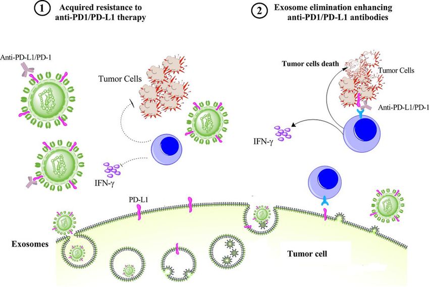
PD-L1 IN EXOSOMES AND THE ACQUIRED RESISTANCE TO
ANTI-PD-1/PD-L1 THERAPY EXOSOME ELIMINATION AS AN ADD-ON THERAPY ENHANCING
There is convincing preclinical and clinical evidence POTENCY OF ANTI-PD-1/PD-L1 ANTIBODIES
that shows the effective role of exosomal PD-L1 in the There is preclinical evidence that shows loss of PD-L1
relatively low response rate of anti-PD-L1/PD-1 therapy. expression from primary tumors along with the elimi-
Clinical significance of plasma circulating exosomal nation of exosome secretion via pharmacological inter-
PD-L1 has been indicated in patients with head and vention or genetic manipulation mitigate the metastatic
neck cancer,71 77 gastric cancer,78 79 NSCLC,72 pancreatic burden and elevate overall survival in a variety of tumor-
cancer,80 and melanoma.69 Among melanoma patients, bearing mice.69 70 73 Combination treatment with GW4869,
the pretreatment levels of circulating exosomal PD-L1 an inhibitor of exosome secretion, and anti-PD-L1 mAb
were found to be negatively correlated with disease demonstrated the highest reduction in primary tumor
response to pembrolizumab. Importantly, a higher level burden in mice bearing 4T-1 tumor, showing a syner-
of circulating exosomal PD-L1 before the treatment was gistic association between immune checkpoint inhibitors
associated with poorer clinical outcomes.69 and exosome elimination.70 Supporting this, anti-PD-L1
There are also preclinical studies that indicate the efficacy was markedly enhanced in mice harboring
removal of exosomal PD-L1 can increase the response rate tetracycline-inducible Rab27 knockdown 4T1 cells.70 Of
of the anti-PD-L1 blockade, supporting the critical impact equal relevance, an Rab27−/− MC38 cell line dramatically
of exosomal PD-L1 in therapeutic resistance to anti-PD-L1 enlarged overall survival in mice receiving anti- PD-
L1
antibody treatment. In the mouse 4T1 breast tumor mAb compared with either monotherapy alone.73 Similar
model, it was shown that the elimination of exosomal results were also observed by an independent group in
PD-L1 by inhibiting exosome secretion using knockdown other tumor models.69 Altogether, these findings suggest
of Rab27a in tumor cells considerably improved the effi- that exosome elimination may act as an effective add-on
ciency of anti-PD-1 therapy and suppressed the tumor therapy to improve the therapeutic efficiency of anti-
growth.70 The TRAMP-C2 model is a syngeneic model PD-1/PD-L1 blockade in patients with cancer. Exosomes
of prostate cancer81 that, like human prostate cancer, can be eliminated through suppressing their generation
is resistant to anti-PD-L1 blockade.82 Removal of tumor and secretion from tumor cells or removal of circulating
exosomal PD- L1 from TRAMP- C2 cells using genetic exosomes from the bloodstream using haemofiltration.
mutations could efficiently enhance the effect of PD-L1
blockade, which included inhibition of tumor growth, Pharmacological approaches for suppressing exosome
elevated cellularity of the spleen, and the activation of a generation and secretion
T cell response in lymph nodes with similar effects on the Elimination of circulating exosomes has emerged as a
various activation, exhaustion, and proliferation markers. novel and potentially useful therapeutic strategy for the
Interestingly, the injection of in vitro collected exosomes development of anticancer drugs.83 Many reports have
carrying PD-L1 was found to reverse all these outcomes in already shown that the reduction of exosome secretion
the TRAMP-C2 model, whereas, in the absence of PD-L1, or secreted exosomes, achieved by using a chemical
the effect of the exogenously introduced exosomes was inhibitor,84 85 genetic manipulation,86 or antibody,87 can
very low.82 Similarly, the removal of exosomal PD-L1 in suppress cancer metastasis and improve the efficiency
the colorectal MC38 model inhibited tumor growth and of cancer chemotherapy. The effective strategies for
extended survival through PD-L1 blockade treatment.73 exosome suppression are the use of pharmacological
Mechanistically, it was suggested that high levels of agents that reduce the level of exosomes by targeting
exosomal PD-L1 might reflect the “exhaustion” of T cells different molecules involving in the generation, pack-
to a stage at which they can no longer be reinvigorated by aging, and release of them. However, since exosomes
anti-PD-1 therapy.73 are implicated in intercellular communications and
Yin Z, et al. J Immunother Cancer 2021;9:e001698. doi:10.1136/jitc-2020-001698 5Open access
J Immunother Cancer: first published as 10.1136/jitc-2020-001698 on 20 January 2021. Downloaded from http://jitc.bmj.com/ on October 17, 2021 by guest. Protected by copyright.
Figure 2 Exosomal PD-L1 induces acquired resistance to anti-PD-1/PD-L1 therapy. Exosomes carrying PD-L1 limit
effectiveness of anti-PD-1/PD-L1 therapy through binding to antibodies and suppression of T-cell activity. However, elimination
of PD-L1 exosomes can improve anti-PD-1/PD-L1 therapy. IFN-γ, interferon-gamma; PD-1, programmed cell death protein 1;
PD-L1, programmed death-ligand 1.
maintaining normal cellular physiology, this represents cargo as well. Notably, cell treatment with sphingomyeli-
the most important limitations to be used as a thera- nase inhibitors was found to markedly reduce the release
peutic strategy, due to the potential toxicity and other of exosomes.93 Moreover, sulfisoxazole, an FDA-approved
side-effects caused by any partial or temporary inhibition oral antibiotic, was also identified as an inhibitor of
of exosome secretion from normal cells when a drug exosome secretion from breast cancer cells through
candidate inhibits the secretion of exosomes from cancer interference with endothelin receptor A, a member of
cells. Using exosome inhibitors developed by drug repur- G protein-coupled receptor (GPCR) family that plays a
posing strategy, a process of finding new indications for critical role in the biogenesis and secretion of exosome
existing FDA-approved drugs, can reduce concerns about in breast cancer cells.88 Of note, sulfisoxazole exhibited
safety/toxicity, because these agents have already passed significant antitumor and anti- metastatic impacts in
toxicity and safety tests in humans.85 88 For example, FDA- mouse models of breast cancer xenografts, the decreased
approved drugs, dimethyl amiloride (DMA) and omepra- expression of proteins participated in exosome biogen-
zole reduce exosome secretion through targeting H+/ esis and secretion, and induced lysosomal degradation of
Na+ and Na+/Ca2+ channels89 that are known to involve multivesicular endosomes.88 Interestingly, a quantitative
in exosome release.90 DMA has been found to reduce high throughput screen assay of many pharmacologi-
the production of exosomes and eliminate their immune cally active compounds approved for clinical application
suppressive effects in tumor-bearing hosts and enhance uncovers the lead compounds tipifarnib, neticonazole,
the antitumor efficacy of the cyclophosphamide as a climbazole, ketoconazole, and triademenol that inhibit
chemotherapeutic drug.89 Another possible option for exosome biogenesis and/or release by aggressive pros-
inhibiting exosome secretion is the use of proton pump tate cancer calls.85 Hence, utility of drug- repurposing
inhibitors (PPIs), which are widely prescribed for miti- for preventing exosome secretion may provide a useful
gating gastric acid.91 Vacuolar H+-ATPases can regulate and safe approach for unmasking the inhibitory effects
pH of extracellular microenvironments and involve in of exosomal PD-L1 on immune function and therapeutic
tumor progression. PPIs through inhibiting vacuolar H+- responses in cancer.
ATPase-driven efflux pumps impair the release of acidic
vesicles and vesicle- like structures by tumor cells and Exosome removal using extracorporeal haemofiltration
enhance the efficacy of chemotherapeutic agents.91 92 In Another alternative promising approach is exosome
addition, ceramide is known to involve in biogenesis and removal from the blood circulation by extracorporeal
release of exosomes, and sphingomyelinases, enzymes haemofiltration, which would not represent the possible
promoting ceramide synthesis, participate in sorting the drug toxicity or interactions, thereby suggesting an
6 Yin Z, et al. J Immunother Cancer 2021;9:e001698. doi:10.1136/jitc-2020-001698Open access
J Immunother Cancer: first published as 10.1136/jitc-2020-001698 on 20 January 2021. Downloaded from http://jitc.bmj.com/ on October 17, 2021 by guest. Protected by copyright.
advantage over pharmacological strategies. Aethlon models. These findings are all consistent with exosomal
ADAPT (adaptive dialysis- like affinity platform tech- PD-L1-mediated resistance to current anti-PD-L1/PD-1
nology) therapy is a therapeutic haemofiltration approach therapies. Although the mechanism underlying thera-
with a mechanism of action similar to kidney dialysis peutic resistance of exosomal PD- L1 is still unknown,
or continuous renal replacement therapy method. In some possible mechanisms are proposed. Importantly,
this approach, plasma and blood cells pass through the circulating exosomal PD-L1 can interact with anti-PD-1/
hollow‐fiber dialysis cartridges (Open access
J Immunother Cancer: first published as 10.1136/jitc-2020-001698 on 20 January 2021. Downloaded from http://jitc.bmj.com/ on October 17, 2021 by guest. Protected by copyright.
8 Ahmadzadeh M, Johnson LA, Heemskerk B, et al. Tumor antigen- 37 Thommen DS, Koelzer VH, Herzig P, et al. A transcriptionally and
specific CD8 T cells infiltrating the tumor express high levels of PD-1 functionally distinct PD-1+ CD8+ T cell pool with predictive potential
and are functionally impaired. Blood 2009;114:1537–44. in non-small-cell lung cancer treated with PD-1 blockade. Nat Med
9 Dong H, Strome SE, Salomao DR, et al. Tumor-Associated B7- 2018;24:994–1004.
H1 promotes T-cell apoptosis: a potential mechanism of immune 38 Sade-Feldman M, Yizhak K, Bjorgaard SL, et al. Defining T cell
evasion. Nat Med 2002;8:793–800. states associated with response to checkpoint immunotherapy in
10 Blank C, Brown I, Peterson AC, et al. PD-L1/B7H-1 Inhibits the melanoma. Cell 2018;175:e1020:998–1013.
Effector Phase of Tumor Rejection by T Cell Receptor (TCR) 39 Tran E, Ahmadzadeh M, Lu Y-C, et al. Immunogenicity of
Transgenic CD8 + T Cells. Cancer Res 2004;64:1140–5. somatic mutations in human gastrointestinal cancers. Science
11 Iwai Y, Ishida M, Tanaka Y, et al. Involvement of PD-L1 on 2015;350:1387–90.
tumor cells in the escape from host immune system and tumor 40 Tran E, Turcotte S, Gros A, et al. Cancer immunotherapy based on
immunotherapy by PD-L1 blockade. Proc Natl Acad Sci U S A mutation-specific CD4+ T cells in a patient with epithelial cancer.
2002;99:12293–7. Science 2014;344:641–5.
12 Rittmeyer A, Barlesi F, Waterkamp D, et al. Atezolizumab versus 41 Monach PA, Meredith SC, T.Siegel C, et al. A unique tumor
docetaxel in patients with previously treated non-small-cell lung antigen produced by a single amino acid substitution. Immunity
cancer (oak): a phase 3, open-label, multicentre randomised 1995;2:45–59.
controlled trial. Lancet 2017;389:255–65. 42 Robbins PF, El-Gamil M, Li YF, et al. A mutated beta-catenin
13 Baxi S, Yang A, Gennarelli RL, et al. Immune-Related adverse events gene encodes a melanoma-specific antigen recognized by tumor
for anti-PD-1 and anti-PD-L1 drugs: systematic review and meta- infiltrating lymphocytes. J Exp Med 1996;183:1185–92.
analysis. BMJ 2018;360:k793. 43 Dubey P, Hendrickson RC, Meredith SC, et al. The immunodominant
14 Friedman CF, Proverbs-Singh TA, Postow MA. Treatment of the antigen of an ultraviolet-induced regressor tumor is generated by a
immune-related adverse effects of immune checkpoint inhibitors. somatic point mutation in the DEAD box helicase p68. J Exp Med
JAMA Oncology 2016;2:1346–53. 1997;185:695–706.
15 Kwok G, Yau TCC, Chiu JW, et al. Pembrolizumab (Keytruda). Hum 44 Lennerz V, Fatho M, Gentilini C, et al. The response of autologous T
Vaccin Immunother 2016;12:2777–89. cells to a human melanoma is dominated by mutated neoantigens.
16 Peters S, Kerr KM, Stahel R. Pd-1 blockade in advanced NSCLC: a Proc Natl Acad Sci U S A 2005;102:16013–8.
focus on pembrolizumab. Cancer Treat Rev 2018;62:39–49. 45 Rizvi NA, Hellmann MD, Snyder A, et al. Mutational landscape
17 Sim F, Leidner R, Bell RB. Immunotherapy for head and neck cancer. determines sensitivity to PD-1 blockade in non–small cell lung
Oral Maxillofac Surg Clin North Am 2019;31:85–100. cancer. Science 2015;348:124–8.
18 Bellmunt J, de Wit R, Vaughn DJ, et al. Pembrolizumab as second- 46 Chen Y, Liu Q, Chen Z, et al. Pd-L1 expression and tumor mutational
line therapy for advanced urothelial carcinoma. N Engl J Med burden status for prediction of response to chemotherapy and
Overseas Ed 2017;376:1015–26. targeted therapy in non-small cell lung cancer. J Exp Clin Cancer Res
19 Herbst RS, Soria J-C, Kowanetz M, et al. Predictive correlates of 2019;38:193.
response to the anti-PD-L1 antibody MPDL3280A in cancer patients. 47 Hellmann MD, Nathanson T, Rizvi H, et al. Genomic features of
Nature 2014;515:563–7. response to combination immunotherapy in patients with advanced
20 Tumeh PC, Harview CL, Yearley JH, et al. Pd-1 blockade induces non-small-cell lung cancer. Cancer Cell 2018;33:e844:843–52.
responses by inhibiting adaptive immune resistance. Nature 48 Hellmann MD, Ciuleanu T-E, Pluzanski A, et al. Nivolumab plus
2014;515:568–71. ipilimumab in lung cancer with a high tumor mutational burden. N
21 Im SJ, Hashimoto M, Gerner MY, et al. Defining CD8+ T cells Engl J Med 2018;378:2093–104.
that provide the proliferative burst after PD-1 therapy. Nature 49 Rosenberg JE, Hoffman-Censits J, Powles T, et al. Atezolizumab in
2016;537:417–21. patients with locally advanced and metastatic urothelial carcinoma
22 Huang AC, Postow MA, Orlowski RJ, et al. T-Cell invigoration to who have progressed following treatment with platinum-based
tumour burden ratio associated with anti-PD-1 response. Nature chemotherapy: a single-arm, multicentre, phase 2 trial. The Lancet
2017;545:60–5. 2016;387:1909–20.
23 Forde PM, Chaft JE, Smith KN, et al. Neoadjuvant PD-1 50 Riaz N, Havel JJ, Makarov V, et al. Tumor and microenvironment
blockade in resectable lung cancer. N Engl J Med Overseas Ed evolution during immunotherapy with nivolumab. Cell
2018;378:1976–86. 2017;171:e916:934–49.
24 Bengsch B, Johnson AL, Kurachi M, et al. Bioenergetic 51 Hugo W, Zaretsky JM, Sun L, et al. Genomic and transcriptomic
Insufficiencies due to metabolic alterations regulated by the inhibitory features of response to anti-PD-1 therapy in metastatic melanoma.
receptor PD-1 are an early driver of CD8 + T cell exhaustion. Cell 2016;165:35–44.
Immunity 2016;45:358–73. 52 Goodman AM, Kato S, Bazhenova L, et al. Tumor mutational burden
25 Gubin MM, Zhang X, Schuster H, et al. Checkpoint blockade cancer as an independent predictor of response to immunotherapy in
immunotherapy targets tumour-specific mutant antigens. Nature diverse cancers. Mol Cancer Ther 2017;16:2598–608.
2014;515:577–81. 53 Legrand FA, Gandara DR, Mariathasan S, et al. Association of high
26 Loke P'ng, Allison JP. Pd-L1 and PD-L2 are differentially regulated by tissue TMB and atezolizumab efficacy across multiple tumor types.
Th1 and Th2 cells. Proc Natl Acad Sci U S A 2003;100:5336–41. JCO 2018;36:12000.
27 Dahan R, Sega E, Engelhardt J, et al. FcγRs modulate the anti-tumor 54 Yarchoan M, Hopkins A, Jaffee EM. Tumor mutational burden
activity of antibodies targeting the PD-1/PD-L1 axis. Cancer Cell and response rate to PD-1 inhibition. N Engl J Med Overseas Ed
2015;28:285–95. 2017;377:2500–1.
28 Jenkins RW, Barbie DA, Flaherty KT. Mechanisms of resistance to 55 Le DT, Durham JN, Smith KN, et al. Mismatch repair deficiency
immune checkpoint inhibitors. Br J Cancer 2018;118:9–16. predicts response of solid tumors to PD-1 blockade. Science
29 Polk A, Svane I-M, Andersson M, et al. Checkpoint inhibitors in 2017;357:409–13.
breast cancer – current status. Cancer Treat Rev 2018;63:122–34. 56 Le DT, Uram JN, Wang H, et al. Pd-1 blockade in tumors with
30 Sheng Z, Zhu X, Sun Y, et al. The efficacy of anti-PD-1/PD-L1 mismatch-repair deficiency. N Engl J Med 2015;372:2509–20.
therapy and its comparison with EGFR-TKIs for advanced non-small- 57 Germano G, Lamba S, Rospo G, et al. Inactivation of DNA repair
cell lung cancer. Oncotarget 2017;8:57826–35. triggers neoantigen generation and impairs tumour growth. Nature
31 Ribas A. Adaptive immune resistance: how cancer protects from 2017;552:116–20.
immune attack. Cancer Discov 2015;5:915–9. 58 Yi M, Jiao D, Xu H, et al. Biomarkers for predicting efficacy of PD-1/
32 Reck M, Rodríguez-Abreu D, Robinson AG, et al. Pembrolizumab PD-L1 inhibitors. Mol Cancer 2018;17:1–14.
versus chemotherapy for PD-L1–positive non–small-cell lung cancer. 59 Kalluri R. The biology and function of exosomes in cancer. J Clin
N Engl J Med Overseas Ed 2016;375:1823–33. Invest 2016;126:1208–15.
33 Ding W, LaPlant BR, Call TG, et al. Pembrolizumab in patients 60 Zhang L, Yu D. Exosomes in cancer development, metastasis, and
with CLL and Richter transformation or with relapsed CLL. Blood immunity. Biochim Biophys Acta Rev Cancer 2019;1871:455–68.
2017;129:3419–27. 61 Tavasolian F, Moghaddam AS, Rohani F. Exosomes: Effectual players
34 Patel SP, Kurzrock R. Pd-L1 expression as a predictive biomarker in in rheumatoid arthritis, 2020.
cancer immunotherapy. Mol Cancer Ther 2015;14:847–56. 62 Moghaddam AS, Afshari JT, Esmaeili S-A, et al. Cardioprotective
35 Yagi T, Baba Y, Ishimoto T, et al. Pd-L1 expression, tumor-infiltrating microRNAs: lessons from stem cell-derived exosomal microRNAs to
lymphocytes, and clinical outcome in patients with surgically treat cardiovascular disease. Atherosclerosis 2019;285:1–9.
resected esophageal cancer. Ann Surg 2019;269:471–8. 63 Moghiman T, Barghchi B, Esmaeili S-A, et al. Therapeutic
36 Teng MWL, Ngiow SF, Ribas A, et al. Classifying cancers based on angiogenesis with exosomal microRNAs: an effectual approach for
T-cell infiltration and PD-L1. Cancer Res 2015;75:2139–45. the treatment of myocardial ischemia, 2020: 1–9.
8 Yin Z, et al. J Immunother Cancer 2021;9:e001698. doi:10.1136/jitc-2020-001698Open access
J Immunother Cancer: first published as 10.1136/jitc-2020-001698 on 20 January 2021. Downloaded from http://jitc.bmj.com/ on October 17, 2021 by guest. Protected by copyright.
64 Zhou R, Wang L, Zhao G. Circulating exosomal microRNAs as 81 Foster BA, Gingrich JR, Kwon ED, et al. Characterization of prostatic
emerging non‐invasive clinical biomarkers in heart failure: Mega bio‐ epithelial cell lines derived from transgenic adenocarcinoma of the
roles of a nano bio‐particle;72. mouse prostate (TRAMP) model. Cancer Res 1997;57:3325–30.
65 Greening DW, Gopal SK, Xu R, et al. Exosomes and their roles in 82 Yu P, Steel JC, Zhang M, et al. Simultaneous inhibition of two
immune regulation and cancer. Semin Cell Dev Biol 2015;40:72–81. regulatory T-cell subsets enhanced interleukin-15 efficacy in a
66 Yi M, Xu L, Jiao Y, et al. The role of cancer-derived microRNAs in prostate tumor model. Proc Natl Acad Sci U S A 2012;109:6187–92.
cancer immune escape. J Hematol Oncol 2020;13:1–14. 83 EL Andaloussi S, Mäger I, Breakefield XO, et al. Extracellular
67 Sun Z, Shi K, Yang S, et al. Effect of exosomal miRNA on cancer vesicles: biology and emerging therapeutic opportunities. Nat Rev
biology and clinical applications. Mol Cancer 2018;17:147. Drug Discov 2013;12:347–57.
68 McAndrews KM, Kalluri R. Mechanisms associated with biogenesis 84 Datta A, Kim H, Lal M, et al. Manumycin a suppresses exosome
of exosomes in cancer. Mol Cancer 2019;18:52. biogenesis and secretion via targeted inhibition of Ras/Raf/ERK1/2
69 Chen G, Huang AC, Zhang W, et al. Exosomal PD-L1 contributes signaling and hnRNP H1 in castration-resistant prostate cancer cells.
to immunosuppression and is associated with anti-PD-1 response. Cancer Lett 2017;408:73–81.
Nature 2018;560:382–6. 85 Datta A, Kim H, McGee L, et al. High-Throughput screening identified
70 Yang Y, Li C-W, Chan L-C, et al. Exosomal PD-L1 harbors active selective inhibitors of exosome biogenesis and secretion: a drug
defense function to suppress T cell killing of breast cancer cells and repurposing strategy for advanced cancer. Sci Rep 2018;8:8161.
promote tumor growth. Cell Res 2018;28:862–4. 86 Bobrie A, Krumeich S, Reyal F, et al. Rab27A supports exosome-
71 Theodoraki M-N, Yerneni SS, Hoffmann TK, et al. Clinical dependent and -independent mechanisms that modify the tumor
Significance of PD-L1+ Exosomes in Plasma of Head and Neck microenvironment and can promote tumor progression. Cancer Res
Cancer Patients. Clin Cancer Res 2018;24:896–905. 2012;72:4920–30.
72 Li C, Li C, Zhi C, et al. Clinical significance of PD-L1 expression in 87 Nishida-Aoki N, Tominaga N, Takeshita F, et al. Disruption of
serum-derived exosomes in NSCLC patients. J Transl Med 2019;17. circulating extracellular vesicles as a novel therapeutic strategy
73 Poggio M, Hu T, Pai C-C, et al. Suppression of exosomal PD- against cancer metastasis. Mol Ther 2017;25:181–91.
L1 induces systemic anti-tumor immunity and memory. Cell 88 Im E-J, Lee C-H, Moon P-G, et al. Sulfisoxazole inhibits the secretion
2019;177:e413:414–27. of small extracellular vesicles by targeting the endothelin receptor a.
74 Ricklefs FL, Alayo Q, Krenzlin H, et al. Immune evasion mediated Nat Commun 2019;10:1387.
by PD-L1 on glioblastoma-derived extracellular vesicles. Sci Adv 89 Chalmin F, Ladoire S, Mignot G, et al. Membrane-Associated
2018;4:eaar2766. HSP72 from tumor-derived exosomes mediates STAT3-dependent
75 Kim DH, Kim H, Choi YJ, et al. Exosomal PD-L1 promotes tumor immunosuppressive function of mouse and human myeloid-derived
growth through immune escape in non-small cell lung cancer. Exp suppressor cells. J Clin Invest 2010;67:457–71.
Mol Med 2019;51:1–13. 90 Savina A, Furlán M, Vidal M, et al. Exosome release is regulated
76 Razzo BM, Ludwig N, Hong C-S, et al. Tumor-Derived exosomes by a calcium-dependent mechanism in K562 cells. J Biol Chem
promote carcinogenesis of murine oral squamous cell carcinoma. 2003;278:20083–90.
Carcinogenesis 2020;41:625–33. 91 Spugnini EP, Citro G, Fais S. Proton pump inhibitors as anti vacuolar-
77 Theodoraki M-N, Yerneni S, Gooding WE, et al. Circulating exosomes ATPases drugs: a novel anticancer strategy. J Exp Clin Cancer Res
measure responses to therapy in head and neck cancer patients 2010;29:44.
treated with cetuximab, ipilimumab, and IMRT. Oncoimmunology 92 Luciani F, Spada M, De Milito A, et al. Effect of proton pump inhibitor
2019;8:e1593805. pretreatment on resistance of solid tumors to cytotoxic drugs. J Natl
78 Fan Y, Liu Y, Qu X. ASO author reflections: the prognostic role of Cancer Inst 2004;96:1702–13.
exosomal PD-L1 in patients with gastric cancer. Ann Surg Oncol 93 Trajkovic K, Hsu C, Chiantia S, et al. Ceramide triggers budding
2019;26:851–2. of exosome vesicles into multivesicular endosomes. Science
79 Fan Y, Che X, Qu J, et al. Exosomal PD-L1 retains 2008;319:1244–7.
immunosuppressive activity and is associated with gastric cancer 94 Tullis RH, Duffin RP, Handley HH, et al. Reduction of hepatitis C virus
prognosis. Ann Surg Oncol 2019;26:3745–55. using lectin affinity plasmapheresis in dialysis patients. Blood Purif
80 Lux A, Kahlert C, Grützmann R, et al. C-Met and PD-L1 on circulating 2009;27:64–9.
exosomes as diagnostic and prognostic markers for pancreatic 95 Marleau AM, Chen C-S, Joyce JA, et al. Exosome removal as a
cancer. Int J Mol Sci 2019;20:3305. therapeutic adjuvant in cancer. J Transl Med 2012;10:134.
Yin Z, et al. J Immunother Cancer 2021;9:e001698. doi:10.1136/jitc-2020-001698 9You can also read