LncRNA SNHG22 sponges miR 128 3p to promote the progression of colorectal cancer by upregulating E2F3
←
→
Page content transcription
If your browser does not render page correctly, please read the page content below
INTERNATIONAL JOURNAL OF ONCOLOGY 59: 71, 2021

               lncRNA SNHG22 sponges miR‑128‑3p to promote the
               progression of colorectal cancer by upregulating E2F3
                          JIANNING YAO*, CHUNFENG WANG*, XUYANG DONG, YANZHEN ZHANG,
                                    YANLE LI, HAINING ZHOU and LIANFENG ZHANG

    Department of Gastroenterology, The First Affiliated Hospital, Zhengzhou University, Zhengzhou, Henan 450052, P.R. China

                                      Received November 15, 2020; Accepted June 14, 2021

                                                     DOI: 10.3892/ijo.2021.5251

Abstract. The long non‑coding RNA (lncRNA) small                     knockdown markedly inhibited CRC cell proliferation, apop‑
nucleolar RNA host gene 22 (SNHG22) has been reported as a           tosis resistance, migration and invasion in vitro, and hindered
crucial regulator in several types of human cancer. The present      tumor growth in vivo. The mechanistic study revealed that
study evaluated the function and mechanism of SNHG22 in              SNHG22 bound to miR‑128‑3p and attenuated its inhibi‑
colorectal cancer (CRC) progression. SNHG22 expression               tory effects on E2F transcription factor 3 (E2F3) expression
was detected in colorectal adenoma, CRC tumor tissues (TTs)          levels and activity. Rescue experiments demonstrated that
and adjacent non‑cancerous tissues (ANTs) using reverse              inhibiting miR‑128‑3p or upregulating E2F3 offset the effects
transcription‑quantitative PCR (RT‑qPCR). The biological             of SNHG22 knockdown on CRC cells. The present findings
behaviors of SNHG22 in CRC cell lines were explored                  support the existence of an interactive regulatory network
in vitro using Cell Counting Kit‑8, flow cytometry, wound            involving SNHG22, miR‑128‑3p and E2F3 in CRC cell lines,
scratch assay and Transwell assay, and in vivo using a nude          indicating that the SNHG22/miR‑128‑3p/E2F3 axis may be
mouse xenograft model. The interaction between SNHG22                considered a novel diagnostic and therapeutic target in CRC.
and microRNA‑128‑3p (miR‑128‑3p), and the target genes
of miR‑128‑3p were explored using online tools, RT‑qPCR,             Introduction
western blotting and a dual‑luciferase reporter assay. The
present study revealed that SNHG22 expression was most               Colorectal cancer (CRC) represents one of the most common
highly expressed in TTs followed by adenoma tissues and              malignancies worldwide. Every year, >1.3 million people are
ANTs. In addition, high SNHG22 expression levels were                diagnosed with CRC globally, of which ~0.7 million succumb
significantly associated with advanced clinicopathological           to this disease (1). Only ~50% of patients with CRC survive for
factors and worse survival in patients with CRC. SNHG22              5 years after diagnosis in Europe (2), despite some improve‑
                                                                     ments being made in early diagnosis and systemic therapies.
                                                                     The pathogenesis and progression of CRC is a complex
                                                                     process, and the potential mechanism remains unclear.
                                                                         Long non‑coding RNAs (lncRNAs) are a class of
Correspondence to: Dr Lianfeng Zhang, Department of
Gastroenterology, The First Affiliated Hospital, Zhengzhou           non‑encoding RNAs, which are >200 nucleotides long.
University, 1 Jianshe Dong Road, Zhengzhou, Henan 450052,            Recent research has indicated that numerous lncRNAs serve
P.R. China                                                           an important role in the majority of physiological and patho‑
E‑mail: 1900932217@qq.com                                            logical processes, including embryonic stem cell self‑renewal,
                                                                     carcinogenesis and cancer metastasis (3,4). In addition,
Abbreviations: CRC, colorectal cancer; EOC, epithelial ovarian       lncRNAs function as oncogenes or tumor suppressor genes,
carcinoma; PTC, papillary thyroid cancer; lncRNA, long non‑coding    which participate in CRC tumorigenesis and progression. For
RNA; snoRNA, small nucleolar RNA; SNHG, small nucleolar RNA          example, Xu et al (5) demonstrated that the lncRNA microRNA
host gene; siRNA, small interfering RNA; UTR, untranslated region;   (miRNA/miR)‑17‑92a‑1 cluster host gene (MIR17HG) drove
mut, mutant; RIP, RNA immunoprecipitation; CCK‑8, Cell Counting
                                                                     tumor development and metastasis in CRC cells by enhancing
Kit‑8; TT, tumor tissue; ANT, adjacent non‑cancerous tissue; TCGA,
                                                                     the expression of NF‑κ B/RELA. In addition, HOX transcript
The Cancer Genome Atlas; DFS, disease‑free survival; OS, overall
survival; ceRNA, competing endogenous RNA; HR, hazard ratio;         antisense RNA has been revealed to enhance CRC cell
CI, confidence interval                                              migration and drug resistance via a miR‑203a‑3p‑dependent
                                                                     Wnt/β ‑catenin signaling pathway (6). As an oncogene, the
*
    Contributed equally                                              lncRNA H19 has been shown to accelerate cell proliferation
                                                                     and metastasis in CRC by acting on Wnt signaling (7).
Key words: small nucleolar RNA host gene 22, E2F transcription           Small nucleolar RNAs (snoRNAs) are a subgroup of
factor 3, microRNA‑128‑3p, colorectal cancer                         ncRNAs that are 60‑300 nucleotides in length, which contribute
                                                                     to tumorigenesis and metastasis in diverse types of human
                                                                     cancer. Most snoRNAs are encoded in the introns of snoRNA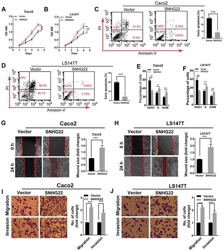
2                                 YAO et al: SNHG22 AND COLORECTAL CANCER PROGRESSION

host genes (SNHGs) (8). Specifically, there are numerous         (E2F3) were purchased from Shanghai GenePharma Co., Ltd..
SNHGs associated with CRC carcinogenesis and progression;        A non‑targeting sequence was used as the negative control
for example, SNHG7 has been shown to be overexpressed in         (siNC; Shanghai GenePharma Co. Ltd.). The miRNA mimics,
CRC tumor tissues (TTs) compared with its expression in adja‑    inhibitor and negative controls (NC mimic and NC inhibitor)
cent non‑cancerous tissues (ANTs). This high expression has      were purchased from Guangzhou RiboBio Co., Ltd.. For the
been revealed to be related to aggressive pathological char‑     in vivo experiments, vectors containing short hairpin RNAs
acteristics, such as tumor size, tumor‑node‑metastasis (TNM)     (shRNAs) targeting SNHG22 (shSNHG22, i.e., sh#1 and sh#2)
stage, distant metastasis and poor survival (9,10). Located on   or a non‑targeting sequence (shNC) were subcloned into Lv5
chromosome 8q21.1, SNHG22 is 2,157 nucleotides long (11).        lentiviruses (Shanghai GenePharma Co., Ltd.) and infected
Previous studies have reported that SNHG22 contributes to cell   into LoVo cells to generate Lv‑sh#1 and Lv‑sh#2. For LoVo
growth, migration, invasion and chemotherapy resistance in       cells infection, cells (5x10⁴ cell/well) were cultured for 24 h,
epithelial ovarian carcinoma (EOC), papillary thyroid cancer     and then recombinant lentivirus in serum‑free growth medium
(PTC) and breast cancer (12‑14). Notably, in these three types   was added at a multiplicity of infection of 50 at 37˚C for
of cancer, SNHG22 expression was revealed to be upregulated      2 days. For transient transfection, Caco2, LS174T and LoVo
in TTs compared with that in ANTs, thus indicating its asso‑     cells (3x10⁴ cell/well) in 6‑well plates were transfected with
ciation with poor prognosis. To the best of our knowledge, the   vectors/sequences at a concentration of 20 µg/ml at 37˚C.
biological role and expression patterns of SNHG22 have not       After culturing for 24 h, cells were harvested for subsequent
been examined in human CRC.                                      experiments. Transfection experiments were conducted
    The present study investigated SNHG22 expression in          with Lipofectamine® 3000 transfection reagent (Invitrogen;
adenoma and CRC tissues, and evaluated its clinical signifi‑     Thermo Fisher Scientific, Inc.) according to the manufacturer's
cance. Furthermore, the biological behaviors of SNHG22           instructions. All the aforementioned sequences are shown in
were explored in CRC cell lines in vitro and in vivo. The        Table SII.
present study also investigated the mechanisms underlying the
pro‑oncogenic effects of SNHG22 on CRC.                          Cell proliferation. Cell proliferation was detected using Cell
                                                                 Counting Kit‑8 (CCK‑8; Beyotime Institute of Biotechnology)
Materials and methods                                            according to the manufacturer's instructions. The cells were
                                                                 transfected with the indicated vectors or sequences, and then
Tissue samples and cell lines. A total of 93 paired CRC TTs      cultured in a 96‑well plate for 1, 2, 3 and 4 days after trans‑
and matched ANTs were collected from patients who had            fection. Subsequently, ~10 µl CCK‑8 reagent was added per
undergone surgery between January 2012 and October 2012          well, and incubated at 37˚C for 2 h in a 5% CO2 humidified
(Zhengzhou cohort). Additional fresh specimens were collected    chamber. The absorbance was measured at 490 nm in each
from patients with colorectal adenoma (CRA) (n=33) who had       well using a microplate reader (Bio‑Rad Laboratories, Inc.).
undergone colonoscopy. The CRA or CRC diagnoses were             The experiment was repeated at least three times.
based on histopathological evaluation using the 7th edition of
the American Joint Committee on Cancer staging system (15).      Flow cytometry. Cell cycle distribution was detected using
All specimens were quickly snap‑frozen in liquid nitrogen and    flow cytometry. After the CRC cells had been harvested
stored at ‑80˚C until required. The present study was approved   and washed, cells were detected with a Cell Cycle and
by the Ethics Committee of Zhengzhou University (approval        Apoptosis Analysis Kit (cat. no. C1052M; Beyotime Institute
no. 2011110402; Zhengzhou, China). Patients with any history     of Biotechnology) according to the manufacturer's protocol.
of other types of cancer, and who had received preoperative      Apoptosis was detected using an Annexin V‑fluorescein
radiotherapy or chemotherapy were excluded. The clinical         isothiocyanate (FITC)/propidium iodide (PI)‑Apoptosis
characteristics of all patients are listed in Table SI.          Detection kit [MultiSciences (Lianke) Biotech Co., Ltd.]. CRC
    Human colon cancer cell lines (Caco2, LS174T, LoVo,          cells (5x10 4/well) were seeded and resuspended in 12‑well
SW480, SW620) and a CRC cell line (HT‑29) were all obtained      plates, and Annexin V‑FITC (5 µl/well) and PI (100 µl/well)
from the Cell Bank of the Chinese Academy of Sciences. The       were added to each reaction system for 6 min. Immediately
cells were maintained in RPMI‑1640 medium (Gibco; Thermo         after staining, flow cytometric assays were conducted using
Fisher Scientific, Inc.) containing 10% fetal bovine serum       a flow cytometer (EPICS; Beckman Coulter, Inc.). Analysis
(FBS; Gibco; Thermo Fisher Scientific, Inc.) in a humidified     of flow cytometry results was conducted using FlowJo
incubator (37˚C, 5% CO2). The FHC human normal colon             software7.6.1 (FlowJo, LLC).
epithelial cell line was purchased from Mingzhou Company
(cat. no. MZ‑0713) and was cultured in 90% Dulbecco's            Wound scratch assay. Cell migration was determined using
modified Eagle's medium (DMEM; Gibco; Thermo Fisher              a wound scratch assay. Briefly, 1x10⁶ transfected cells/well
Scientific, Inc.) supplemented with 20% FBS. All cells tested    were seeded into a 24‑well plate. When cells reached 100%
negative for mycoplasma contamination and this result was        confluence, a sterile 20‑µl pipette tip was used to produce a
verified by short tandem repeat fingerprinting before use.       clear line in the wells. After washing with phosphate‑buffered
                                                                 saline (PBS), the cells were grown in serum‑free medium at
Cell transfection. Small interfering RNAs (siRNAs) against       37˚C for 24 h. Under a light microscope, images of the cells
SNHG22 (si#1 and si#2) or E2F transcription factor 3 (E2F3)      were captured to record the wound width at 0 h and more
(siE2F3), and the overexpression vectors pcDNA3.1/Control        images were captured after 24 h. The migration distance was
(Vector), pcDNA3.1/SNHG22 (SNHG22) and pcDNA3.1/E2F3             determined as the distance covered between 0 and 24 h.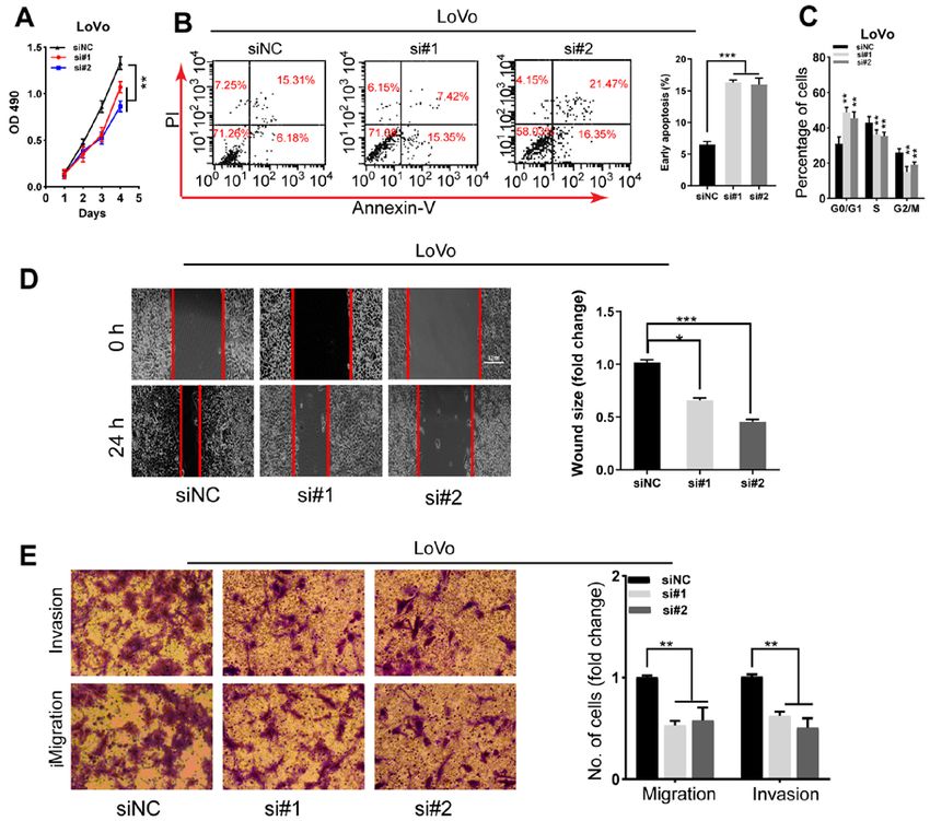
INTERNATIONAL JOURNAL OF ONCOLOGY 59: 71, 2021                                                    3

Table I. Cox proportional hazards regression models for overall and recurrence‑free survival among patients with colorectal
cancer.

                                                              Univariate analysis                  Multivariate analysis
	--------------------------------------------------------------	-------------------------------------------------------------
Parameter                                                HR (95% CI)           P‑value          HR (95% CI)          P‑value

Disease‑free survival
 Location, rectum vs. colon                      1.98 (0.85‑4.60) 0.112 1.67 (0.71‑3.90)                                            0.237
 T stage, T3‑4 vs. T1‑2                          2.13 (1.05‑4.30) 0.035 1.66 (1.02‑2.70)                                            0.041
 Node involvement, yes vs. no                    2.35 (1.07‑5.13) 0.033 1.27 (1.00‑1.61)                                            0.048
 M stage, M1 vs. M0                              2.56 (1.23‑5.33) 0.012 1.51 (1.04‑2.20)                                            0.031
 Tumor differentiation, poor vs. well + moderate 1.49 (0.95‑2.34) 0.084 1.14 (0.88‑1.48)                                            0.321
 CEA, ≥5 vs.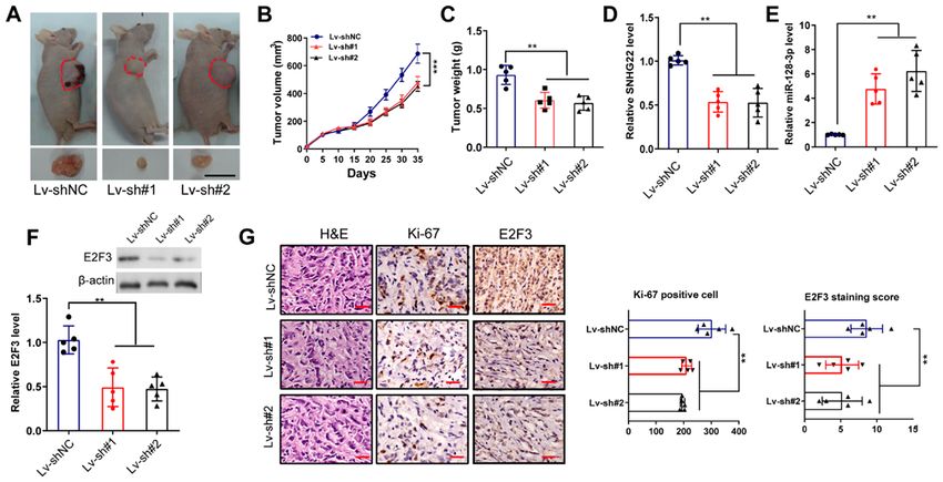
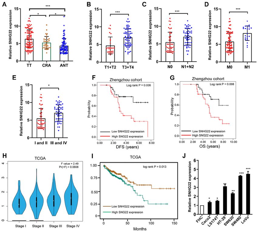
4                                        YAO et al: SNHG22 AND COLORECTAL CANCER PROGRESSION

Figure 1. Differential expression of SNHG22 in CRC tissues and cell lines. (A) RT‑qPCR analysis of the expression levels of SNHG22 in TTs (n=93), ANTs
(n=93) and CRA (n=33) colon tissues. Differential expression of SNHG22 in CRC tissues was analyzed according to (B) T stage, (C) node status, (D) distant
metastasis and (E) TNM stage. Kaplan‑Meier survival analysis of SNHG22 expression and (F) DFS and (G) OS in patients with CRC. (H) Expression of
SNHG22 in patients with CRC split according to TNM stage in TCGA‑COAD dataset. (I) OS in patients with CRC in TCGA‑COAD dataset. (J) RT‑qPCR
analysis of the expression levels of SNHG22 in CRC cell lines and the FHC cell line, a human normal colon epithelial cell line. Data are presented as the
mean ± SD from triplicate experiments. *P
INTERNATIONAL JOURNAL OF ONCOLOGY 59: 71, 2021                                                               5

Figure 2. Overexpression of SNHG22 promotes colorectal cancer cell proliferation, apoptosis resistance, migration and invasion in vitro. Cell Counting
Kit‑8 assay analysis of the proliferative ability of (A) Caco2 and (B) LS147T cells transfected with the indicated vectors. Flow cytometric analysis of the
(C and D) apoptotic rates and (E and F) cell cycle progression of Caco2 and LS147T cells transfected with the indicated vectors. Wound scratch analysis of
the migration of (G) Caco2 and (H) LS147T cells transfected with the indicated vectors (magnification, x200). Transwell assay analysis of the migration and
invasion of (I) Caco2 and (J) LS147T cells transfected with the indicated vectors (magnification, x200). Data are presented as the mean ± SD from triplicate
experiments. *P
6                                          YAO et al: SNHG22 AND COLORECTAL CANCER PROGRESSION

Figure 3. Knockdown of SNHG22 inhibits colorectal cancer cell proliferation, apoptosis resistance, migration and invasion in vitro. (A) Cell Counting Kit‑8
assay analysis of the proliferative ability of LoVo cells after transfection with the indicated sequences. Flow cytometric analysis of the (B) apoptotic rates
and (C) cell cycle progression of LoVo cells after transfection with the indicated sequences. (D) Wound scratch analysis of the migration of LoVo cells after
transfection with the indicated sequences (magnification, x200). (E) Transwell invasion assay analysis of the invasion of LoVo cells after transfection with the
indicated sequences (magnification, x200). Data are presented as the mean ± SD from triplicate experiments. *P
INTERNATIONAL JOURNAL OF ONCOLOGY 59: 71, 2021                                                             7

Figure 4. SNHG22 functions as a competing endogenous RNA to regulate miR‑128‑3p expression in CRC. (A) Subcellular localization of SNHG22 in CRC
cell lines (Caco2, LS147T and LoVo). β‑Actin and U6 served as cytoplasmic and nuclear localization markers, respectively. (B) Predicted binding sites of
miR‑128‑3p to the SNHG22 sequence. (C) RNA immunoprecipitation assay was performed with an antibody against Ago2 or IgG in Caco2 and LS147T cell
lines. (D) Luciferase activity of 293T, Caco2 and LS147T cell lines co‑transfected with miR‑128‑3p mimic (or NC mimic) and luciferase reporters containing
SNHG22 wt or SNHG22 mut transcript was analyzed. (E and F) Reverse transcription‑quantitative PCR analysis of the expression levels of miR‑128‑3p in
CRC cells after transfection with the indicated vectors and sequences. (G) Negative correlation between SNHG22 and miR‑128‑3p expression in human CRC
tissues from the Zhengzhou cohort (Spearman's correlation analysis; R=‑0.4649, P=0.002). Data are presented as the mean ± SD from triplicate experiments.
**
  P
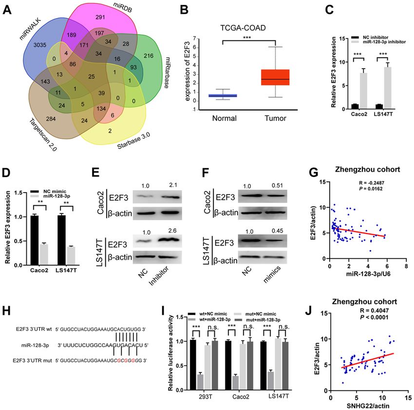
8                                         YAO et al: SNHG22 AND COLORECTAL CANCER PROGRESSION

Figure 5. miR‑128‑3p targets and regulates E2F3. (A) A Venn diagram showing the number of genes identified as potential targets of miR‑128‑3p. (B) Expression
levels of E2F3 in tumor and unmatched normal tissues in TCGA‑COAD dataset. (C and D) Reverse transcription‑quantitative PCR and (E and F) western blot
analysis of the mRNA and protein expression levels of E2F3 in Caco2 and LS147T cells after transfection with the indicated sequences. Protein expression
levels were semi‑quantified by normalization against β‑actin with ImageJ. (G) A significant inverse association was detected between miR‑128‑3p and E2F3
mRNA expression in human CRC tissues from the Zhengzhou cohort (R=‑0.2487, P=0.016). (H) miR‑128‑3p putative binding sites and corresponding mut
sequence of E2F3. (I) Dual‑luciferase reporter assays in CRC cell lines co‑transfected with miR‑128‑3p mimic and luciferase reporters containing wt or mut
E2F3 transcripts. The relative luciferase activity was normalized to the Renilla luciferase activity. (J) A positive correlation between SNHG22 and E2F3
expression in human CRC tissues from Zhengzhou cohort (R=‑0.4047, P
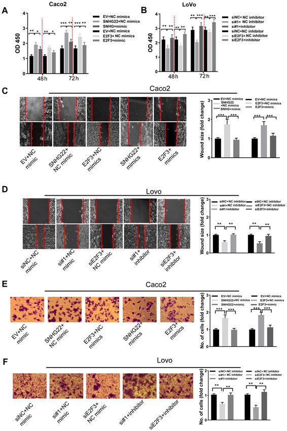
INTERNATIONAL JOURNAL OF ONCOLOGY 59: 71, 2021                                                                 9

Figure 6. SNHG22 exerts its function by inhibiting the miR‑128‑3p/E2F3 axis in CRC cells. Cell Counting Kit‑8 assay analysis of the proliferative ability of
(A) Caco2 and (B) LoVo cells after transfection with the indicated vectors and sequences. Wound scratch analysis of the migratory ability of (C) Caco2 and
(D) LoVo cells after transfection with the indicated vectors and sequences (magnification, x200). Transwell assay analysis of the invasive ability of (E) Caco2
and (F) LoVo cells after transfection with the indicated vectors and sequences (magnification, x200). Data are presented as the mean ± SD from triplicate
experiments. *P
10                                        YAO et al: SNHG22 AND COLORECTAL CANCER PROGRESSION

Figure 7. Knockdown of SNHG22 inhibits tumor xenograft growth of CRC in vivo. (A) LoVo cells were inoculated in BALB/c nude mice (n=5/group) to
establish subcutaneous xenograft tumors, and images of the dissected tumors were captured. Scale bar, 10 mm. Effects of SNHG22 knockdown in LoVo cells
on (B) tumor volume and (C) tumor weight in the subcutaneous xenograft mouse models. (D and E) Reverse transcription‑quantitative PCR and (F) western
blot analysis of the expression levels of SNHG22, miR‑128‑3p and E2F3 in the subcutaneous xenograft mouse models. (G) Immunohistochemical analysis of
the expression levels of Ki‑67 and E2F3 in the subcutaneous xenograft mouse models. Scale bar, 100 µm. Data are presented as the mean ± SD from triplicate
experiments. **P
INTERNATIONAL JOURNAL OF ONCOLOGY 59: 71, 2021                                           11

CRC cells (Fig. S6A and B). SNHG22 or E2F3 overexpres‑             SNHG22 expression may indicate poor prognosis in these
sion stimulated Caco2 cell proliferation, apoptosis resistance,    three human malignancies (12,14,24). The present study first
migration and invasion, whereas upregulating miR‑128‑3p            explored the expression patterns of SNHG22 in CRC TTs
reversed these effects. By contrast, knocking down SNHG22 or       and ANTs, and in CRA tissues, and revealed that SNHG22
E2F3 significantly weakened the LoVo cell malignant pheno‑         expression was upregulated in the order of ANTs > CRA
type; nevertheless, these inhibitory effects were attenuated by    tissues > TTs. High SNHG22 expression levels were associ‑
co‑transfection with miR‑128‑3p inhibitors (Figs. 6A‑F, and        ated with advanced T stage, node involvement, metastasis
S6C and D). These results indicated that the miR‑128‑3p/E2F3       and poor survival in patients with CRC. Therefore, SNHG22
axis reversed the stimulatory effects of SNHG22 on the prolif‑     may serve as an independent prognostic indicator in patients
erative, migratory and invasive capacity of CRC cells.             with CRC.
                                                                       As an oncogene, SNHG22 may serve critical roles in
Knockdown of SNHG22 inhibits CRC tumor xenograft growth            tumor advancement processes; for example, it was shown to
in vivo. A tumor xenograft assay was performed to confirm the      be highly expressed in EOC, and to stimulate EOC cell prolif‑
roles of SNHG22 in CRC growth in vivo. As shown in Fig. 7A‑C,      eration, invasion and chemotherapy resistance by sponging
compared with in the Lv‑shNC group, the tumor size, volume         miR‑2467 to facilitate galectin‑1 expression in EOC cells (11).
and weight in mice in the Lv‑sh#1 and Lv‑sh#2 groups were          Fang et al (13) observed that, in breast cancer, SNHG22
markedly reduced (P
12 YAO et al: SNHG22 AND COLORECTAL CANCER PROGRESSION
could be a direct target of miR‑128‑3p in CRC cells. Further References
functional rescue experiments validated the hypothesis that
SNHG22 may regulate CRC proliferation and invasion by 1. Bray F, Ferlay J, Soerjomataram I, Siegel RL, Torre LA and
Jemal A: Global cancer statistics 2018: GLOBOCAN estimates
competitively sponging miR‑128‑3p and restoring E2F3 of incidence and mortality worldwide for 36 cancers in 185 coun‑
activity. Recent studies have revealed that E2F3 may serve an tries. CA Cancer J Clin 68: 394‑424, 2018.
essential role in regulating human cancer cell proliferation, 2. De Angelis R, Sant M, Coleman MP, Francisci S, Baili P,
Pierannunzio D, Trama A, Visser O, Brenner H, Ardanaz E, et al:
apoptosis and chemosensitivity (33,34). Cancer survival in Europe 1999‑2007 by country and age: Results
In conclusion, the results of the present study revealed that, of EUROCARE‑5‑a population‑based study. Lancet Oncol 15:
as an oncogenic lncRNA in CRC, SNHG22 was upregulated 23‑34, 2014.
3. He Q, Long J, Yin Y, Li Y, Lei X, Li Z and Zhu W: Emerging
and related to poor survival in patients with CRC. Functional roles of lncRNAs in the formation and progression of colorectal
and mechanistic analyses demonstrated that SNHG22 cancer. Front Oncol 9: 1542, 2020.
promoted CRC tumorigenesis and metastasis by sponging 4. Luo Y, Yang J, Yu J, Liu X, Yu C, Hu J, Shi H and Ma X: Long
non‑coding RNAs: Emerging roles in the immunosuppressive
miR‑128‑3p, leading to elevated expression of E2F3. These tumor microenvironment. Front Oncol 10: 48, 2020.
findings may provide novel insights into the development of 5. Xu J, Meng Q, Li X, Yang H, Xu J, Gao N, Sun H, Wu S,
therapeutics for CRC. Familiari G, Relucenti M, et al: Long noncoding RNA MIR17HG
promotes colorectal cancer progression via miR‑17‑5p. Cancer
Res 79: 4882‑4895, 2019.
Acknowledgements 6. Xiao Z, Qu Z, Chen Z, Fang Z, Zhou K, Huang Z, Guo X and
Zhang Y: LncRNA HOTAIR is a prognostic biomarker for
the proliferation and chemoresistance of colorectal cancer via
Not applicable. miR‑203a‑3p‑mediated Wnt/ß ‑catenin signaling pathway. Cell
Physiol Biochem 46: 1275‑1285, 2018.
Funding 7. Ding D, Li C, Zhao T, Li D, Yang L and Zhang B: LncRNA
H19/miR‑29b‑3p/PGRN axis promoted epithelial‑mesenchymal
transition of colorectal cancer cells by acting on Wnt signaling.
The present study was supported by the Key R & D Promotion Mol Cells 41: 423‑435, 2018.
Projects of Henan Province 2019 (grant no. 192102310055). 8. Williams GT and Farzaneh F: Are snoRNAs and snoRNA host
genes new players in cancer? Nat Rev Cancer 12: 84‑88, 2012.
9. Shan Y, Ma J, Pan Y, Hu J, Liu B and Jia L: LncRNA SNHG7
Availability of data and materials sponges miR‑216b to promote proliferation and liver metastasis
of colorectal cancer through upregulating GALNT1. Cell Death
Dis 9: 722, 2018.
The datasets used and/or analyzed during the current study are 10. Li Y, Zeng C, Hu J, Pan Y, Shan Y, Liu B and Jia L: Long
available from the corresponding author on reasonable request non‑coding RNA‑SNHG7 acts as a target of miR‑34a to
or from the TCGA repository (https://portal.gdc.cancer.gov/. increase GALNT7 level and regulate PI3K/Akt/mTOR
pathway in colorectal cancer progression. J Hematol Oncol 11:
89, 2018.
Authors' contributions 11. Ota T, Suzuki Y, Nishikawa T, Otsuki T, Sugiyama T, Irie R,
Wakamatsu A, Hayashi K, Sato H, Nagai K, et al: Complete
sequencing and characterization of 21,243 full‑length human
LFZ, JNY, CFW and XYD conceived and designed the present cDNAs. Nat Genet 36: 40‑45, 2004.
study. JNY, YLL, HNZ and XYD performed the experiments. 12. Zhang PF, Wu J, Luo JH, Li KS, Wang F, Huang W, Wu Y,
JNY, CFW, XYD and YZZ analyzed and interpretated the Gao SP, Zhang XM and Zhang PN: SNHG22 overexpression
indicates poor prognosis and induces chemotherapy resistance
data. JNY, CFW and LFZ wrote, reviewed and/or revised the via the miR‑2467/Gal‑1 signaling pathway in epithelial ovarian
manuscript. JNY and LFZ confirm the authenticity of all the carcinoma. Aging (Albany NY) 11: 8204‑8216, 2019.
raw data. All authors read and approved the final manuscript. 13. Fang X, Zhang J, Li C, Liu J, Shi Z and Zhou P: Long
non‑coding RNA SNHG22 facilitates the malignant phenotypes
in triple‑negative breast cancer via sponging miR‑324‑3p and
Ethics approval and consent to participate upregulating SUDS3. Cancer Cell Int 20: 252, 2020.
14. Gao H, Sun X, Wang H and Zheng Y: Long noncoding RNA
SNHG22 increases ZEB1 expression via competitive binding
The present study involving human tissues was approved by with microRNA‑429 to promote the malignant development of
the Ethics Committee of Zhengzhou University (approval papillary thyroid cancer. Cell Cycle 19: 1186‑1199, 2020.
no. 2011110402). All patients provided written informed 15. Edge S, Byrd DR, Compton CC, Fritz AG, Greene F and
Trotti A (eds): AJCC cancer staging handbook. 7th edition.
consent prior to their inclusion within the study. The Springer, New York, NY, 2010.
research has been carried out in accordance with the World 16. Livak KJ and Schmittgen TD: Analysis of relative gene expres‑
Medical Association Declaration of Helsinki. All animal sion data using real‑time quantitative PCR and the 2(‑Delta Delta
C(T)) method. Methods 25: 402‑408, 2001.
experiments were approved by the Committee of the Ethics 17. Du Y, Wei N, Hong J and Pan W: Long non‑coding RNASNHG17
of Animal Experiments of Zhengzhou University (approval promotes the progression of breast cancer by sponging
no. 20180376). miR‑124‑3p. Cancer Cell Int 20: 40, 2020.
18. National Research Council (US) Committee for the Update of
the Guide for the Care and Use of Laboratory Animals: Guide
Patient consent for publication for the care and use of laboratory animals. 8th edition. National
Academies Press (US), Washington, DC, 2011.
All patients included in the present study have provided 19. Cao Z, Pan X, Yang Y, Huang Y and Shen HB: The lncLocator:
A subcellular localization predictor for long non‑coding RNAs
consent for both participation and publication. based on a stacked ensemble classifier. Bioinformatics 34:
2185‑2194, 2018.
Competing interests 20. Lan Y, Xiao X, He Z, Luo Y, Wu C, Li L and Song X: Long
noncoding RNA OCC‑1 suppresses cell growth through desta‑
bilizing HuR protein in colorectal cancer. Nucleic Acids Res 46:
The authors declare that they have no competing interests. 5809‑5821, 2018.INTERNATIONAL JOURNAL OF ONCOLOGY 59: 71, 2021 13
21. Wang Y, Lu JH, Wu QN, Jin Y, Wang DS, Chen YX, Liu J, 29. Liu X, Dong C, Ma S, Wang Y, Lin T, Li Y, Yang S, Zhang W,
Luo XJ, Meng Q, Pu HY, et al: LncRNA LINRIS stabilizes Zhang R and Zhao G: Nanocomplexes loaded with miR‑128‑3p
IGF2BP2 and promotes the aerobic glycolysis in colorectal for enhancing chemotherapy effect of colorectal cancer through
cancer. Mol Cancer 18: 174, 2019. dual‑targeting silence the activity of PI3K/AKT and MEK/ERK
22. Wang D, Zhang H, Fang X, Zhang X and Liu H: Prognostic pathway. Drug Deliv 27: 323‑333, 2020.
value of long non‑coding RNA GHET1 in cancers: A systematic 30. Liu T, Zhang X, Du L, Liu X, Tian H, Wang L, Li P, Zhao Y,
review and meta‑analysis. Cancer Cell Int 20: 109, 2020. Duan W, Xie Y, et al: Exosome‑transmitted miR‑128‑3p increase
23. Xiao M, Feng Y, Liu C and Zhang Z: Prognostic values of long chemosensitivity of oxaliplatin‑resistant colorectal cancer. Mol
noncoding RNA PVT1 in various carcinomas: An updated Cancer 18: 43, 2019.
systematic review and meta‑analysis. Cell Prolif 51: e12519, 2018. 31. Li X, Lv X, Li Z, Li C, Li X, Xiao J, Liu B, Yang H and Zhang Y:
24. Yang W, Zhang K, Li L, Ma K, Hong B, Gong Y and Gong K: Long noncoding RNA ASLNC07322 functions in VEGF‑C
Discovery and validation of the prognostic value of the lncRNAs expression regulated by smad4 during colon cancer metastasis.
encoding snoRNAs in patients with clear cell renal cell carci‑ Mol Ther Nucleic Acids 18: 851‑862, 2019.
noma. Aging (Albany NY) 12: 4424‑4444, 2020. 32. Fu C, Li D, Zhang X, Liu N, Chi G and Jin X: LncRNA PVT1
25. Li M, Bian Z, Jin G, Zhang J, Yao S, Feng Y, Wang X, Yin Y, facilitates tumorigenesis and progression of glioma via regula‑
Fei B, You Q and Huang Z: LncRNA‑SNHG15 enhances cell tion of MiR‑128‑3p/GREM1 axis and BMP signaling pathway.
proliferation in colorectal cancer by inhibiting miR‑338‑3p. Neurotherapeutics 15: 1139‑1157, 2018.
Cancer Med 8: 2404‑2413, 2019. 33. Wang H, Wang L, Zhang S, Xu Z and Zhang G: Downregulation
26. Yan J, Jia Y, Chen H, Chen W and Zhou X: Long non‑coding of LINC00665 confers decreased cell proliferation and invasion
RNA PXN‑AS1 suppresses pancreatic cancer progression by via the miR‑138‑5p/E2F3 signaling pathway in NSCLC. Biomed
acting as a competing endogenous RNA of miR‑3064 to upregu‑ Pharmacother 127: 110214, 2020.
late PIP4K2B expression. J Exp Clin Cancer Res 38: 390, 2019. 34. Chen J, Liu X, Xu Y, Zhang K, Huang J, Pan B, Chen D, Cui S,
27. Chen DL, Lu YX, Zhang JX, Wei XL, Wang F, Zeng ZL, Pan ZZ, Song H, Wang R, et al: TFAP2C‑activated MALAT1 modulates
Yuan YF, Wang FH, Pelicano H, et al: Long non‑coding RNA the chemoresistance of docetaxel‑resistant lung adenocarcinoma
UICLM promotes colorectal cancer liver metastasis by acting cells. Mol Ther Nucleic Acids 14: 567‑582, 2019.
as a ceRNA for microRNA‑215 to regulate ZEB2 expression.
Theranostics 7: 4836‑4849, 2017.
28. Zhao J, Li D and Fang L: MiR‑128‑3p suppresses breast This work is licensed under a Creative Commons
cancer cellular progression via targeting LIMK1. Biomed Attribution-NonCommercial-NoDerivatives 4.0
Pharmacother 115: 108947, 2019. International (CC BY-NC-ND 4.0) License.You can also read


