Endoplasmic Reticulum (ER) Stress and Unfolded Protein Response (UPR) in Mammalian Oocyte Maturation and Preimplantation Embryo Development - MDPI
←
→
Page content transcription
If your browser does not render page correctly, please read the page content below
International Journal of
           Molecular Sciences

Review
Endoplasmic Reticulum (ER) Stress and Unfolded
Protein Response (UPR) in Mammalian Oocyte
Maturation and Preimplantation
Embryo Development
Tao Lin, Jae Eun Lee, Jung Won Kang, Hyeon Yeong Shin, Ju Bin Lee and Dong Il Jin *
 Division of Animal & Dairy Science, Chungnam National University, Daejeon 34134, Korea;
 ailuomansi@gmail.com (T.L.); lje9090@hanmail.net (J.E.L.); rinstein@naver.con (J.W.K.);
 sin-hy@cnu.ac.kr (H.Y.S.); dlwnqlssla@naver.com (J.B.L.)
 * Correspondence: dijin@cnu.ac.kr; Tel.: +82-42-821-5876

 Received: 7 December 2018; Accepted: 9 January 2019; Published: 18 January 2019

 Abstract: Mammalian oocytes and early embryos derived from in vitro production are highly
 susceptible to a variety of cellular stresses. During oocyte maturation and preimplantation embryo
 development, functional proteins must be folded properly in the endoplasmic reticulum (ER) to
 maintain oocyte and embryo development. However, some adverse factors negatively impact ER
 functions and protein synthesis, resulting in the activation of ER stress and unfolded protein response
 (UPR) signaling pathways. ER stress and UPR signaling have been identified in mammalian oocytes
 and embryos produced in vitro, suggesting that modulation of ER stress and UPR signaling play
 very important roles in oocyte maturation and the development of preimplantation embryos. In this
 review, we briefly describe the current state of knowledge regarding ER stress, UPR signaling
 pathways, and their roles and mechanisms in mammalian (excluding human) oocyte maturation and
 preimplantation embryo development.

 Keywords: endoplasmic reticulum stress; unfolded protein response; oocytes; embryos; apoptosis

1. Introduction
     In eukaryotic cells, the endoplasmic reticulum (ER) is the major intracellular organelle responsible
for protein synthesis [1,2]. The accumulation of misfolded or unfolded proteins in the ER lumen
can disrupt ER homeostasis and activate ER stress [3,4]. The unfolded protein response (UPR) is an
adaptive mechanism of the ER; its activation allows cells to respond to ER stress conditions [5,6].
Although the precise molecular mechanisms of the UPR remain poorly described in mammalian
embryo development [4], it is generally accepted that the UPR is regulated by three major sensors:
Double-stranded RNA-activated protein kinase-like ER kinase (PERK), activating transcription factor
6 (ATF6) and inositol-requiring enzyme 1 (IRE1) [7]. As a pro-survival response, the UPR works to
alleviate the accumulation of misfolded proteins and restore ER function [8]. However, in cases where
ER stress becomes prolonged or too severe for UPR-based mitigation, apoptosis is triggered.
     New protein synthesis due to the translation of maternal mRNA is extremely important for oocyte
maturation and embryo development [9,10]. During oocyte maturation and preimplantation embryo
development, the ER acts as a major site for the biosynthesis of proteins, lipids and secretory proteins,
and thus plays a key role in meeting the oocyte’s increased demand for new proteins. These functional
proteins must be folded properly in the ER to maintain appropriate oocyte maturation and embryo
development. Thus, regulation of ER stress/homeostasis is likely to be an important mechanism
during these processes [9].

Int. J. Mol. Sci. 2019, 20, 409; doi:10.3390/ijms20020409                          www.mdpi.com/journal/ijms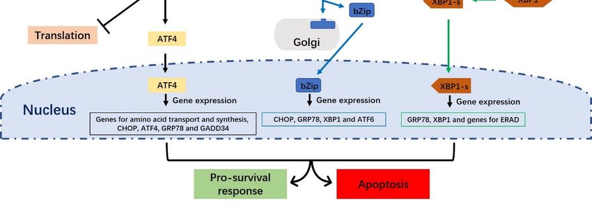
Int. J.
Int. J. Mol.
        Mol. Sci.
             Sci. 2019, 20, x409
                  2019, 20,                                                                                           22of
                                                                                                                        of 18
                                                                                                                           19

     Developing gametes and embryos may encounter various types of exogenous stress in an in
     Developing gametes and embryos may encounter various types of exogenous stress in an in vitro
vitro culture system [11,12]. It is becoming more and more apparent that some of these adverse factors
culture system [11,12]. It is becoming more and more apparent that some of these adverse factors
negatively impact ER functions and protein synthesis, resulting in the activation of ER stress and the
negatively impact ER functions and protein synthesis, resulting in the activation of ER stress and the
UPR signaling pathways [11]. Indeed, ER stress and UPR signaling appear to play critical roles during
UPR signaling pathways [11]. Indeed, ER stress and UPR signaling appear to play critical roles during
oocyte meiotic resumption and preimplantation embryo development. Inhibition of UPR signaling
oocyte meiotic resumption and preimplantation embryo development. Inhibition of UPR signaling by ER
by ER stress inhibitors has been shown to improve oocyte maturation and early embryo development
stress inhibitors has been shown to improve oocyte maturation and early embryo development in pigs,
in pigs, mice, bovines, etc. [4,13–16], suggesting that ER stress is detrimental to mammalian oocyte
mice, bovines, etc. [4,13–16], suggesting that ER stress is detrimental to mammalian oocyte maturation
maturation and preimplantation embryo development. Thus, understanding the mechanistic
and preimplantation embryo development. Thus, understanding the mechanistic relationships between
relationships between ER stress and in vitro development could help the improvement of the
ER stress and in vitro development could help the improvement of the maturation of oocytes and early
maturation of oocytes and early development of embryos.
development of embryos.
     This review examines what currently is known regarding the involvement, potential impacts
      This review examines what currently is known regarding the involvement, potential impacts and
and mechanisms of ER stress and UPR signaling in mammalian (excluding human) oocyte
mechanisms of ER stress and UPR signaling in mammalian (excluding human) oocyte maturation and
maturation and preimplantation embryonic development.
preimplantation embryonic development.
2. Endoplasmic Reticulum Stress and Unfolded Protein Response Signaling Pathways
2. Endoplasmic Reticulum Stress and Unfolded Protein Response Signaling Pathways
2.1.
2.1. Endoplasmic
     Endoplasmic Reticulum
                 Reticulum Stress
                           Stress and the Unfolded
                                  and the Unfolded Protein
                                                   Protein Response
                                                           Response
      The
      The ERER isis aa multifunctional   essential organelle
                       multifunctional essential     organelle found
                                                                 found in in eukaryotic
                                                                             eukaryotic cells.
                                                                                            cells. It
                                                                                                   It is
                                                                                                      is aa major
                                                                                                            major site
                                                                                                                  site for
                                                                                                                       for the
                                                                                                                           the
synthesis    of  transmembrane      proteins    and  lipids, and   is involved     in  maintaining
synthesis of transmembrane proteins and lipids, and is involved in maintaining intracellular calcium  intracellular  calcium
homeostasis       [1,2,17].The
homeostasis [1,2,17].        TheERERquality
                                      quality     control
                                               control     (ERQC)
                                                       (ERQC)          system
                                                                  system   is in is   in charge
                                                                                  charge          of identifying
                                                                                           of identifying           properly
                                                                                                             properly  folded
folded   proteins,     which  are  channeled     for transport   to  the Golgi    complex,    versus
proteins, which are channeled for transport to the Golgi complex, versus misfolded proteins, which are  misfolded   proteins,
which
retained areinretained
                the ER to in undergo
                             the ER tocorrect
                                         undergo      correct
                                                  folding   or folding   or befor
                                                               be targeted        targeted   for degradation
                                                                                      degradation                 by the ER-
                                                                                                     by the ER-associated
associated    degradation     (ERAD)    machinery      [18]. Correct    protein    folding  is
degradation (ERAD) machinery [18]. Correct protein folding is one of the most important steps  one   of the  most  important
                                                                                                                       during
steps  during     protein  synthesis,  and    the  accumulation     of  misfolded      proteins in
protein synthesis, and the accumulation of misfolded proteins in the ER lumen disturbs ER functions the  ER  lumen   disturbs
ER
andfunctions
      leads to ER  andstress.
                        leads Activation
                               to ER stress.ofActivation     of ER
                                                 ER stress can       stressthe
                                                                  trigger    canUPR,trigger the UPR,
                                                                                         which           which is
                                                                                                 is a cascade    ofaadaptive
                                                                                                                     cascade
of  adaptive    pathways     that seek  to maintain     cellular  homeostasis        and  normal
pathways that seek to maintain cellular homeostasis and normal ER function. However, if ER stress  ER   function.  However,
if
is ER  stress
   severe   orisprolonged,
                  severe or prolonged,
                              the UPR is the     UPR is of
                                           incapable     incapable    of re-establishing
                                                            re-establishing                  ER homeostasis
                                                                                ER homeostasis        and normal andfunction,
                                                                                                                      normal
function,
and apoptosisand apoptosis
                    may occurmay     occur
                                 (Figure  1).(Figure 1).

       Figure 1. The role of the unfolded protein response (UPR) in addressing Endoplasmic Reticulum (ER)
       Figure 1. The role of the unfolded protein response (UPR) in addressing Endoplasmic Reticulum (ER)
       stress. Under ER stress, misfolded or unfolded proteins accumulate in the ER. This triggers activation
       stress. Under ER stress, misfolded or unfolded proteins accumulate in the ER. This triggers activation
       of the UPR, which is a pro-survival response designed to alleviate the accumulation of misfolded
       of the UPR, which is a pro-survival response designed to alleviate the accumulation of misfolded
       proteins, restore ER homeostasis and re-establish normal ER function. However, if the UPR fails to
       proteins, restore ER homeostasis and re-establish normal ER function. However, if the UPR fails to
       restore ER homeostasis, cell death (apoptosis) is induced.
       restore ER homeostasis, cell death (apoptosis) is induced.

2.2. The Unfolded Protein Response Signaling Pathway
   Three ER transmembrane proteins (PERK, ATF6 and IRE1) and the ER molecular chaperone
immunoglobulin-binding protein, BiP (also known as glucose-regulated protein 78 or GRP78), trigger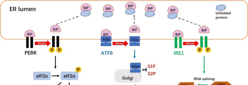
Int. J. Mol. Sci. 2019, 20, 409 3 of 19
2.2. The Unfolded Protein Response Signaling Pathway
Int. J. Mol. Sci. 2019, 20, x 3 of 18
Three ER transmembrane proteins (PERK, ATF6 and IRE1) and the ER molecular chaperone
immunoglobulin-binding
the UPR response to ER stressprotein,
[17].BiP
The(also knownresponse
canonical as glucose-regulated protein 78
to the accumulation or GRP78),proteins
of unfolded trigger
thethe
in UPRERresponse
involves to
theER stress [17].(and
association Thethus
canonical responseof
sequestration) toGRP78/BiP,
the accumulation
PERK, of unfolded
ATF6 proteins
and IRE1 [11].
in the ER involves the association (and thus sequestration) of GRP78/BiP, PERK, ATF6
Under normal physiological (unstressed) conditions, GRP78/BiP directly interacts with PERK, ATF6 and IRE1 [11].
Under normal physiological (unstressed) conditions, GRP78/BiP directly interacts
and IRE1. However, an increase in misfolded/unfolded proteins separates GRP78/BiP from these with PERK, ATF6
and IRE1. to
inducers, However,
activateanthe
increase
UPR in misfolded/unfolded
signaling proteins
pathways (Figure 2).separates GRP78/BiP
PERK signaling from these
decreases the
inducers, to activate the UPR signaling pathways (Figure 2). PERK signaling decreases
translocation of new proteins into the ER lumen and prevents protein overloading, while the ATF6the translocation
of new
and proteins
IRE1 into the
pathways ER lumen
regulate and prevents protein
the transcriptional overloading,
activation of variouswhile the including
genes, ATF6 and those
IRE1
pathways
responsibleregulate the transcriptional
for increasing activation
the translocation, of variousexport,
protein-folding, genes,degradation,
including those
and responsible for
other functions
increasing the
of the ER [19]. translocation, protein-folding, export, degradation, and other functions of the ER [19].
Figure 2.
Figure The unfolded
2. The unfolded protein
protein response
response (UPR)(UPR) signaling
signaling pathways.
pathways. ThereThere areare three
three distinct
distinct UPR
UPR
signaling pathways
signaling pathwaysin in mammalian
mammalian cells:cells:
The PERK,
The PERK,ATF6 and
ATF6 IRE1andpathways. Under normal
IRE1 pathways. Under conditions,
normal
the endoplasmic
conditions, reticulum (ER)
the endoplasmic molecular
reticulum (ER) chaperone, BiP, directlyBiP,
molecular chaperone, interacts
directlywith PERK,with
interacts ATF6 and
PERK,
IRE1. Upon activation of the UPR, PERK undergoes dimerization
ATF6 and IRE1. Upon activation of the UPR, PERK undergoes dimerization and and autophosphorylation, and then
phosphorylates eIF-2α to
autophosphorylation, prevent
and then the initiation of translation
phosphorylates eIF-2α toand block new
prevent proteins from
the initiation being produced
of translation and
in the cytoplasm.
block new proteins Thefrom
PERK-phosphorylated
being produced in eIF2α
the also activatesThe
cytoplasm. ATF4, which translocates to the
PERK-phosphorylated nucleus
eIF2α also
and triggers
activates the which
ATF4, transcription of genes
translocates to required
the nucleus to restore ER homeostasis.
and triggers When BiP
the transcription is separated
of genes requiredfromto
ATF6, the latter factor moves to the Golgi apparatus, where it is cleaved into
restore ER homeostasis. When BiP is separated from ATF6, the latter factor moves to the Golgi its active form by S1P and S2P.
Cleavage ofwhere
apparatus, ATF6 produces a soluble
it is cleaved into basic leucine
its active zipper
form (bZip)
by S1P andtranscription
S2P. Cleavage factor.
of Active
ATF6 ATF6 (bZip)
produces a
translocates to the nucleus and induces the transcription of ER stress-response genes
soluble basic leucine zipper (bZip) transcription factor. Active ATF6 (bZip) translocates to the nucleus via ERSE-1 and -2.
The
and IRE1 endoribonuclease
induces is activated
the transcription of ER through dimerizationgenes
stress-response and transphosphorylation.
via ERSE-1 and -2.This Theleads
IRE1to
the removal of a 26-nucleotide intron from the premature unspliced XBP1 (XBP1-u)
endoribonuclease is activated through dimerization and transphosphorylation. This leads to the gene form to produce
the spliced
removal of XBP1 (XBP1-s) form.
a 26-nucleotide intronXBP1-s
from the moves to the nucleus
premature unspliced andXBP1
induces UPR-responsive
(XBP1-u) gene form genes.
to produce
the spliced XBP1 (XBP1-s) form. XBP1-s moves to the nucleus and induces UPR-responsive genes.
2.2.1. The PERK Signaling Pathway
PERK is an ER transmembrane sensor protein that binds to BiP and is found in the ER lumen.
Under ER stress, PERK releases BiP and triggers the dimerization and phosphorylation of eukaryotic
translation initiation factor 2 alpha (eIF2α), leading to inhibition of protein synthesis (Figure 2). The
PERK-mediated phosphorylation of eIF2α at ser51 initiates translation [20]. Phosphorylated eIF2α
binds the guanine nucleotide exchange factor, eIF2B, to interfere with the cell’s ability to exchange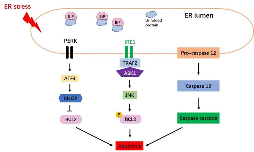
Int. J. Mol. Sci. 2019, 20, 409                                                                4 of 19

2.2.1. The PERK Signaling Pathway
     PERK is an ER transmembrane sensor protein that binds to BiP and is found in the ER lumen.
Under ER stress, PERK releases BiP and triggers the dimerization and phosphorylation of eukaryotic
translation initiation factor 2 alpha (eIF2α), leading to inhibition of protein synthesis (Figure 2).
The PERK-mediated phosphorylation of eIF2α at ser51 initiates translation [20]. Phosphorylated eIF2α
binds the guanine nucleotide exchange factor, eIF2B, to interfere with the cell’s ability to exchange
guanosine diphosphate (GDP) with guanosine triphosphate (GTP) to form eIF2-GTP tRNA [18,21].
Thus, phosphorylation of eIF2α is thought to be an important step in reducing the synthesis of new
proteins in the ER, releasing ER stress and restoring ER homeostasis [5,21]. PERK-phosphorylated
eIF2α also activates ATF4 (activating transcription factor 4), which is key UPR-mediated gene
expression. The transcriptional activity of ATF4 induces both cell survival and cell death programs.
Under mild ER stress, ATF4 induces cell survival by triggering genes involved in stress responses,
protein secretion and amino-acid metabolism [22]. Under prolonged stress, however, PERK signaling
promotes cell apoptosis by increasing ATF4 and C/EBP homologous protein (CHOP) [1,23].

2.2.2. The ATF6 Signaling Pathway
     ATF6 is an ER type II transmembrane protein; it comprises an N-terminal cytoplasmic region that
includes bZip and DNA transcriptional activation domains, and a C-terminal region that is exposed to
the lumen of the ER [19]. There are two mammalian homologs of ATF6: ATF6-α and ATF6-β [19,24].
Under normal conditions, ATF6 maintains its resting form in the ER and is associated with BiP.
When the accumulation of unfolded or misfolded proteins occurs, however, ATF6 is sequestered away
from BiP and is translocated from the ER to the Golgi apparatus, where it is cleaved into its active
form by site-1 protease (S1P) and site-2 protease (S2P) (Figure 2). This cleavage of ATF6 produces a
soluble basic leucine zipper (bZip) transcription factor (i.e., cleaved ATF6). This active ATF6 moves
to the nucleus, where it induces transcriptional activation of ER stress-response genes together with
ER stress-response elements 1 and 2 (ERSE-1 and 2). The important target genes of ATF6 during
the UPR include GRP78/BiP, XBP1 (X-box binding protein-1) and CHOP. Over-expression of cleaved
ATF6 has also been shown to activate the transcription of XBP1 [19], which is an important product in
IRE1 signaling.

2.2.3. The IRE1 Signaling Pathway
     As an ER-resident transmembrane protein, IRE1 has a serine-threonine kinase domain and an
endoribonuclease domain [25]. Under non-stressed conditions, IRE1 interacts with BiP and maintains
an inactive form. Under ER stress, however, IRE1 is sequestered away from BiP and shifts to its
active form. The endonuclease activity of IRE1, which is activated through its dimerization and
transphosphorylation, removes a 26-nucleotide intron from the prematurely unspliced XBP1 gene
form (XBP1-u) to produce the spliced XBP1-s form (Figure 2). XBP1-s is usually regarded as an
appropriate marker for the induction of the IRE1 arm of the UPR, because XBP1 is spliced exclusively
under ER stress conditions [26]. Unlike the XBP1-u protein, which is quickly degraded, XBP1-s has
a transcriptional activation domain in its C-terminus [27,28]. The XBP1-s protein translocates to the
nucleus and binds to the specific promoter elements, such as the ERSE (ER stress response element)
and unfolded protein response elements, to trigger transactivation of UPR target genes, such as those
whose products are involved in protein folding, secretion and degradation (Figure 2).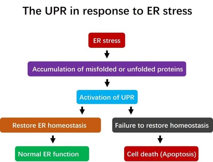
Int. J. Mol. Sci. 2019, 20, 409                                                                     5 of 19

3. Endoplasmic Reticulum Stress and the Unfolded Protein Response Are Intrinsic in Mammalian
Oocytes and Preimplantation Embryos

3.1. Endoplasmic Reticulum Stress and the Unfolded Protein Response in Oocytes
      XBP1 plays a critical role as a potent coordinator of ER stress and may assist in porcine oocyte
maturation, early embryo development and embryonic genome activation [4]. It had been reported
that the ER stress signaling pathway was essential for mouse oocyte maturation [16,29]. In mice,
endogenously expressed XBP1 protein was reported to localize principally in the nucleus and weakly
in the cytoplasm of germinal vesicle (GV)-stage oocytes. Assessment along the meiotic stages revealed
that XBP1 localized to the spindle microtubules in M I (metaphase I)-stage oocytes, but the signal at
the spindle microtubules weakened gradually in M II (metaphase II)-stage oocytes [16]. The classic
ER stress marker genes, ATF4, ATF6, XBP1, HSPA 5 and GRP78, have been detected in immature
or mature oocytes of in vitro and in vivo derivations [29,30]. Porcine XBP1 protein was found to be
weakly expressed in M II-stage oocytes, whereas it was localized mainly in the nucleus and weakly
in the cytoplasm of GV-stage oocytes [4]. RT-PCR showed that the mRNAs for porcine XBP1-u and
XBP1-s were clearly detected in GV-stage oocytes, but that only the mRNA for XBP1-u was detected
in M I- and M II-stage oocytes [4]. This suggests that porcine XBP1 may play a significant role in
oocyte maturation. In a recent study examining the expression pattern of UPR markers during porcine
oocyte maturation in vitro, Park et al. [3] reported that the BiP/GRP78 mRNA expression level was
significantly increased in cumulus-oocyte complexes (COCs) at 44 h of maturation culture relative
to 22 h of maturation culture. In addition, the protein expression levels of UPR signaling markers
(e.g., BiP/GRP78, ATF4, p50ATF6 and CHOP) were significantly higher in COCs at 44 h than at 22 h.
These results suggest that ER stress and UPR signaling are intrinsic in mammalian oocytes, and that
their proper function could be essential for oocyte maturation and oocyte quality.

3.2. Endoplasmic Reticulum Stress and the Unfolded Protein Response in Preimplantation Embryos
     It had been reported that the mechanisms responsible for controlling ER stress affect preimplantation
embryo development [4]. For example, the ER stress-mediated activation of UPR signaling
impairs preimplantation embryo development in vitro [31] and influences post-implantation embryo
development [32,33]. In mice, knockout of UPR-associated genes, such as GRP78/BiP and Ppp1r15/Gadd34,
was reported to have negative effects on embryo development, suggesting that ER chaperones and ER
stress signaling play important roles in preimplantation embryos [34,35]. In mouse embryos, ER stress
signaling was found at the 1-cell stage and was very abundant at the blastocyst stage [36]. XBP1 is a
key transcription factor that regulates a subset of genes that are active during ER stress [16]. Zhang and
colleagues reported that the XBP1 signal was higher in the nucleus than the cytoplasm at the 2-, 4-,
8-cell, morula, and blastocyst stages, as assessed by fluorescence staining. RT-PCR analysis showed
that the mRNAs for XBP1-u and XBP1-s were found at the 2-cell to blastocyst stages, whereas only
XBP1-u was detected at the 1-cell stage [16]. In an analysis of ER stress response-associated genes
throughout preimplantation development, Abraham et al. reported that the mRNAs for PERK, ASK1,
ATF4, ATF6, GRP78/BiP, CHOP, GADD34 and IRE1 were detected at all stages of mouse preimplantation
development [26]. In addition, the mRNAs for BiP, ATF6 and PERK pathway-related genes (e.g., PERK,
ATF4 and CHOP) were also detected in blastocyst-stage mouse embryos [37].
     In pig embryos derived from parthenogenetic activation (PA) [4], the mRNAs for XBP1-s and
XBP1-u were clearly detected at the 4-cell, morula and blastocyst stages, whereas only XBP1-u was
found at the 1- and 2-cell stages. The porcine XBP1 protein was localized strongly in the nucleus
and weakly in the cytoplasm during the 4-cell, morula and blastocyst stages. Moreover, the evidence
suggests that ER stress-induced XBP1 splicing may regulate early embryonic genome activation in
pigs [4]. Dicks et al. evaluated ER stress in both early- and late-cleaving embryos by measuring the
RNA abundance of XBP1 and GRP78 using real time PCR and immunofluorescence technologies [38].
In buffalo in vitro fertilization (IVF)-derived embryos, the mRNAs for the ER chaperone-encoding
Int. J. Mol. Sci. 2019, 20, 409 6 of 19
genes, GRP78 and GRP94, were detected at the blastocyst stage [14]. ER stress-associated mRNAs,
including GRP78, EDEM, ATF6, IRE1, ATF4, CHOP and XBP1, were also reportedly expressed in
bovine IVF-derived embryos [12,39].
ER stress and UPR signaling can also be detected in embryos derived from somatic cell nuclear
transfer (SCNT). It appears that ER stress is stronger in electrofusion-derived SCNT embryos compared
to IVF-derived embryos and Sendai virus-mediated SCNT embryos [39]. RT-PCR revealed that
electrofusion- or Sendai virus-mediated SCNT embryos clearly express the mRNAs for XBP1-u and
XBP1-s. The relative transcript levels of XBP1-s to total XBP1 were reported to be significantly higher
in electrofusion-mediated SCNT embryos than in Sendai virus-mediated SCNT embryos [39] or
IVF-derived embryos [15]. The mRNA expression levels of ER stress-associated genes, such as GRP78,
EDEM and ATF6, were markedly higher in electrofusion-mediated SCNT embryos than IVF-derived
or Sendai virus-mediated SCNT embryos [39]. The expression of functional XBP1 protein was detected
at all stages in porcine SCNT-derived embryos, and the mRNAs for XBP1-u and XBP1-s were detected
in porcine SCNT-derived blastocysts [13]. Overall, these findings reveal that ER stress and the UPR
pathways are intrinsic in IVF-, PA- and SCNT-derived embryos of many species, and thus are very
likely to play important roles in preimplantation embryo development. The intrinsic ER stress and UPR
signaling pathway markers that have been identified in mammalian oocytes and/or preimplantation
embryos are presented in Table 1.
Table 1. Intrinsic endoplasmic reticulum stress markers identified in mammalian oocytes and
preimplantation embryos.
ER Stress Markers Refs
Species Stage (Derivation)
Genes Proteins
Mouse Oocytes ATF4, ATF6 and BiP/GRP78 [30]
Mouse Oocytes XBP1, ATF4, ATF6 and HSPA5 [29]
Mouse Oocytes (vitrified) XBP1 [40]
Mouse Oocytes/blastocysts XBP1-u and XBP1-s XBP1 [16]
ATF4, ATF6, BiP, CHOP
Mouse Blastocysts [37]
and PERK
XBP1, BIP, IRE1, ASK1, ATF4,
Mouse Blastocysts [26]
ATF6, CHOP and PERK
Mouse Blastocysts XBP1 BiP [31]
BiP, ATF4, XBP1-u, XBP1-s ATF4, ATF6, BiP/GRP78,
Pig Oocytes [3]
and CHOP P90ATF6, P50ATF6 and CHOP
Oocytes and 1-cell to
Pig XBP1-u and XBP1-s XBP1 [4]
blastocyst-stage embryos (PA)
Embryos (day 5)/
Pig XBP1 and GRP78 [38]
blastocysts (PA)
Pig Blastocysts (SCNT) XBP1-u and XBP1-s XBP1 [13]
XBP1-u, XBP1-s, IRE1, ATF4,
Bovine Blastocysts (IVF) [12]
ATF6 and CHOP
Bovine Blastocysts (IVF and SCNT) XBP1-u, XBP1-s and CHOP [15]
XBP1, BiP/GRP78, EDEM,
Bovine Blastocysts (IVF and SCNT) [39]
ATF4, ATF6, CHOP and IRE1
Bovine Blastocysts (SCNT) BiP, CHOP and IRE1 [41]
Buffalo Blastocysts (IVF) GRP78 and GRP94 [14]
ATF4, ATF6, GRP78
Sheep Oocytes [42]
and CHOP
Refs: References.Int. J. Mol. Sci. 2019, 20, 409 7 of 19
4. Activation and Induction of Endoplasmic Reticulum Stress in Oocytes and Preimplantation Embryos
Mammalian oocytes and preimplantation embryos are usually sensitive to variations in many
exogenous factors, including temperature, osmotic stress, shear stress, chemical exposure, oxidative stress,
etc. [11,43]. All of these factors are known to induce ER stress [17], and reduce embryo developmental
potential through alterations in gene expression, epigenetic mechanisms and metabolism [44,45].
During oocyte maturation and early embryo development, various enzymes and metabolic
pathways produce endogenous reactive oxygen species (ROS) [46]. The accumulation of ROS in cells
leads to cell membrane lipid peroxidation, blockade of RNA transcription, damage of DNA and decreases
in protein synthesis [47–49]. It has been reported that oxidative stress can induce ER stress and UPR
signaling by impeding correct protein folding and calcium homeostasis [50]. Oxidative stress can decrease
the formation rate of bovine IVF-derived blastocysts by upregulating the mRNA expression levels of IRE1,
ATF4, ATF6 and CHOP [12]. ER stress and oxidative stress are usually found together [43,51]; ER stress
can produce ROS [52], whereas ROS can induce ER stress by disturbing correct protein folding/transport
and altering calcium homeostasis [50].
Tunicamycin (TM) is a chemical reagent that inhibits N-glycosylation, which is often essential for
protein folding; TM is generally used to induce ER stress [53,54]. TM has been reported to negatively
affect oocyte maturation and embryo development by promoting ER protein misfolding and inducing
apoptosis in mice, pig and cattle [4,13,14,16,37,40]. TM-induced ER stress usually has a negative influence
on embryo development. For example, mouse embryos treated with more than 5 µg/mL TM were
completely blocked at the 2-cell stage and failed to develop into blastocysts [16], while bovine and pig
PA- or IVF-derived embryos failed to develop to the blastocyst stage when exposed to 5 µg/mL TM
during in vitro culture [4,14,55]. In porcine SCNT-derived embryos, in contrast, as little as 1 µg/mL TM
was found to block the ability of embryos to develop to the blastocyst stage [13]. This suggests that
SCNT-derived embryos are more sensitive to TM than IVF-derived embryos, perhaps because cloned
embryos exposed to electrofusion-mediated activation are subject to an increased ER stress response [13].
Electrofusion, which is the most common method employed to create a cloned embryo, typically
increases intracellular calcium ions, decreases maturation-promoting factor activity and enhances
improper nuclear remodeling [39,56]. SCNT-derived embryos derived from electrofusion reportedly
undergo activation of ER stress and UPR signaling via upregulation of the mRNAs for XBP1-s, GRP78
and ATF6, leading to decreases in the developmental competence and quality of SCNT embryos [39].
Similarly, the author of a porcine SCNT study speculated that the nuclear transfer process used during
SCNT (e.g., electrofusion) could induce extra ER stress [57]. Therefore, it may be useful to decrease
ER stress during SCNT embryo production by bypassing electrofusion, as a means to improve SCNT
embryo development.
Osmotic stress, cryopreservation and shear stress during embryo handling can also induce the
ER stress response. Culture media of an appropriate osmolality has been shown to improve oocyte
maturation and embryo development [58,59], whereas hyperosmolarity of the culture media has
been shown to decrease embryo developmental capacity by inducing ER stress and apoptosis [16].
For example, the addition of 50 mM sorbitol to the culture medium completely arrested mouse embryos
at the 2-cell stage, while the addition of 25 mM sorbitol significantly decreased the blastocyst formation
rate by increasing XBP1 protein expression [16]. Cryopreservation can also induce ER stress and UPR
pathway activation. Zhao et al. reported that the protein level of XBP1 was significantly higher in
vitrified-warmed mouse oocytes than in fresh mouse oocytes, suggesting that the freeze-thawing
procedure triggers ER stress in mouse oocytes [40]. Similarly, sheep COCs subjected to vitrification
showed increased expression levels of ER stress markers, such as the mRNAs for ATF4, ATF6, GRP78
and CHOP, when compared to controls, suggesting that sheep oocyte cryopreservation is associated
with ER stress and activation of the UPR signaling pathways [42]. Embryo pipetting often produces
shear stress, which can damage oocytes and negatively impact embryo development [60]. Although
transient shear stress may not negatively influence embryos, prolonged or repeated handling can
have such effects [61]. The IRE1 arm of the UPR pathway is reportedly activated in response toInt. J. Mol. Sci. 2019, 20, 409 8 of 19
embryo collection techniques [26]. Overall, the studies described above indicate that ER stress and
the UPR signaling pathways are inducible and activated in mammalian oocytes and preimplantation
embryos. The impacts of common ER stress activators on mammalian oocyte maturation and embryo
development are summarized in Table 2.
Table 2. The impact of endoplasmic reticulum stress inducers on mammalian oocyte maturation and
embryo development.
Inducer Treatment Species Results Refs
Reduces porcine SCNT-derived blastocyst
0.1–1 µM formation rate, decreases total cell and ICM
TM Pig [13]
during IVC cell numbers, increases XBP1-s mRNA
expression and apoptosis
Reduces blastocyst formation rate, increases
TM 0.5 µM during IVC Mouse [37]
ROS and apoptosis
Prevents blastocyst formation, increases the
TM 0.5 µM during IVC Mouse levels of cleaved caspase 3 protein and the [31]
mRNA for XBP1-s
1 µM in freezing Increases XBP1 and caspase 12 protein
TM Mouse [40]
medium expression levels
Decreases blastocyst formation rate and cell
TM 1–2 µM during IVC Buffalo [14]
numbers, increases apoptosis
Induces active XBP1 in nuclei of 4-cell stage
PA-derived embryos, reduces cleavage rate,
TM 2 µM during IVC Pig [4]
blastocyst formation rate, and cell numbers
in blastocysts
Reduces porcine IVF-derived embryo
TM 1–5 µM during IVC Pig development by reducing the rates of [55]
cleavage and blastocyst formation
Reduces oocyte maturation in pig by
1–5 µM
TM Pig increasing the expression levels of BiP, ATF4 [3]
during IVM
and ATF6
1–10 µM Reduces blastocyst formation rate, increases
TM Mouse [16]
during IVC apoptotic index
10–75 mM Reduces blastocyst formation rate, increases
Sorbitol Mouse [16]
during IVC apoptotic index
Reduces bovine IVF-derived blastocyst
Oxidative Embryos cultured formation rate; increases XBP1-s, ATF4, ATF6,
Bovine [12]
stress under 20% O2 CHOP and IRE1 gene expression levels and
the apoptotic index
Triggers transient activation of the XBP1 arm
Shear stress Embryo collection Mouse [26]
of ER stress
Reduces SCNT-derived embryonic
development by increasing XBP1-s, CHOP,
SCNT embryos
GRP78 and EDEM gene expression levels and
Electro-fusion produced by Bovine [39]
the apoptotic index, while reducing the
electrofusion
blastocyst formation rate and the total and
ICM cell numbers
Refs: References.Int. J. Mol. Sci. 2019, 20, 409 9 of 19
5. Endoplasmic Reticulum Stress Induces Apoptosis
5.1. Three Main Canonical Apoptosis Pathways Are Induced by Endoplasmic Reticulum Stress
UPR signaling is extremely important for the restoration of ER homeostasis and the maintenance of
normal ER function. When misfolded or unfolded proteins accumulate in the ER lumen, UPR signaling
is activated to ameliorate this source of ER stress. However, if misfolded or unfolded protein
accumulation is persistent and the stress cannot be relieved, apoptosis is induced by activation
of the CHOP, Jun N-terminal kinase (JNK), and caspase 12 [25,62,63] pathways (Figure 3).
5.1.1. The CHOP Pathway
CHOP, which is encoded by growth arrest- and DNA damage-inducible gene 153 (GADD153),
was identified as a factor that responds to DNA damage [25]. Under conditions of severe or prolonged
ER stress, all three arms of the UPR can induce CHOP transcription; however, the PERK-eIF2α-ATF4
pathway has been shown to be essential for upregulating CHOP protein expression. BiP is released
from PERK to trigger ATF4, leading to the upregulation of CHOP expression (Figure 3). CHOP mainly
induces
Int. J. Mol. Sci. 2019, 20, xthrough downregulation of the anti-apoptotic gene, BCL2 [19,25].
apoptosis 9 of 18
Schematicdiagram
Figure3.3.Schematic
Figure diagramofofapoptosis
apoptosisinduced
inducedbybyEndoplasmic
EndoplasmicReticulum
Reticulum(ER)
(ER)stress.
stress.Three
Threemain
main
pathwaysofofER
pathways ERstress-mediated
stress-mediated pro-apoptotic
pro-apoptotic signaling
signaling areare shown,
shown, namely
namely the CHOP
the CHOP pathway,
pathway, the
the JNK
JNK pathway
pathway andcaspase
and the the caspase 12 pathway.
12 pathway.
5.1.2. The JNK Pathway
5.1.2. The JNK Pathway
JNK is activated by the IRE1-TRAF2-ASK1 pathway (Figure 3). IRE1 binds to TRAF2 (TNF
JNK is activated by the IRE1-TRAF2-ASK1 pathway (Figure 3). IRE1 binds to TRAF2 (TNF
receptor-associated factor 2) and ASK1 (apoptosis signal-regulating kinase 1), leading to the activation
receptor-associated factor 2) and ASK1 (apoptosis signal-regulating kinase 1), leading to the
of JNK and ASK1 [62]. ER stress-activated JNK triggers apoptosis mainly by phosphorylating
activation of JNK and ASK1 [62]. ER stress-activated JNK triggers apoptosis mainly by
ER-localized BCL2 [64].
phosphorylating ER-localized BCL2 [64].
5.1.3. The Caspase 12 Pathway
5.1.3. The Caspase 12 Pathway
Caspase 12 is an ER membrane-localized pro-apoptotic cysteine protease [19] that acts as a key
Caspase 12 is an ER membrane-localized pro-apoptotic cysteine protease [19] that acts as a key
mediator of ER stress-induced apoptosis [65] (Figure 3). Caspase 12 is activated by m-Calpain in the
mediator of ER stress-induced apoptosis [65] (Figure 3). Caspase 12 is activated by m-Calpain in the
cytoplasm [66], or potentially via caspase 7-mediated cleavage [67]. After its release from the ER membrane,
cytoplasm [66], or potentially via caspase 7-mediated cleavage [67]. After its release from the ER
caspase 12 associates with caspase 9, which may lead to caspase 3 activation and subsequent apoptosis [19].
membrane, caspase 12 associates with caspase 9, which may lead to caspase 3 activation and
subsequent apoptosis [19].
5.2. Endoplasmic Reticulum Stress-Mediated Apoptosis in Oocytes and Preimplantation Embryos
Apoptosis plays important roles in mammalian oocyte maturation and preimplantation
embryonic development [68]. In the latter context, apoptosis may contribute to embryonic loss [69]
and affect some cellular responses [70] in embryos produced both in vitro and in vivo. ER stress-Int. J. Mol. Sci. 2019, 20, 409 10 of 19
5.2. Endoplasmic Reticulum Stress-Mediated Apoptosis in Oocytes and Preimplantation Embryos
Apoptosis plays important roles in mammalian oocyte maturation and preimplantation embryonic
development [68]. In the latter context, apoptosis may contribute to embryonic loss [69] and affect
some cellular responses [70] in embryos produced both in vitro and in vivo. ER stress-induced
apoptosis has been reported in mammalian oocytes and IVM-derived embryos. The apoptotic index
in blastocyst-stage mouse embryos treated with 5 µg/mL TM was found to be significantly higher
than that in the TM-untreated group [16]. In addition, a higher concentration of ER stress inducer (e.g.,
50 mM sorbitol) was also reported to significantly increase the apoptosis rate in mouse blastocysts
compared with the control group, as assessed by the TUNEL assay [16]. Mouse oocyte cryopreservation
is associated with ER stress, and the level of caspase 12 protein was reported to be significantly higher
in both vitrified oocytes and vitrified oocytes treated with TM, compared to non-vitrified group [40].
In buffalo IVF-derived blastocysts, TM treatment increased the relative mRNA expression of BAX
and the TUNEL-positive cell rate compared to the TM-untreated group [14]. Treatment with 20%
O2 significantly increased the apoptosis rate of bovine early embryos through the activation of ROS
and ER stress [12]. In porcine SCNT-derived embryos, blastocysts derived from the TM-treated
group exhibited a significantly increased apoptosis rate along with upregulation of the mRNA for
pro-apoptotic BAX and downregulation of the mRNA for anti-apoptotic BCL2 [13]. The higher
apoptotic index of SCNT-derived embryos compared to IVF-derived embryos may be attributed to the
increased activation of ER stress seen in the former [15,39]. Taken together, these studies show that ER
stress (e.g., those induced by TM treatment, hyperosmosis or cooling) can trigger apoptosis in oocytes
and/or preimplantation embryos.
6. Relief of Endoplasmic Reticulum Stress Reduces Apoptosis, Improves Oocyte Maturation and
Enhances the Developmental Potential of Embryos
The ER stress inhibitor-induced decrease of ER stress-induced UPR signaling not only
improves oocyte maturation and preimplantation embryo development potential, it also prevents ER
stress-mediated apoptosis (Table 3, Figure 4). Tauroursodeoxycholic acid (TUDCA), which is a bile
acid that acts as a potent chemical chaperone to inhibit ER stress in vitro [71], has been widely used to
alleviate ER stress during in vitro oocyte maturation and/or embryo development. The beneficial role
of TUDCA is typically attributed to suppression of the UPR [72,73].
Table 3. The positive influence of Endoplasmic Reticulum stress inhibitor supplementation during
IVM/IVC on mammalian oocyte maturation and/or embryo development.
Inhibitor Treatment * Species Results Refs
Improves embryo development and increases
TUDCA 50 µM during IVC Mouse the implantation and live birth rates of [57]
transferred mouse embryos
Improves the blastocyst formation rate and
TUDCA 50 µM during IVC Mouse [16]
reduces apoptosis
Enhances the viability and embryonic
50 µM in the
TUDCA Mouse developmental capacity of vitrified-warmed [40]
freezing medium
mouse oocytes by reducing ER stress
Reduces ER stress and ROS levels; improves
TUDCA 50 µM during IVC Bovine [12]
bovine embryo development
Improves bovine SCNT embryo development
TUDCA 50 µM during IVC Bovine by increasing cell numbers and reducing ER [39]
stress and apoptosis
Attenuates apoptosis and ER stress in buffalo
TUDCA 50 µM during IVC Buffalo [14]
IVF-derived embryosInt. J. Mol. Sci. 2019, 20, 409 11 of 19
Table 3. Cont.
Inhibitor Treatment * Species Results Refs
Enhances porcine oocyte maturation and
50 µM during IVM,
TUDCA Pig PA-derived embryo developmental potential [4]
50 µM during IVC
by preventing ER stress
Enhances DNA damage repair and improves
TUDCA 50 µM during IVC Pig porcine preimplantation embryo development [38]
by reducing ER stress
Improves porcine SCNT embryonic
TUDCA 100 µM during IVC Pig development by attenuating ER stress and [57]
reducing apoptosis
100 µM (treatment Improves the development of bovine
TUDCA bovine [41]
of donor cells) SCNT-derived embryos by reducing ER stress
Improves the development of porcine
TUDCA 200 µM during IVC Pig IVF-derived embryos by modulating ER [55]
stress-induced apoptosis
200 µM during Improves the quality and maturation of
TUDCA Pig [3]
IVM porcine oocytes
Improves cumulus cell expansion and oocyte
Melatonin 0.1 µM during IVM Pig [3]
maturation by combating ER stress
Improves the development of bovine
Valproic acid 3 mM during IVC Bovine SCNT-derived embryos by reducing ER stress [15]
and apoptosis
Increases the mouse blastocyst formation rate
GSH 1 mM during IVC Mouse [37]
and alleviates ER stress
Reduces ROS levels and increases the
GSH 1 mM during IVC Bovine [12]
blastocyst formation rate
Improves pentraxin-3 secretion, mitochondrial
100 nM during
Salubrinal Mouse membrane potential, and embryonic [29]
IVM
development by reducing ER stress
Refs: References. * The optimal concentrations of the ER stress inhibitors are listed.
TUDCA was reported to significantly improve porcine oocyte maturation by triggering the MAPK
pathway and enhance the developmental capacity of early PA-derived embryos by preventing ER
stress-induced apoptosis [4]. Inhibition of ER stress by supplementation with TUDCA reportedly
reduces the incidence of DNA double-strand breaks (DSBs) and improves preimplantation embryo
development [38]. Incubation of porcine IVF-derived embryos with TUDCA was shown to improve
the blastocyst formation rate and total cell and inner cell mass (ICM) cell numbers, while decreasing
the apoptosis rate and the mRNA level of pro-apoptotic BAX [55]. Similarly, in cattle, TUDCA
supplementation of the culture medium can enhance the blastocyst formation rate, trophectoderm
proportion and cell survival [12]. TUDCA (50 µM) improved the cleavage rate of buffalo IVF-derived
embryos and attenuated TM-induced apoptosis by decreasing the expression levels of BAX and
ER chaperones [14]. In mice, the addition of TUDCA to the culture medium improved the rate at
which 2-cell embryos developed to blastocysts, reduced apoptosis [16] and improved the implantation
and live birth rates of transferred embryos [57]. TUDCA was also shown to improve the viability
and subsequent embryo developmental potential of vitrified-warmed mice oocytes by reducing
cryopreservation-induced ER stress [40].
The blastocyst formation rate, total cell number, ICM cell number and apoptotic rate were
significantly lower and higher in electrofusion-mediated SCNT embryos compared to Sendai
virus-mediated SCNT embryos or IVF-derived embryos, respectively; this reflects the increased
activation of ER stress by the electrofusion process, suggesting that ER stress plays a negative role in
early SCNT-derived embryos [39]. However, TUDCA treatment significantly enhanced the formation
rate and quality of electrofusion-mediated SCNT blastocysts by reducing apoptosis and increasingInt. J. Mol. Sci. 2019, 20, 409 12 of 19
cell numbers [39]. The presence of TUDCA in porcine in vitro maturation (IVM) medium did not
Int. J. Mol. Sci. 2019, 20, x 11 of 18
improve the cleavage or blastocyst formation rates of SCNT embryos, but inclusion of TUDCA in
the in vitro culture (IVC) medium appeared to enhance the blastocyst formation rate and quality
Increases the mouse blastocyst formation
of porcine GSH SCNT-derived 1 mM during
embryosIVC by alleviating
Mouse ER stress, [37] the
ratereducing the ER
and alleviates ROS level, increasing
stress
GSH level and decreasing apoptosis [13]. A recent study in bovine
Reduces SCNT-derived
ROS levels and increasesembryos
the showed
GSH 1 mM during IVC Bovine [12]
that TUDCA treatment of the donor cell can also significantly enhance
blastocyst the fusion
formation rate rate, cleavage rate,
blastocyst formation rate and total cell number while decreasing Improves thepentraxin-3
apoptotic secretion,
index [41]. These studies
collectively suggest100 that ER stressIVM
is a common mitochondrial membrane potential, and
Salubrinal nM during Mouseevent in mammalian oocyte IVM and/or embryo [29] IVC
embryonic development by reducing ER
systems, and that relieving ER stress by TUDCA treatment can improve oocyte maturation, enhance
stress
embryo developmental potential and reduce apoptosis.
Refs: References. * The optimal concentrations of the ER stress inhibitors are listed.
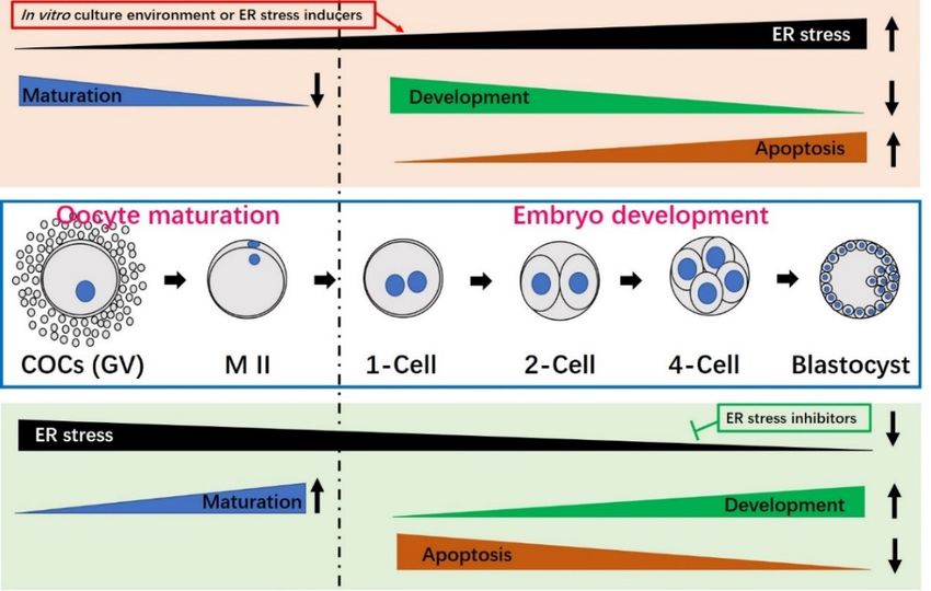
Figure
Figure 4. influences
4. The The influences of endoplasmic
of endoplasmic reticulumreticulum
(ER) stress(ER) stressmaturation
on oocyte on oocyteand maturation and
preimplantation
preimplantation
embryo development. embryo development.
Oocyte maturation Oocyte
and/or maturation and/or embryo
embryo culture culture associated
environments environmentswith
associated with prolonged or severe ER stress (e.g., treatment with the ER stress inducer,
prolonged or severe ER stress (e.g., treatment with the ER stress inducer, TM) can significantly reduce TM) can
significantly
oocyte maturation,reduce oocyteembryo
decrease maturation, decrease embryo
developmental potentialdevelopmental potential and
and increase apoptosis (top increase
box with
apoptosis (top box with light pink color). When ER stress is inhibited by an ER stress
light pink color). When ER stress is inhibited by an ER stress inhibitor (e.g., TUDCA), maturationinhibitor (e.g.,
and
TUDCA), maturation and development improve and the apoptotic index decreases (bottom box with
development improve and the apoptotic index decreases (bottom box with light green color). Oocyte
light green color). Oocyte maturation is shown to the left of the broken black line, while embryo
maturation is shown to the left of the broken black line, while embryo development is presented on the
development is presented on the right. COCs, cumulus oocyte-complexes; GV, germinal vesicle; M II,
right. COCs, cumulus oocyte-complexes; GV, germinal vesicle; M II, metaphase II.
metaphase II.
Melatonin (N-acetyl-5methoxytryptamine), which is an indole that is mainly synthesized
TUDCA was reported to significantly improve porcine oocyte maturation by triggering the
from tryptophan by the pineal gland in animals, contributes to many important physiological
MAPK pathway and enhance the developmental capacity of early PA-derived embryos by
functions, such as sleep, temperature regulation, metabolism, the circadian rhythm and seasonal
preventing ER stress-induced apoptosis [4]. Inhibition of ER stress by supplementation with TUDCA
reproduction [74–78]. It is also a free radical scavenger, anti-oxidant and anti-apoptotic factor [79].
reportedly reduces the incidence of DNA double-strand breaks (DSBs) and improves
Unlike other free radical scavengers, melatonin is multifunctional and universal: It can directly
preimplantation embryo development [38]. Incubation of porcine IVF-derived embryos with TUDCA
obliterate toxic oxygen
was shown to improve derivatives, prevent
the blastocyst ROS generation,
formation rate andand induce
total anti-oxidative
cell and enzymes
inner cell mass (ICM)[80–82].
cell
Melatonin
numbers, reportedly has beneficial
while decreasing influencesrate
the apoptosis whenandadded to the culture
the mRNA level ofmedium duringBAX
pro-apoptotic the oocyte
[55].
maturation
Similarly,andin embryo development
cattle, TUDCA of pigs, cattle
supplementation ofand
themice [80,83,84].
culture mediumAscan mentioned
enhance above, oxidative
the blastocyst
stress (e.g., ROS)
formation rate,contributes
trophectoderm to UPR activation;
proportion thus,
and cellmelatonin can exert
survival [12]. TUDCAits functions on cell viability
(50 μM) improved the
by regulating
cleavage ratetheofUPR signaling
buffalo pathways
IVF-derived or reducing
embryos ER stressTM-induced
and attenuated via its anti-oxidant andbyanti-apoptotic
apoptosis decreasing
properties. Although
the expression melatonin
levels of BAX and has ER
repeatedly
chaperonesbeen used
[14]. to reduce
In mice, ER stressofinTUDCA
the addition cells [85,86],
to therelatively
culture
fewmedium
studies improved
have focused on at
the rate thewhich
mechanisms through
2-cell embryos which melatonin
developed decreases
to blastocysts, reduced ER stress during
apoptosis [16]
and improved
mammalian oocyte the implantation
maturation and preimplantation
and/or live birth rates ofembryo
transferred embryos [57].
development. OneTUDCA
study inwas also
a porcine
shown
oocyte to improve
maturation the viability
system revealed and subsequent
that embryo developmental
adding melatonin to the maturation potential
medium of vitrified-warmed
improved meiotic
mice oocytes by reducing cryopreservation-induced ER stress [40].
maturation by reducing ER stress, suggesting that melatonin critically modulates UPR signaling and
The blastocyst formation rate, total cell number, ICM cell number and apoptotic rate were
significantly lower and higher in electrofusion-mediated SCNT embryos compared to Sendai virus-Int. J. Mol. Sci. 2019, 20, 409 13 of 19
reduces ER stress during oocyte IVM in pig [3]. Numerous factors involved in in vitro culture can
induce ER stress, which damages oocyte maturation and embryo development. The use of melatonin
as an anti-oxidant and anti-apoptotic factor could relieve ER stress by modulating UPR signaling
pathways, which could yield improved oocyte maturation and/or embryo developmental potential.
The mechanisms through which melatonin acts on ER stress and the UPR signaling pathways during
mammalian oocyte maturation and preimplantation embryo development await further clarification.
In addition to TUDCA and melatonin, the HDAC (histone deacetylase) inhibitor, valproic acid,
or glutathione (GSH) can also reduce ER stress by regulating the UPR pathways. In bovine SCNT
embryos, valproic acid was reported to significantly reduce the mRNA levels of XBP1-s (an ER
stress marker), CHOP and BAX (a pro-apoptotic gene) while increasing those of the ER molecular
chaperone, BiP, and the anti-apoptotic gene, BCL-xl; these findings suggested that valproic acid could
improve the developmental potential of bovine SCNT-derived embryos by alleviating ER stress and
reducing apoptosis [15]. GSH reportedly inhibits ER stress during mouse embryo development [37].
It was also found to significantly decrease the mRNA expression levels of ATF6, ATF4, BiP, CHOP
and PERK, and increase the cell numbers and apoptosis index in blastocysts; thus, GSH appears
to improve the developmental capacity and quality of mouse embryos by alleviating ER stress and
apoptosis [37]. Salubrinal, a selective eIF2α dephosphorylation inhibitor, has been reported to protect
cells from ER stress [87]. In mouse COCs, palmitic acid-mediated ER stress reportedly impairs
mitochondrial membrane potential, pentraxin-3 secretion and embryonic development; however,
salubrinal treatment was shown to significantly improve these deficiencies by reducing ATF4, ATF6
and XBP1 gene expression levels, suggesting that salubrinal could reverse the cellular dysfunctions
induced by ER stress and improve oocyte development [29].
The studies above show that supplementation of the in vitro culture medium with chemical
inhibitors of ER stress could serve as a beneficial approach to preventing ER stress-induced oocyte
damage and alterations of embryo development. The effects of various ER stress inhibitors on
mammalian oocyte maturation and embryo development are summarized in Table 3. ER stress often
occurs alongside other stress responses, particularly oxidative stress [11]. ER stress itself induces ROS
generation, whereas ROS contributes to the activation of ER stress and UPR signaling, so it is possible
that some anti-oxidants may hold promise as potential inhibitors of ER stress.
7. Conclusions
Oocyte IVM and embryo IVC systems are essential for the successful production of live animals
via in vitro-produced (IVP) embryos. In vitro culture conditions can increase numerous stresses that
have the ability to damage the oocyte maturation and preimplantation embryo development of IVP
embryos. Mammalian oocytes and embryos are highly sensitive to these diverse exogenous stresses.
Many factors can negatively impact the ER, new protein synthesis and protein processing, initiating
ER stress and the UPR signaling responses. As an adaptive response to ER stress, the UPR can facilitate
clearance of the unfolded or misfolded proteins and cell survival. However, under prolonged stress or
failure of the UPR, apoptosis is induced. ER stress usually plays a negative role in oocyte maturation
and/or early embryo development (Table 2, Figure 4). Fortunately, oocyte quality and embryonic
developmental potential may be improved by the addition of ER stress-reducing chemicals to the
in vitro culture medium (Table 3, Figure 4). The ER stress inhibitor, TUDCA, has been widely used to
attenuate ER stress during mammalian oocyte maturation and embryo development. In the future,
we need to search for more ER stress inhibitors that can improve oocyte and embryo quality and
continue studying the mechanisms through which ER stress and UPR signaling affect mammalian
oocyte maturation and preimplantation embryo development.
In addition, although success rate of human IVF and intracytoplasmic sperm injection (ICSI)
have improved progressively, the efficiency of assisted reproductive technology (ARTs), based on
practical embryo production, is still low [88]. Embryos must be manipulated in vitro during human
ARTs, and adverse in vitro condition mentioned above in mammalian embryo development in vitroInt. J. Mol. Sci. 2019, 20, 409 14 of 19 could induce ER stress and UPR signaling pathways that may negatively impact human embryo development. Thus, understanding the mechanistic relationships between ER stress and in vitro development during human embryo production in vitro could help the improvement of human ARTs. Author Contributions: Conceptualization, T.L. and D.I.J.; investigation, T.L., J.E.L., J.W.K.; data curation, T.L. and D.I.J.; writing—original draft preparation, T.L.; writing—review and editing, D.I.J.; visualization, H.Y.S. and J.B.L.; funding acquisition, D.I.J. Funding: This research was funded by D.I.J., grant number PJ011851012017, Cooperative Research Program for Agriculture Science & Technology Development in Rural Development Administration, Republic of Korea. Conflicts of Interest: The authors declare no conflicts of interest. Abbreviations ASK1 Apoptosis signal-regulating kinase 1 ATF4 Activating transcription factor 4 ATF6 Activating transcription factor 6 BiP Immunoglobulin-binding protein bZip Basic leucine zipper CHOP C/EBP homologous protein COCs Cumulus-oocyte complexes DNA Deoxyribonucleic acid eIF2α Eukaryotic translation initiation factor 2 alpha ER Endoplasmic reticulum ERAD ER-associated degradation ERQC Endoplasmic reticulum quality control ERSE ER stress response element GADD153 Growth arrest- and DNA damage-inducible gene 153 GDP Guanosine diphosphate GRP78 Glucose-regulated protein 78 GSH Glutathione GTP Guanosine triphosphate GV Germinal vesicle ICM Inner cell mass IRE1 Inositol-requiring enzyme 1 IVC In vitro culture IVF In vitro fertilization IVM In vitro maturation IVP In vitro-produced JNK Jun N-terminal kinase MI Metaphase I M II Metaphase II mRNA Messenger ribonucleic acid PA Parthenogenetic activation PERK Double-stranded RNA-activated protein kinase-like ER kinase ROS Reactive oxygen species S1P Site-1 protease S2P Site-2 protease SCNT Somatic cell nuclear transfer TM Tunicamycin TRAF2 TNF receptor-associated factor 2 TUDCA Tauroursodeoxycholic acid UPR Unfolded protein response XBP1 X-box binding protein-1 XBP1-s Spliced XBP1 XBP1-u Unspliced XBP1
Int. J. Mol. Sci. 2019, 20, 409                                                                                       15 of 19

References
1.    Tabas, I.; Ron, D. Integrating the mechanisms of apoptosis induced by endoplasmic reticulum stress.
      Nat. Cell Biol. 2011, 13, 184–190. [CrossRef] [PubMed]
2.    Kaufman, R.J. Regulation of mRNA translation by protein folding in the endoplasmic reticulum.
      Trends Biochem. Sci. 2004, 29, 152–158. [CrossRef] [PubMed]
3.    Park, H.J.; Park, J.Y.; Kim, J.W.; Yang, S.G.; Jung, J.M.; Kim, M.J.; Kang, M.J.; Cho, Y.H.; Wee, G.;
      Yang, H.Y.; et al. Melatonin improves the meiotic maturation of porcine oocytes by reducing endoplasmic
      reticulum stress during in vitro maturation. J. Pineal Res. 2018, 64. [CrossRef] [PubMed]
4.    Zhang, J.Y.; Diao, Y.F.; Oqani, R.K.; Han, R.X.; Jin, D.I. Effect of endoplasmic reticulum stress on porcine
      oocyte maturation and parthenogenetic embryonic development in vitro. Biol. Reprod. 2012, 86, 128.
      [CrossRef] [PubMed]
5.    Kroeger, H.; Chiang, W.C.; Felden, J.; Nguyen, A.; Lin, J.H. ER stress and unfolded protein response in ocular
      health and disease. FEBS J. 2018. [CrossRef] [PubMed]
6.    Walter, P.; Ron, D. The unfolded protein response: From stress pathway to homeostatic regulation. Science
      2011, 334, 1081–1086. [CrossRef] [PubMed]
7.    Ghemrawi, R.; Battaglia-Hsu, S.F.; Arnold, C. Endoplasmic Reticulum Stress in Metabolic Disorders. Cells
      2018, 7. [CrossRef]
8.    Schroder, M.; Kaufman, R.J. The mammalian unfolded protein response. Annu. Rev. Biochem. 2005, 74,
      739–789. [CrossRef]
9.    Guzel, E.; Arlier, S.; Guzeloglu-Kayisli, O.; Tabak, M.S.; Ekiz, T.; Semerci, N.; Larsen, K.; Schatz, F.;
      Lockwood, C.J.; Kayisli, U.A. Endoplasmic Reticulum Stress and Homeostasis in Reproductive Physiology
      and Pathology. Int. J. Mol. Sci. 2017, 18, 792. [CrossRef]
10.   Zeng, F.; Schultz, R.M. RNA transcript profiling during zygotic gene activation in the preimplantation mouse
      embryo. Dev. Biol. 2005, 283, 40–57. [CrossRef]
11.   Latham, K.E. Stress signaling in mammalian oocytes and embryos: A basis for intervention and improvement
      of outcomes. Cell Tissue Res. 2016, 363, 159–167. [CrossRef] [PubMed]
12.   Yoon, S.B.; Choi, S.A.; Sim, B.W.; Kim, J.S.; Mun, S.E.; Jeong, P.S.; Yang, H.J.; Lee, Y.; Park, Y.H.; Song, B.S.;
      et al. Developmental competence of bovine early embryos depends on the coupled response between
      oxidative and endoplasmic reticulum stress. Biol. Reprod. 2014, 90. [CrossRef]
13.   Lin, T.; Lee, J.E.; Oqani, R.K.; Kim, S.Y.; Cho, E.S.; Jeong, Y.D.; Baek, J.J.; Jin, D.I. Tauroursodeoxycholic
      acid improves pre-implantation development of porcine SCNT embryo by endoplasmic reticulum stress
      inhibition. Reprod. Biol. 2016, 16, 269–278. [CrossRef]
14.   Sharma, A.; Agrawal, H.; Mullani, N.; Sandhu, A.; Singh, M.K.; Chauhan, M.S.; Singla, S.K.; Palta, P.;
      Manik, R.S. Supplementation of tauroursodeoxycholic acid during IVC did not enhance in vitro development
      and quality of buffalo IVF embryos but combated endoplasmic reticulum stress. Theriogenology 2015, 84,
      200–207. [CrossRef] [PubMed]
15.   Song, B.S.; Yoon, S.B.; Sim, B.W.; Kim, Y.H.; Cha, J.J.; Choi, S.A.; Jeong, K.J.; Kim, J.S.; Huh, J.W.; Lee, S.R.; et al.
      Valproic acid enhances early development of bovine somatic cell nuclear transfer embryos by alleviating
      endoplasmic reticulum stress. Reprod. Fertil. Dev. 2014, 26, 432–440. [CrossRef] [PubMed]
16.   Zhang, J.Y.; Diao, Y.F.; Kim, H.R.; Jin, D.I. Inhibition of endoplasmic reticulum stress improves mouse embryo
      development. PLoS ONE 2012, 7, e40433. [CrossRef]
17.   Michalak, M.; Gye, M.C. Endoplasmic reticulum stress in periimplantation embryos. Clin. Exp. Reprod. Med.
      2015, 42, 1–7. [CrossRef] [PubMed]
18.   Ali, I.; Shah, S.Z.; Jin, Y.; Li, Z.S.; Ullah, O.; Fang, N.Z. Reactive oxygen species-mediated unfolded protein
      response pathways in preimplantation embryos. J. Vet. Sci. 2017, 18, 1–9. [CrossRef] [PubMed]
19.   Shen, X.H.; Zhang, K.Z.; Kaufman, R.J. The unfolded protein response—A stress signaling pathway of the
      endoplasmic reticulum. J. Chem. Neuroanat. 2004, 28, 79–92. [CrossRef] [PubMed]
20.   Oslowski, C.M.; Urano, F. Measuring ER stress and the unfolded protein response using mammalian tissue
      culture system. Methods Enzymol. 2011, 490, 71–92.
21.   Harding, H.P.; Zhang, Y.H.; Bertolotti, A.; Zeng, H.Q.; Ron, D. Perk is essential for translational regulation
      and cell survival during the unfolded protein response. Mol. Cell 2000, 5, 897–904. [CrossRef]
You can also read