Doxorubicin-induced DNA damage causes extensive ubiquitination of ribosomal proteins associated with a decrease in protein translation.
←
→
Page content transcription
If your browser does not render page correctly, please read the page content below
MCP Papers in Press. Published on February 14, 2018 as Manuscript RA118.000652

Doxorubicin-induced DNA damage causes extensive ubiquitination of ribosomal proteins
associated with a decrease in protein translation.

Vincentius A. Halim1,2,3*, Iraia García-Santisteban3,4*, Daniel O. Warmerdam3,5, Bram van den
Broek3, Albert J.R. Heck1,2, Shabaz Mohammed1,2,6,7, René H. Medema3†

1
    Biomolecular Mass Spectrometry and Proteomics Group, Bijvoet Center for Biomolecular
Research and Utrecht Institute for Pharmaceutical Sciences, Utrecht University, 3584 CH
                              2
Utrecht, The Netherlands.         Netherlands Proteomics Centre, 3584 CH Utrecht, The
Netherlands. 3Division of Cell Biology and Cancer Genomics Center, Netherlands Cancer
                                                       4
Institute, 1066 CX Amsterdam, The Netherlands.             Department of Genetics, Physical

                                                                                                Downloaded from http://www.mcponline.org/ by guest on September 28, 2018
Anthropology and Animal Physiology, University of the Basque Country (UPV/EHU), Leioa,
Spain. 5European Research Institute for the Biology of Ageing, University Medical Center
Groningen, 9713 AV Groningen, The Netherlands, 6Department of Biochemistry, University of
Oxford, OX13QU Oxford, United Kingdom. 7Chemistry Research Laboratory, Department of
Chemistry, University of Oxford, OX13TA Oxford, United Kingdom.
* Shared first author
†Corresponding author. E-mail: r.medema@nki.nl

                                              1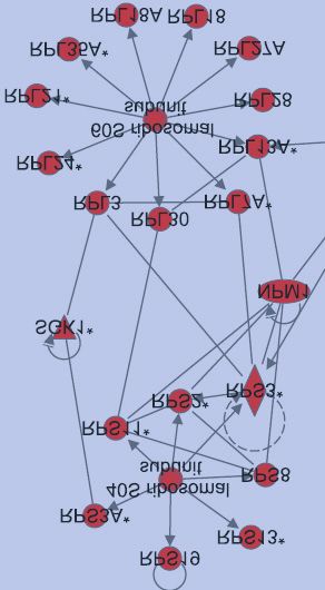
Abstract
Protein post-translational modifications (PTMs) play a central role in the DNA damage
response. In particular, protein phosphorylation and ubiquitination have been shown to be
essential in the signalling cascade that coordinates break repair with cell cycle progression.
Here, we performed whole-cell quantitative proteomics to identify global changes in protein
ubiquitination that are induced by DNA double-strand breaks. In total, we quantified more
than 9400 ubiquitin sites and found that the relative abundance of approximately 10% of
these sites was altered in response to DNA double-strand breaks. Interestingly, a large
proportion of ribosomal proteins, including those from the 40S as well as the 60S subunit,
were ubiquitinated in response to DNA damage. In parallel, we discovered that DNA damage
leads to the inhibition of ribosome function. Taken together, these data uncover the ribosome

                                                                                                 Downloaded from http://www.mcponline.org/ by guest on September 28, 2018
as a major target of the DNA damage response.

                                              2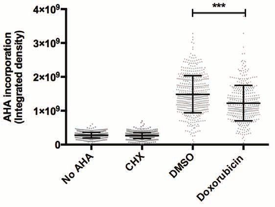
Introduction
The genome of a cell is frequently damaged by insults generated by internal (reactive cell
metabolites) and external (irradiation, UV) sources (1-3). This causes a threat to the stability
of the genome and can contribute to cancer development. To protect themselves against this
potential threat, cells are equipped with powerful surveillance mechanisms that detect and
repair the damage before it is propagated to subsequent daughter cells. This response is
collectively referred to as the DNA damage checkpoint or the DNA damage response (DDR) (1-
3). Notwithstanding these harmful effects of DNA damage, several DNA damaging agents are
widely applied to treat cancer, as they can cause a non-reversible checkpoint arrest or trigger
cell death, thus curbing the rapid proliferation in cancer cells. For example, doxorubicin, which
induces DNA double-strand breaks (DSBs), is a potent anti-cancer drug that is commonly used

                                                                                                    Downloaded from http://www.mcponline.org/ by guest on September 28, 2018
in the clinic.
Execution of the DDR requires damage detection, and initates a signaling cascade that halts
further progression through the cell cycle, whilst promoting repair. This signaling cascade is
mainly driven by posttranslational modifications (PTMs) (4, 5). The phosphatidylinositol 3-
kinase-related kinases (PIKKs) ataxia-telangiectasia mutated (ATM) and ataxia- and Rad3-
related (ATR) play a central role in the initiation of DDR signaling. ATM and ATR are recruited
to sites of DNA damage and subsequently phosphorylate over 700 substrates (6). Important
downstream targets of ATM and ATR are Chk2 and Chk1 kinases, respectively (3).
Phosphorylation of these effector kinases results in their activation, causing a subsequent
wave of protein phosphorylations that are essential for the function of the DNA damage
checkpoint and promotes cell cycle arrest (3, 6, 7).
A recent proteomics study showed that ubiquitination events following DNA damage are as
common as phosphorylation (8). It is well established that in response to a DSB, ubiquitin-
mediated signaling is initiated by the ubiquitin ligases RNF8 and RNF168 (9-13). Whereas
phosphorylation events spread rapidly throughout the nucleus after damage, ubiquitination
seems mostly limited to the proximity of the break-site and more controlled (10, 12). Protein
ubiquitination is essential for the buildup of the checkpoint, and plays an important role in
various repair pathways. Excessive protein ubiquitination can be detrimental for the
maintenance of the DNA damage checkpoint and DNA repair pathway choice (14-16).
There are several reports showing that individual ribosomal proteins play a role in the DNA
damage response, most notably in the activation of p53 (17, 18), but so far there are few

                                               3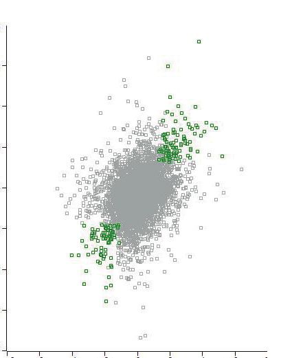
reports that connect the DNA damage response with ribosomal function. DNA damage is
known to affect mRNA translation by disruption of cap initiation complexes that are required
for the recruitment of mRNAs to ribosomes (19, 20). In addition, DNA damaging agents have
been shown to affect signaling through the mTOR pathway, and in consequence also protein
translation. In fact, the effects of irradiation on translation are much more pronounced than
on transcription (21). These changes in translation are at least in part mediated through
altered recruitment of mRNAs to polysomes (21), but how this is established is not known.
The eukaryotic 80S ribosome consists of four ribosomal RNAs (rRNAs) and 80 ribosomal
proteins (RPs) (22-24). After their transcription in the nucleoli, rRNAs associate with RPS and
RPL proteins in the nucleus, forming the small (40S) and large (60S) ribosomal subunits,
respectively (22-24). The small and large subunits will be assembled together in the cytoplasm

                                                                                                  Downloaded from http://www.mcponline.org/ by guest on September 28, 2018
to make a mature ribosome (or monosome) (22-24). Importantly, several ribosomes can
simultaneously translate a single mRNA molecule to synthesize the same protein, forming the
so-called polysomes (22, 23).
In this study, we investigate how specific ubiquitination events change after DNA damage and
identify the ribosome as a target of the DNA damage checkpoint.

Materials and Methods
Cell culture, transfection and drugs
U2OS cells were grown in Dulbecco's modified Eagle's medium (DMEM) supplemented with
6% fetal bovine serum (FBS) and penicillin/streptomycin. Thymidine and doxorubicin were
purchased from Sigma and used at 2.5 mM and 1 μM respectively. ATM inhibitor KU55933
from Calbiochem and ATR inhibitor VE-821 from Axon Medchem were used at concentration
10 μM each. MG132 was purchased from Millipore and used at 5 µM. Cycloheximide was
purchased from Sigma and used between 50 and 100 µg/ml. DUB inhibitor PR-619 was from
Tebu-bio and used in the lysis buffer at the concentration of 50 µM. Cell synchronization and
DNA damage application were performed as previously described (25) and outlined
schematically in Fig. 1A and Fig. S3.

Antibodies
The antibody for diglycyl-remnant peptide enrichment was obtained from Cell Signaling
Technology and used according to the standard protocol from the company. The following

                                              4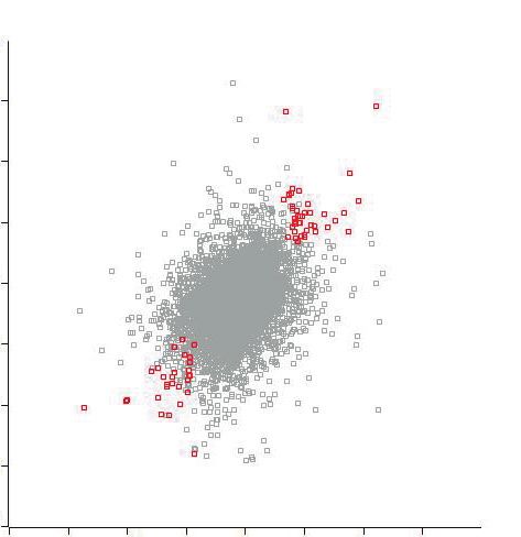
antibodies were used for immunofluorescence and western blotting experiments: anti-
nucleophosmin 1 and anti-nucleolin (Abcam, 1:1000), anti-RPL24 (Thermo Scientific, 1:1000),
anti-RPS27/27L (Thermo Scientific, 1:200), anti-RPL26 (Bethyl Laboratories, 1:1000), anti-
RPL27a (Novus, 1:1000), anti-RPS6 (Cell Signaling Technology, 1:500), anti-pS139-H2AX
(Millipore, 1:1000) and anti-α-tubulin (1:5000, Sigma).

Nascent Protein Synthesis Analysis
G2-synchronized U2OS cells were washed with PBS and cultured in methionine-free DMEM
(Invitrogen) for 30 minutes to deplete the intracellular methionine reserves. Cells were then
treated with a pulse of doxorubicin for 1 hour to induce DSBs. After washout, cells were
incubated with the methionine analog L-azidohomoalanine (AHA; Invitrogen) for 2 hours; cells

                                                                                                Downloaded from http://www.mcponline.org/ by guest on September 28, 2018
were treated with the translation inhibitor cycloheximide (CHX) or with no AHA as negative
controls. Cells were fixed in 3.7% formaldehyde, permeabilized with 0.5% Triton X-100 in PBS
and blocked using 3% bovine serum albumin (BSA) in PBS. Proteins containing AHA were
labeled with Alexa Fluor488-alkyne using a click chemistry-based reaction (Click-iT, Life
Technologies). Cells were counterstained with DAPI to stain the nuclei. The amount of nascent
protein synthesis in each condition was quantified by measuring AHA fluorescence intensity
per cell using a macro developed for that purpose in ImageJ.

Sucrose gradients
U2OS cells were lysed in lysis buffer (20 mM Tris–HCl, pH 7.5, 10 mM MgCl2, 100 mM KCl, 1%
NP40) supplemented with 2 mM DTT, 100 μg/ml cycloheximide, EDTA-free protease inhibitor
cocktail (Roche) and RiboLockRNAse inhibitor (40 U/ml, Life Technologies). Lysates were
centrifuged at 1300 g and the supernatant was fractionated on a linear sucrose gradient (7–
47%) using a SW-41Ti rotor at 36,000 rpm for 2 hours. Thirteen fractions were collected, and
samples were analyzed by western blot using the indicated antibodies.

Experimental design and statistical rational
Preparation of cell lysates for proteomics analysis
U2OS cells were synchronized in G2 phase. Subsequently, one-hour doxorubicin pulse was
applied. Upon removal of doxorubicin, cells were incubated in fresh media containing 5 µM
MG132 for two and six hours, and subsequently harvested for proteomics analysis.

                                               5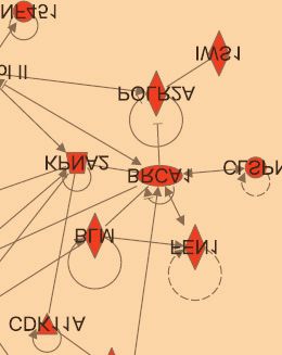
Undamaged cells with MG132 treatment are the control for this experiment. Time scale of this
experiment is presented in Fig. 1A. To ensure the reproducibility, two independent
experiments were carried out. Each experiment contains two time points and their respective
controls.
For the ATM/ATR inhibitor experiment, G2-synchronized U2OS cells were treated with 10 μM
ATM- and ATR-inhibitors for half an hour before DNA damage induction. DNA damage was
induced by a pulse of doxorubicin. Upon removal of doxorubicin, cells were incubated in fresh
media containing 5 µM MG132 for two hours, and cells were subsequently harvested for
proteomics analysis. Doxorubicin- and MG132-treated cells without ATM and ATR inhibition
are the control for this experiment. Time scale is presented in Fig. S3.
For the MG132 vs DMSO experiment, U2OS cells were synchronized in G2 phase and one-hour

                                                                                                 Downloaded from http://www.mcponline.org/ by guest on September 28, 2018
doxorubicin pulse was applied. Upon removal of doxorubicin, cells were incubated in fresh
media with and without 5 µM MG132 for two hours, and subsequently harvested for
proteomics analysis.

Protein extraction, proteolytic digestion and peptide purification
Harvested cells were lysed using ultrasonicator for 3 times 1 minute at 0.6 cycle and 90%
amplitude and proteins were extracted using 50 mM ammonium bicarbonate buffer
containing 8 M urea, protease inhibitors, and 50 µM deubiquitinase inhibitor PR619. For each
sample, 20 mg protein was reduced and alkylated using 5 mM DTT and 10 mM
chloroacetamide respectively. Subsequently, samples were digested with lys-C (1:50 w/w
enzyme:protein ratio). After buffer dilution (to 2 M urea), samples were digested with trypsin
(1:50 w/w enzyme:protein ratio). The peptide product was then purified using a Seppak C8
column and concentrated using a speedvac. Finally, the purified peptides were reconstituted
in the immunoprecipitation buffer for further enrichment by immunoprecipiation with an
antibody recognizing the diglycyl-remnant. The immunoprecipitation buffer was supplied by
Cell Signaling Technology as part of the enrichment kit. Details on extraction, digestion, and
peptide purification were described previously (26, 27).

Peptide enrichment and MS analysis
Following diglycyl-remnant peptide enrichment, peptides were eluted in two subsequent
washes using a total of 105 µl of 0.15% TFA. Twenty-five microliter of samples were injected

                                               6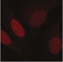
in triplicate into the nano-UPLC Proxeon system (Easy-nLC 1000, Thermo Scientific) coupled
to a Orbitrap Elite mass spectrometer (Thermo Scientific). The injected samples were first
trapped on an in-house packed trap column (ReproSil-Pur C18-AQ, 3 µm (Dr. Maisch GmbH,
Ammerbuch, Germany) 2 cm × 100 µm) before being separated with 2 hour gradient on an in-
house made analytical column (Zorbax SB-C18, 1.8 µm (Agilent Technologies, Baltimore, MD,
USA) 50 cm × 50 µm) at a constant temperature of 40 degrees. For the Orbitrap Elite a voltage
of 1.7 kV was applied to the needle. The survey scan was recorded with a resolution of 60,000.
The 20 most intense precursors were selected for subsequent fragmentation using HCD as the
activation technique. Singly- and doubly-charged ions were excluded in the analysis.

Ubiquitin/peptide-site identification and quantification; data analysis and evaluation

                                                                                                     Downloaded from http://www.mcponline.org/ by guest on September 28, 2018
Raw data were processed using MaxQuant (version 1.4.0.3) (28) and the MS/MS data were
queried against the human UniProt database (23630 entries, released 2013_06). Trypsin/P
was chosen as cleavage specificity allowing 2 missed cleavages. Carbamidomethylation (C)
was set as a fix modification, while oxidation (M) and GlyGly (K) were used as variable
modifications. Peptide identification was based on a search with a mass deviation of the
precursor ion up to 4.5 ppm, and the allowed fragment mass deviation was set to 20 ppm for
FTMS and 0.5 Da for ITMS. Data filtering was carried out using the following parameters:
peptide and protein FDR were set to 1%, minimum peptide length was set to 6, and
Andromeda minimum score was set to 40 (29). MaxQuant label free quantification was used
to quantify the ubiquitin- peptide/ site, with peak area as the output. To analyze the data,
peak area of treated samples was compared with their respective controls such as with and
without damage 2h; with and without damage 6h; with and without ATM-/ATR- inhibitor; with
and without MG132. Log scale 2 was used to present the ratio proportionally. Data imputation
was done using the lowest peak area quantified in the same run. Quantified sites were
evaluated with Perseus (version 1.4.0.8) (30). Significant B is an outlier test provided by
Perseus to calculate the significance of a ratio based on a ratio population (from the total data)
binned by log intensity. Only ubiquitin sites that obtained a p
Results
Profiling of protein ubiquitination in response to DNA double-strand breaks
To analyze global changes of protein ubiquitination in response to DNA damage, G2-
synchronized U2OS cells were either left untreated or treated with a pulse of doxorubicin to
induce DSBs. After doxorubicin washout, cells were cultured in the presence of the
proteasome inhibitor MG132 to prevent the degradation of ubiquitinated proteins, and
harvested two and six hours later to profile protein ubiquitination (Fig. 1A). Proteins were
extracted, digested with trypsin, and the ubiquitinated peptides were enriched using a diglycyl
remnant peptide antibody (31). Subsequently, peptides were analysed by LC-MS and
quantified using MaxQuant Label-Free Quantification (32) (Fig. 1B). Two independent
biological replicates were performed for each experiment.
Downloaded from http://www.mcponline.org/ by guest on September 28, 2018
As expected, treatment with MG132 markedly increased the abundance of diglycine peptides
compared to the DMSO-treated control (Fig. S1A), and allowed us to detect ubiquitinated
proteins that otherwise would have been degraded by the proteasome in the absence of
MG132. Importantly, upon treatment with doxorubicin and MG132, the overall levels of
ubiquitin conjugates did not change (Fig. S1B), and the types of ubiquitin linkages were not
significantly altered (Fig. S1C).
In total, we could identify more than 10000 ubiquitin sites at each timepoint, with an overlap
of approximately 90% of identified and quantified sites between each biological replicate (Fig.
1C). The overlap in sites quantified at each timepoint was also high (87%, Fig. S1D). This leads
to a total of more than 11000 unique ubiquitin sites (Table S1). To validate some of the sites
identified in our large-scale analysis, we expressed a His-tagged variant of ubiquitin in U2OS
cells. After lysis, proteins modified with His-tagged ubiquitin were pulled-down using nickel
charged beads, digested and the ubiquitinated peptides were immunoprecipitated using the
ubiquitin remnant antibody. We identified a total of 836 unique ubiquitinated peptides, 469
of which overlapped with the peptides we identified in our large-scale proteomics (Table S2
and Fig. 1D). Thus, close to 60% of the ubiquitin sites identified by His-ubiquitin tagging were
also identified in the direct ubiquitin remnant isolation (Fig. 1D). While this provides some
validation to our large-scale ubiquitin analysis approach, it also shows that a substantial
amount of protein ubiquitination was missed in the direct ubiquitin remnant pull-downs as
compared to the His-Ubiquitin pull-downs and vice versa.
8As an example of our validation, the highly ubiquitinated nucleophosmin (NPM1) protein is
shown (Fig. 1E). Our initial large-scale proteomics identified six ubiquitin-sites (K32, K239,
K248, K250, K257 and K273) in NPM1. Using His-ubiquitin protein IP as well as endogenous
protein IP, we were able to independently validate the ubiquitination on K239, K248, K257
and K273 in NPM1. Combined, our validation experiments indicate the robustness of our
large-scale ubiquitin analysis approach and thereby strengthen our findings.
Changes in protein ubiquitination after DNA damage
Using label-free quantification of diglycyl remnant peptides, we determined changes in
abundance of ubiquitinated peptides between the damaged and control samples. To obtain
an overview of all of the changes in protein ubiquitination, we subjected all of the unique sites
Downloaded from http://www.mcponline.org/ by guest on September 28, 2018
that were identified at each single timepoint to a Significant B test considering both intensity
and ratio (pBRAP, RAP80, BRCA1, BLM) (Tables S5 and S6). But more strikingly, we noticed a very high
proportion of ribosomal and nucleolar proteins to be enriched in the list of proteins whose
ubiquitination increased upon doxorubicin treatment (Tables S5 and S6).
To confirm the overrepresentation of ribosomal proteins in the ubiquitinated protein list, we
sorted all identified ubiquitin sites based on their ratio (Fig. 3A). In comparison with the sorted
list of all ubiquitin-site changes, where ubiquitination/deubiquitination is more or less equal
(Fig. 3A, right panel), we observed prominent ubiquitination of ribosomal and nucleolar
proteins after the DNA damage pulse (Fig. 3A, left panel; Tables S5 and S6). In addition, protein
deubiquitination is also observed at a few sites in the ribosomal proteins, indicating that
ubiquitination of ribosomal proteins upon damage is not necessarily uniformly regulated. This
is further supported by the differences we observe in the relative ratios of six ubiquitin sites
Downloaded from http://www.mcponline.org/ by guest on September 28, 2018
of different ribosomal proteins at both two and six hours after the DNA damage pulse (Fig.3B).
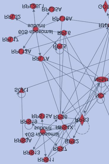
Next, we used Ingenuity Pathway Analysis to confirm that indeed, the ribosomal pathway was
identified as a target of protein ubiquitination after DNA damage. The highest scoring
networks identified by Ingenuity Pathway Analysis for the 2 and 6 hour timepoints respectively
are depicted in Fig. 3C, and strikingly, both contain a high proportion of ribosomal proteins.
To confirm that activation of the DNA damage checkpoint results in increased ubiquitination
of ribosomal proteins, we performed the same ubiquitin profiling described above in the
presence of ATM and ATR inhibitors (Fig. S2A), and the changes in protein ubiquitination were
analyzed. Indeed, we found that the ubiquitination of ribosomal and nucleolar proteins was
mostly dependent on ATM and ATR, since inhibition of these kinases largely prevented the
changes in protein ubiquitination after DNA damage (Fig. S2B). These data suggest that
activation of the DNA damage checkpoint results in a substantial, ATM/ATR-dependent,
change in ubiquitination of ribosomal and nucleolar proteins.
We next compared our results with two recently published large-scale ubiquitin studies (8,
38). We used the 2 hour timepoint from our data set, since this most closely reflected the
timepoints used to obtain the data sets in DTT- and ionizing radiation-treated cells to induce
endoplasmic reticulum (ER)- and DNA damage-stress, respectively (Fig. S4 and Table S7). While
the overlap in the overall data sets was low, DTT-induced ER stress also triggered widespread
site-specific ubiquitination of ribosomal proteins. Further analysis showed that a number of
ribosomal proteins were ubiquitinated in response to ER- as well as after DNA damage-
induced stress (Fig. S4 and Table S7). For example, our analysis identified 5 ubiquitin sites on
10RPS3, all of which were also upregulated in response to ER stress. In addition, while no
common ubiquitin sites of RPS2 and RPS20 were found when comparing ER- and DNA damage-
induced stress, both proteins are ubiquitinated in response to DNA damage and ER stress.
Thus, despite a very limited overall similarity in the outcome of the different screens,
ubiquitination of ribosomal proteins appears to be a common response of cells to different
forms of stress.
DNA damage results in a decrease in general protein translation
The function of nucleoli and ribosomes in protein synthesis is well established (39). Since we
find that a large number of nucleolar and ribosomal proteins are ubiquitinated following DNA
damage, this sparked our interest to study the effect of DNA damage on protein synthesis.
Downloaded from http://www.mcponline.org/ by guest on September 28, 2018
Therefore, we analysed changes in global nascent protein synthesis following DNA damage.
To this end, L-azidohomoalanine (AHA), an analog of methionine amino acid, was added to
the cell culture following a doxorubicin pulse, and incorporation of AHA into newly synthesized
proteins was measured by Click-iT reaction (40, 41). Significantly less AHA incorporation was
observed in doxorubicin-treated cells compared to DMSO-treated cells, indicating a decrease
in nascent protein synthesis following DNA damage (Fig. 4A,B). This decrease in nascent
protein synthesis was also observed when doxorubicin was combined with the proteasome
inhibitor MG132 (Fig. S4A,B), indicating that the effects we observe on ubiquitination of
ribosomal proteins and protein synthesis are primarily caused by the DNA damaging insult.
Next, we aimed to test whether the decrease in global protein synthesis was accompanied by
a change in ribosome activity. Protein synthesis involves translation of mRNA strands by
ribosomes. During active protein synthesis, several ribosomes are attached to a single mRNA
strand simultaneously, forming so-called polysomes. When not translationally active,
polysomes dissociate again into monosomes. Both monosomes and polysomes can be
separated on sucrose gradients and identified by immunoblotting with antibodies against
ribosomal proteins (Fig. 4C). As a control, we inhibited mRNA translation using an mTOR
inhibitor (Torin I) which results in a clear loss of polysomes (Fig. 4D). Similarly, a reduction of
polysomes in the doxorubicin-treated cells is observed (Fig. 4D), indicating a decrease in
ribosome activity in response to DNA damage. Together, these results indicate that protein
translation is affected by DNA damage stress.
11DNA damage affects the subcellular localization of ribosomal proteins
Depending on the type of linkage, ubiquitination can promote protein degradation or alter
protein behaviour. Particularly in the DNA damage response, protein ubiquitination is known
to play an important role in checkpoint signaling and repair by controlling protein function. To
study the consequences of ubiquitination of ribosomal proteins, we first studied if DNA
damage induces degradation of ribosomal proteins. We harvested cells at different timepoints
after doxorubicin treatment and analyzed expression levels of several ribosomal proteins by
western blot using commercially available antibodies. Expression of nucleolin,
nucleophosmin, RPS6, RPL24 and RPL26 remained constant throughout the experiment (Fig.
5A). Expression of RPS27/27L seemed to increase during the course of the experiment,
whereas expression of RPL27A was reduced over time (Fig. 5A). The reduction in RPL27A
Downloaded from http://www.mcponline.org/ by guest on September 28, 2018
expression was not reverted by addition of MG132, indicating that this reduction is not due to
enhanced proteasomal degradation (Fig. 5B). Thus we can conclude that, at least for the
ribosomal proteins analyzed here, DNA damage does not seem to induce increased
proteasomal degradation. This does not exclude the possibility that ubiquitination can induce
degradation of some of the other ribosomal proteins that we find to be ubiquitinated after
DNA damage, but at least this shows that DNA damage does not decrease stability of
ribosomal proteins in general.
Nucleolar stress produced by DNA damage has been shown to promote the translocation of
nucleolin and nucleophosmin from the nucleolus to the nucleoplasm, and therefore we
analyzed if ubiquitination affects subcellular localization of other ribosomal proteins in a
similar manner. As expected, we find that both nucleolin and nucleophosmin are dispersed
from the nucleolus to the nucleoplasm in response to treatment with doxorubicin (Fig. 5C). In
addition to nucleolin and nucloephosmin, both RPL26 and RPS27/27L were dispersed from the
nucleoli after a pulse of doxorubicin (Fig. 5C), but while RPS27/27L accumulated in the
nucleoplasm, RPS26 was found in the the cytoplasm (Fig. 5C). No clear differences in
subcellular localization were observed for RPS6, RPL24 and RPL27A, but none of these latter
ribosomal proteins accumulated in the nucleoli in the untreated cells either (Fig. 5C). These
data indicate that the enhanced ubiquitination of ribosomal proteins that we observe after
doxorubicin coincides with nucleolar stress and disruption of the nucleoli. Based on our data
we cannot discriminate if ubiquitination is involved in the onset of nucleolar disruption, or if
it occurs as a consequence of this disruption. However, the fact that we find a clear increase
12in ubiquitination of ribosomal proteins as early as 2 hours after the damaging insult could be
compatible with a role in the disruption of the nucleolus itself.
Discussion
In the study described here, we have profiled global changes of protein ubiquitination in
response to doxorubicin-induced DNA damage. In a previous study, ionizing radiation was
used to induce DNA damage, and protein ubiquitination during the early stages of DNA
damage signaling was analyzed (8). In turn, our study focuses on the analysis of ubiquitination
not only at early time points, but also at relatively late time points after doxorubicin-induced
DNA damage. The different experimental approaches used in each study highlights the
Downloaded from http://www.mcponline.org/ by guest on September 28, 2018
complementary of our work, and explains the limited overlap between them (Fig. S3).
Similar to other studies on protein ubiquitination, we made use of the proteasomal inhibitor
MG132 to increase the chance to retrieve ubiquitination sites before their degradation (8).
This has the potential caveat that addition of MG132 might also lead to depletion of the free
ubiquitin pool and thereby restrict non-degradative ubiquitination. As such, it is important to
strike a balance between the inhibition of ubiquitin-dependent protein degradation and the
availability of free ubiquitin. This can be achieved by applying relatively low MG132
concentration in a short time period, similar to the conditions used in the present study and
other previous works (34, 36, 42). Importantly, we were able to show that MG132 itself does
not grossly alter DNA damage induced-ubiquitination (Fig. S1). Also, the large amount of
ubiquitination sites identified in each experiment (~9,000), suggests that the free ubiquitin
pool was not overly compromised. Moreover, the detection of well-known
monoubiquitinated sites in proteins such as FANCD2 and FANCI (K561 and K523, respectively;
see Tables S3 and S4) suggests that our MG132 treatment did not severely compromise the
identification of non-degradative ubiquitination sites. On the other hand, it is important to
note that the ubiquitin sites were enriched and identified based on the diglycil remnant on
the peptides following a tryptic digestion. Consequently, we cannot exclude the presence of
some other ubiquitin-like molecules containing C-terminal diglycil motifs in the data set (34,
43). These include neddylated proteins, whose role in DDR has previously been reported (43).
Also, without an analysis at the proteome level we cannot deconvolute the contribution of
change in protein expression on the change in level of ubiquitination observed.
13We find that at 6 hours post-doxorubicin treatment, proteins with a function in the DNA
replication, recombination and repair are substantially ubiquitinated. Network analysis also
showed that a large group of ribosomal and nucleolar proteins are ubiquitinated in response
to DNA damage. Since the main function of the ribosome is to synthesize proteins, we
hypothesized that DNA damage-induced signaling could suppress ongoing protein translation,
as previously reported after other types of DNA damage such as γ-irradiation or UV (44, 45)
The resulting inability to generate new proteins could help preventing further progression
through the cell cycle, allowing more time to repair the damage (46). Indeed we find that DNA
damage results in a rapid inhibition of protein synthesis.
But how does DNA damage control protein synthesis? DNA damage can affect initiation of
protein translation through the mTOR signaling pathway (47-49), but additional mechanisms
Downloaded from http://www.mcponline.org/ by guest on September 28, 2018
that control protein synthesis in response to DNA damage might very well exist. Recently,
Higgins et. al. proposed site-specific regulatory ubiquitination of 40S ribosomal proteins as a
novel mechanism to inhibit protein translation in response to cellular stress, in particular
following ionizing radiation or DTT treatment (8, 38). Similar to those studies, we also find
extensive ubiquitination of ribosomal proteins following doxorubicin treatment, several of
which identical to proteins identified in these earlier studies (RPS2, RPS3, RPS20 (Fig. S3 and
Table S7). Moreover, we have identified many additional sites on ribosomal proteins that are
ubiquitinated in response to doxorubicin, and that could also affect ribosome activity.
Alternatively, DNA damage could affect ribosome biogenesis, and in this way inhibit protein
translation in a more general fashion. Indeed, we observe a clear nucleolar stress response
after doxorubicin treatment, as both nucleolin and nucleophosmin disperse from the
nucleolus. Translocation of nucleophosmin during nucleolar stress was recently shown to
require S-glutathionylation, which occurs within minutes and can promote the activation of
p53 (50). Our measurements of protein ubiquitination were performed as early as 2 hours
after the damage, at a timepoint when the nucleolar dispersment is well underway. Thus,
futher experiments are required to resolve if the ubiquitination plays a role in the
translocation of ribosomal proteins from the nucleolus, or if it is a mere consequence of the
dispersal.
It is tempting to speculate that ubiquitination of ribosomal proteins is involved in the
inhibition of protein translation that we observe in response to doxorubicin treatment. Given
the large number of ubiquitination events, it will be challenging to provide direct evidence for
14this, since each single event could potentially contribute to it. Nonetheless, closer examination
of a selected number ubiquitination sites could prove very informative. RPS6, for example, is
a component of the 40S subunit and localizes at the interface between two ribosomal
subunits. It interacts with mRNA, tRNA and initiation factors (22, 49), indicating that it sits at
an important interface during protein translation. Therefore, an in-depth analysis of RPS6
ubiquitination, combined with their effects on protein translation is likely to generate
interesting insights. In addition, ribosomal proteins can also be highly selective in controlling
protein translation. For example, RPL26 can specifically control p53 translation by interacting
with the p53 mRNA (18). At the same time, p53 has been reported to induce the expression
of RPS27L after the treatment with the DNA damaging agent etoposide (51), a similar increase
to what we observe in response to doxorubicin (Fig. 5A).
Downloaded from http://www.mcponline.org/ by guest on September 28, 2018
We find extensive ubiquitination of ribosomal proteins at 2 and 6 hours after the induction of
DNA damage. Interestingly, Elia and co-workers reported no significant change in
ubiquitination of ribosomal proteins at earlier timepoints after damage (8). Thus, it is possible
that ribosomal protein ubiquitination is part of the intermediate-to-late DNA damage
response. It would therefore be interesting to study the role of ribosomal proteins during
recovery from a DNA damage-induced arrest. In this respect, it is interesting to note that we
find that depletion of several ribosomal proteins results in a substantial decrease in recovery
from a G2 arrest (data not shown). Similarly, depletion of RPS27L resulted in a deficiency in
DNA damage checkpoints, leading to a shift of DNA damage-induced p53 response from cell
cycle arrest to apoptosis (52). How ribosomal proteins or their ubiquitination affects recovery
is not clear. Recently, an E3 ligase complex with a role in nonfunctional rRNA decay has been
identified, and its associated protein Mms1p, identified previously as factor involved in DNA
repair (53). Several studies have linked ribosomal proteins to the activation of p53, controlling
its abundance either by binding to p53 mRNA or by binding to its ubiquitin ligase MDM2 (17,
54-56). Given the crucial role for p53 in the control of cell cycle re-entry in G2 (57, 58) these
extra-ribosomal functions could prove important.
In summary, our proteomics study provides a useful data set of protein ubiquitination events
that occur in response to DNA damage. Our set further expands previously published datasets,
and provides the first global analysis of protein ubiquitination at early and late time points
after treatment with doxorubicin. In addition, our study highlights the extensive effects of
DNA damage on ubiquitination of ribosomal proteins, as well as its effects on protein
15synthesis. While we would like to unravel the molecular details that could link these events,
the large number of ubiquitination events that we and others find represents a huge
challenge. Nonetheless, the fact that many subunits of the ribosome are affected during a
stress response strongly implies that tight control of ribosomal function is crucial for the
cellular response to stress.
Acknowledgments
All raw files and annotated spectra from these experiments are available on PRIDE (Project ID
PXD004445). This work is funded by a TOP-GO grant from the Netherlands Organization for
Scientific Research (NWO ZonMW 912100651 to R.H.M., S.M., and V.A.H.). IGS was supported
Downloaded from http://www.mcponline.org/ by guest on September 28, 2018
with a postdoctoral fellowship from the Basque Country Government (Spain). The authors
thank Christian Frese and Teck Yew Low for fruitful discussions. The authors also thank Teck
Yew Low for submitting the raw files and annotated spectra to PRIDE. We thank Fabricio
Loayza-Puch for his technical help with the sucrose gradients.
16References
1. J. Bartek, J. Lukas, DNA damage checkpoints: from initiation to recovery or adaptation. Curr
Opin Cell Biol 19, 238-245 (2007).
2. S. P. Jackson, J. Bartek, The DNA-damage response in human biology and disease. Nature 461,
1071-1078 (2009).
3. A. Sancar, L. A. Lindsey-Boltz, K. Unsal-Kaçmaz, S. Linn, Molecular mechanisms of mammalian
DNA repair and the DNA damage checkpoints. Annu Rev Biochem 73, 39-85 (2004).
4. A. Ciccia, S. J. Elledge, The DNA damage response: making it safe to play with knives. Mol Cell
40, 179-204 (2010).
5. J. W. Harper, S. J. Elledge, The DNA damage response: ten years after. Mol Cell 28, 739-745
(2007).
6. S. Matsuoka et al., ATM and ATR substrate analysis reveals extensive protein networks
responsive to DNA damage. Science 316, 1160-1166 (2007).
7. M. A. van Vugt, V. A. Smits, R. Klompmaker, R. H. Medema, Inhibition of Polo-like kinase-1 by
DNA damage occurs in an ATM- or ATR-dependent fashion. J Biol Chem 276, 41656-41660
(2001).
8. A. E. Elia et al., Quantitative Proteomic Atlas of Ubiquitination and Acetylation in the DNA
Downloaded from http://www.mcponline.org/ by guest on September 28, 2018
Damage Response. Mol Cell, (2015).
9. S. Bekker-Jensen, N. Mailand, The ubiquitin- and SUMO-dependent signaling response to DNA
double-strand breaks. FEBS Lett 585, 2914-2919 (2011).
10. C. Doil et al., RNF168 binds and amplifies ubiquitin conjugates on damaged chromosomes to
allow accumulation of repair proteins. Cell 136, 435-446 (2009).
11. S. P. Jackson, D. Durocher, Regulation of DNA damage responses by ubiquitin and SUMO. Mol
Cell 49, 795-807 (2013).
12. N. Mailand et al., RNF8 ubiquitylates histones at DNA double-strand breaks and promotes
assembly of repair proteins. Cell 131, 887-900 (2007).
13. T. E. Messick, R. A. Greenberg, The ubiquitin landscape at DNA double-strand breaks. J Cell Biol
187, 319-326 (2009).
14. N. Mailand, S. Bekker-Jensen, J. Bartek, J. Lukas, Destruction of Claspin by SCFbetaTrCP
restrains Chk1 activation and facilitates recovery from genotoxic stress. Mol Cell 23, 307-318
(2006).
15. I. Mamely et al., Polo-like kinase-1 controls proteasome-dependent degradation of Claspin
during checkpoint recovery. Curr Biol 16, 1950-1955 (2006).
16. A. Peschiaroli et al., SCFbetaTrCP-mediated degradation of Claspin regulates recovery from the
DNA replication checkpoint response. Mol Cell 23, 319-329 (2006).
17. M. A. Lohrum, R. L. Ludwig, M. H. Kubbutat, M. Hanlon, K. H. Vousden, Regulation of HDM2
activity by the ribosomal protein L11. Cancer Cell 3, 577-587 (2003).
18. M. Takagi, M. J. Absalon, K. G. McLure, M. B. Kastan, Regulation of p53 translation and
induction after DNA damage by ribosomal protein L26 and nucleolin. Cell 123, 49-63 (2005).
19. V. Kumar et al., Regulation of the rapamycin and FKBP-target 1/mammalian target of
rapamycin and cap-dependent initiation of translation by the c-Abl protein-tyrosine kinase. J
Biol Chem 275, 10779-10787 (2000).
20. S. Paglin et al., Rapamycin-sensitive pathway regulates mitochondrial membrane potential,
autophagy, and survival in irradiated MCF-7 cells. Cancer Res 65, 11061-11070 (2005).
21. X. Lü, L. de la Peña, C. Barker, K. Camphausen, P. J. Tofilon, Radiation-induced changes in gene
expression involve recruitment of existing messenger RNAs to and away from polysomes.
Cancer Res 66, 1052-1061 (2006).
22. O. Nygård, L. Nilsson, Translational dynamics. Interactions between the translational factors,
tRNA and ribosomes during eukaryotic protein synthesis. Eur J Biochem 191, 1-17 (1990).
23. A. de Las Heras-Rubio, L. Perucho, R. Paciucci, J. Vilardell, M. E. LLeonart, Ribosomal proteins
as novel players in tumorigenesis. Cancer Metastasis Rev 33, 115-141 (2014).
1724. S. Robledo et al., The role of human ribosomal proteins in the maturation of rRNA and
ribosome production. RNA 14, 1918-1929 (2008).
25. L. Macůrek et al., Polo-like kinase-1 is activated by aurora A to promote checkpoint recovery.
Nature 455, 119-123 (2008).
26. P. J. Boersema, R. Raijmakers, S. Lemeer, S. Mohammed, A. J. Heck, Multiplex peptide stable
isotope dimethyl labeling for quantitative proteomics. Nat Protoc 4, 484-494 (2009).
27. S. Gauci et al., Lys-N and trypsin cover complementary parts of the phosphoproteome in a
refined SCX-based approach. Anal Chem 81, 4493-4501 (2009).
28. J. Cox, M. Mann, MaxQuant enables high peptide identification rates, individualized p.p.b.-
range mass accuracies and proteome-wide protein quantification. Nat Biotechnol 26, 1367-
1372 (2008).
29. J. Cox et al., Andromeda: a peptide search engine integrated into the MaxQuant environment.
J Proteome Res 10, 1794-1805 (2011).
30. J. Cox, M. Mann, 1D and 2D annotation enrichment: a statistical method integrating
quantitative proteomics with complementary high-throughput data. BMC Bioinformatics 13
Suppl 16, S12 (2012).
31. G. Xu, J. S. Paige, S. R. Jaffrey, Global analysis of lysine ubiquitination by ubiquitin remnant
Downloaded from http://www.mcponline.org/ by guest on September 28, 2018
immunoaffinity profiling. Nat Biotechnol 28, 868-873 (2010).
32. J. Cox et al., Accurate proteome-wide label-free quantification by delayed normalization and
maximal peptide ratio extraction, termed MaxLFQ. Mol Cell Proteomics 13, 2513-2526 (2014).
33. S. Tikoo et al., Ubiquitin-dependent recruitment of the Bloom syndrome helicase upon
replication stress is required to suppress homologous recombination. EMBO J 32, 1778-1792
(2013).
34. W. Kim et al., Systematic and quantitative assessment of the ubiquitin-modified proteome.
Mol Cell 44, 325-340 (2011).
35. L. K. Povlsen et al., Systems-wide analysis of ubiquitylation dynamics reveals a key role for
PAF15 ubiquitylation in DNA-damage bypass. Nat Cell Biol 14, 1089-1098 (2012).
36. S. A. Wagner et al., A proteome-wide, quantitative survey of in vivo ubiquitylation sites reveals
widespread regulatory roles. Mol Cell Proteomics 10, M111.013284 (2011).
37. P. Mertins et al., Integrated proteomic analysis of post-translational modifications by serial
enrichment. Nat Methods 10, 634-637 (2013).
38. R. Higgins et al., The Unfolded Protein Response Triggers Site-Specific Regulatory
Ubiquitylation of 40S Ribosomal Proteins. Mol Cell 59, 35-49 (2015).
39. T. A. Steitz, A structural understanding of the dynamic ribosome machine. Nat Rev Mol Cell
Biol 9, 242-253 (2008).
40. D. C. Dieterich, A. J. Link, J. Graumann, D. A. Tirrell, E. M. Schuman, Selective identification of
newly synthesized proteins in mammalian cells using bioorthogonal noncanonical amino acid
tagging (BONCAT). Proc Natl Acad Sci U S A 103, 9482-9487 (2006).
41. D. C. Dieterich et al., Labeling, detection and identification of newly synthesized proteomes
with bioorthogonal non-canonical amino-acid tagging. Nat Protoc 2, 532-540 (2007).
42. N. D. Udeshi et al., Methods for quantification of in vivo changes in protein ubiquitination
following proteasome and deubiquitinase inhibition. Mol Cell Proteomics 11, 148-159 (2012).
43. J. S. Brown et al., Neddylation promotes ubiquitylation and release of Ku from DNA-damage
sites. Cell Rep 11, 704-714 (2015).
44. M. Guerra-Rebollo et al., Nucleolar exit of RNF8 and BRCA1 in response to DNA damage. Exp
Cell Res 318, 2365-2376 (2012).
45. I. R. Powley et al., Translational reprogramming following UVB irradiation is mediated by DNA-
PKcs and allows selective recruitment to the polysomes of mRNAs encoding DNA repair
enzymes. Genes Dev 23, 1207-1220 (2009).
46. A. Mazumder, L. Q. Pesudo, S. McRee, M. Bathe, L. D. Samson, Genome-wide single-cell-level
screen for protein abundance and localization changes in response to DNA damage in S.
cerevisiae. Nucleic Acids Res 41, 9310-9324 (2013).
1847. S. Braunstein, M. L. Badura, Q. Xi, S. C. Formenti, R. J. Schneider, Regulation of protein
synthesis by ionizing radiation. Mol Cell Biol 29, 5645-5656 (2009).
48. A. R. Tee, C. G. Proud, DNA-damaging agents cause inactivation of translational regulators
linked to mTOR signalling. Oncogene 19, 3021-3031 (2000).
49. I. Ruvinsky, O. Meyuhas, Ribosomal protein S6 phosphorylation: from protein synthesis to cell
size. Trends Biochem Sci 31, 342-348 (2006).
50. K. Yang et al., A redox mechanism underlying nucleolar stress sensing by nucleophosmin. Nat
Commun 7, 13599 (2016).
51. H. He, Y. Sun, Ribosomal protein S27L is a direct p53 target that regulates apoptosis. Oncogene
26, 2707-2716 (2007).
52. J. Li et al., Ribosomal protein S27-like, a p53-inducible modulator of cell fate in response to
genotoxic stress. Cancer Res 67, 11317-11326 (2007).
53. K. Fujii, M. Kitabatake, T. Sakata, A. Miyata, M. Ohno, A role for ubiquitin in the clearance of
nonfunctional rRNAs. Genes Dev 23, 963-974 (2009).
54. M. S. Dai et al., Ribosomal protein L23 activates p53 by inhibiting MDM2 function in response
to ribosomal perturbation but not to translation inhibition. Mol Cell Biol 24, 7654-7668 (2004).
55. S. Kurki et al., Nucleolar protein NPM interacts with HDM2 and protects tumor suppressor
Downloaded from http://www.mcponline.org/ by guest on September 28, 2018
protein p53 from HDM2-mediated degradation. Cancer Cell 5, 465-475 (2004).
56. J. R. Warner, K. B. McIntosh, How common are extraribosomal functions of ribosomal
proteins? Mol Cell 34, 3-11 (2009).
57. L. Krenning, F. M. Feringa, I. A. Shaltiel, J. van den Berg, R. H. Medema, Transient activation of
p53 in G2 phase is sufficient to induce senescence. Mol Cell 55, 59-72 (2014).
58. A. Lindqvist et al., Wip1 confers G2 checkpoint recovery competence by counteracting p53-
dependent transcriptional repression. EMBO J 28, 3196-3206 (2009).
19Figure Legends
Figure 1. Experimental setting, proteomics analysis and validation
(A) U2OS cells were synchronized in G2 using a thymidine block, followed by a 6 hour release.
Subsequently, DNA damage was induced by a 1 hour doxorubicin pulse. MG132 was added
after the pulse to inhibit proteasomal degradation. Cells were harvested two and six hours
after DNA damage treatment for proteomics analysis. Two biological replicates were
generated (B) Proteomics platform. Following the harvest, cells were lysed and proteins were
digested with trypsin. Dyglycil (Di-Gly) peptides were enriched with ubiquitin remnant peptide
IP. Peptides were analyzed with LC-MS, followed by MaxQuant label-free quantification. Three
MS runs were performed and combined for each biological replicate. (C) Venn diagrams show
the overlap between both biological replicates with respect to ubiquitin sites identified in the
Downloaded from http://www.mcponline.org/ by guest on September 28, 2018
2 hours post damage (left) and 6 hours post damage (right) timepoints. (D) Independent
validation of identified ubiquitin sites. Venn diagram shows the number of ubiquitin sites
identified following ubiquitin remnant peptide IP (blue) and His-ubiquitin protein IP (red). The
overlap shows the number of ubiquitin sites that were identified after both enrichment
methods. (E) Schematic representation of ubiqutin sites identified in nucleophosmin (NPM)
protein following di-Gly peptide IP, His-ubiquitin IP and endogenous protein IP.
Figure 2. Ubiquitome data evaluation.
(A) Pie charts showing the proportion of doxorubicin-regulated ubiquitin sites over the total
number of quantified ubiquitin sites at each timepoint. The ubiquitin sites whose abundance
increased or decreased at least two fold following doxorubicin treatment were considered
doxorubicin-regulated. (B) Scatter plots represent the correlation of the log2 ratios of
doxorubicin-regulated ubiquitin sites between the two independent experiments. Colored
dots indicate sites that are significantly regulated in both experiments (pRPL26L1, RPL27A, RPS6 and RPS27, both at 2 and 6 hours post-damage timepoints. The
ubiquitinated site is indicated after the name of the protein. (C) Interaction networks of
proteins with ubiquitination sites showing a significant change two (left) and six (right) hours
after the DNA damage pulse. The highest scoring network (according to Ingenuity Pathway
Analysis) for each timepoint is plotted. Arrows indicate an interaction, and lines without
arrowheads indicate binding. Ribosomal and nucleolar proteins are highlighted in blue and
DNA damage response proteins, which are enriched in the 6 hour timepoint, are highlighted
in orange.
Figure 4. DNA damage affects ribosomal function.
(A) U2OS cells were synchronized in G2 as described for Fig. 1A, and treated with doxorubicin
Downloaded from http://www.mcponline.org/ by guest on September 28, 2018
for 1 hour in methionine-depleted media. After washout, cells were incubated with L-
azidohomoalanine (AHA) for 2 hours; cells with no AHA or treated with cycloheximide (CHX)
were used as negative controls. Cells were fixed and incubated with Alexa Fluor 488 alkyne to
label AHA incorporation into nascent proteins; cells were counterstained with DAPI to show
the nuclei. ɣH2AX staining is used as a marker for DNA break formation. Panels show
representative confocal images from each condition. (B) Scatter-plot of individual AHA levels
in No AHA, CHX-, DMSO- and doxorubicin-treated samples from one out of the six
experiments. The amount of nascent protein synthesis in each condition was quantified by
measuring AHA fluorescence intensity per cell using a macro developed for this purpose. Each
bar represents mean ± SD from each condition. Statistical significance was determined using
nonparametric Kruskal-Wallis test (***PU2OS cells were synchronized in G2 and treated with a pulse of doxorubicin for one hour. Cells
were harvested at the indicated hours post damage (hpd), and the expression of several
proteins was analyzed with the indicated antibodies. Tubulin and ponceau S were used as
loading controls. Both in A and C γH2AX was used as a marker for DNA damage. (B) Same as
in (A), but with the inclusion of MG132 after the damaging insult. (C) Cellular localization of
ribosomal and nucleolar proteins after DNA damage. U2OS cells were synchronized in G2,
fixed 2 hours after doxorubicin pulse and stained with the indicated antibodies using
immunofluorescence. Cells were counterstained with DAPI to show the nuclei.
Downloaded from http://www.mcponline.org/ by guest on September 28, 2018
22Figure 1
A ± Doxorubicin
Thymidine MG132
24h 24h 6h 1h 2h 4h
Wash Wash
Harvest 2 hpd Harvest 6 hpd
(Control vs Doxorub.) (Control vs Doxorub.)
B C
1 2 1 2
Downloaded from http://www.mcponline.org/ by guest on September 28, 2018
Control Doxorubicin
(+MG132) (+MG132) 851 808 1228 1211 8583 959
2 hpd
6 hpd
GGR-Ubi GGR-Ubi 2 hpd 6 hpd
GGR-Ubi GGR-Ubi
Trypsinize D
GG GG
Di-Gly His-ubiquitin
GG GG
peptide IP protein IP
(11172 ub sites) (8 ub sites)
GG GG
Di-Gly 10703 469 3
GG peptide IP GG
E
LC-MS analysis
Ubiquitination site validation for N
Di-Gly peptide IP K248 K257
K32 K239 K250 K273
1-
-294
His-ubiquitin IP K248
MaxQuant 1-
K202 K239 K257
Label-Free Quantification -294
Endogenous protein IP K248 K257
K212 K215 K273
1-
Ubiquitome -294Figure 2
A Quantified ub. sites
2 hpd 6 hpd
(8084 sites) (8583 sites)
1080 710
7004 7873
Doxorubicin regulated Doxorubicin regulated
Downloaded from http://www.mcponline.org/ by guest on September 28, 2018
B
Quantified ub. sites with sig. B (2 hpd) Quantified ub. sites with sig. B (6 hpd)
8 8
6 6
-4
89
-4 157
Log ratio experiment 2
Log ratio experiment 2
2 2
0 0
-2 -2
-4 -4
81 134
-6 -6
-8 -8
-8 -6 -4 -2 0 2 4 6 8 -8 -6 -4 -2 0 2 4 6 8
Log ratio experiment 1 Log ratio experiment 1Figure 3
A
B
Downloaded from http://www.mcponline.org/ by guest on September 28, 2018
5
Ratio damage/ control (log 2)
4
3 2 hpd
6 hpd
2
1
0
RPL24-35
RPL26L1-36
RPL27A-55
RPS6-14
RPS6-58
-1
RPS27-16
-2
C
increased (2 hpd) increased (6 hpd)
Ribosomal related proteins
DNA damage response proteinsFigure
A B
no AHA CHX DMSO Doxorubicin
Click-iT*(AHA) DAPI
H2AX
Downloaded from http://www.mcponline.org/ by guest on September 28, 2018
C D
Fractions Monosomes Polysomes
Input
1 2 3 4 5 6 7 8 9 10 11 12 13 (Fraction 6) (Fraction 11) Input
Doxorub .
Doxorub .
Doxorub .
Anti-RPL10
Torin I
Torin I
Torin I
DMSO
DMSO
DMSO
Polysomes
Monosomes
RPL27A
RPL10
RPS27/27L
RPS6
RPL24
RPL26Figure 5
C
DMSO Doxorubicin DMSO Doxorubicin
Doxorubicin (hpd)
- 0 1 2 4 6
DAPI
DAPI
Nucleolin
Nucleophosmin
Nucleophosmin
RPL26
RPS6
DMSO Doxorubicin DMSO Doxorubicin
DAPI
DAPI
Nucleolin
Downloaded from http://www.mcponline.org/ by guest on September 28, 2018
Tubulin
Ponceau
DMSO Doxorubicin DMSO Doxorubicin
DAPI
DAPI
B
Doxorubicin
+ MG132 (hpd)
RPL24
RPS6
1 2 4 6
DMSO Doxorubicin DMSO Doxorubicin
Tubulin
DAPI
DAPI
Ponceau
RPS27/ 27L
RPL27AYou can also read