Fabrication and characterization of 3D-printed gellan gum/starch composite scaffold for Schwann cells growth
←
→
Page content transcription
If your browser does not render page correctly, please read the page content below
Nanotechnology Reviews 2021; 10: 50–61
Research Article
Liling Zhang, Tiantian Zheng, Linliang Wu, Qi Han, Shiyu Chen, Yan Kong, Guicai Li*, Lei Ma,
Hong Wu, Yahong Zhao, Yinxian Yu, and Yumin Yang*
Fabrication and characterization of 3D-printed
gellan gum/starch composite scaffold for
Schwann cells growth
https://doi.org/10.1515/ntrev-2021-0004 scaffolds were evaluated using L929 fibroblasts and RSC96
received November 27, 2020; accepted January 22, 2021 Schwann cells, respectively. The results displayed that
Abstract: Peripheral nerve injury has seriously affected the GG/ST scaffold exhibited a porous network structure.
patient’s health and life. Schwann cells play an important The cross-sectional pore density of the hydrogel had a
role in peripheral nerve regeneration. However, the effect tendency to increase with the ascending printing gap.
of the current tissue engineered scaffolds for promoting The swelling rate and degradation rate of the hydrogel
Schwann cells growth is still not as good as that of auto- gradually increased and eventually reached an equilibrium
logous graft. In this study, new developed three-dimen- state. The rheological test results showed that the scaffolds
sional gellan gum/starch (GG/ST) scaffolds with various had good printability. MTT cytotoxicity test and CCK-8 cell
printing gap for Schwann cells growth were prepared by proliferation test displayed that the scaffold was nontoxic,
3D printing technology. Various physiochemical character- and Schwann cells could grow well on the scaffold after 5
izations of the printed scaffolds were performed including days of culture, whereas the number of cells on the scaffold
morphology, rheological behavior, swelling ratio, and degra- with the printing gap of 3 mm was the largest. These results
dation behavior. The cytotoxicity and biocompatibility of the indicated that the GG/ST scaffold prepared by 3D printing
technology may have a potential application in peripheral
nerve regeneration.
Keywords: 3D printing, gellan gum, starch, peripheral
nerve, scaffolds
* Corresponding author: Guicai Li, Key laboratory of
Neuroregeneration of Jiangsu and Ministry of Education, Nantong
University, 226001, Nantong, China; Co-innovation Center of
Neuroregeneration, Nantong University, 226001, Nantong, China; 1 Introduction
Key Laboratory of Organ Regeneration & Transplantation of the
Ministry of Education, Jilin University, 130061, Changchun, China, Peripheral nerve injury (PNI) is one of the global clinical
e-mail: gcli1981@ntu.edu.cn, tel: +86-0513-8505-1800
diseases that seriously affects human health and life [1].
* Corresponding author: Yumin Yang, Key laboratory of
Neuroregeneration of Jiangsu and Ministry of Education, Nantong
Autologous nerve transplantation is the golden standard
University, 226001, Nantong, China; Co-innovation Center of for clinical repair of PNI, but the inadequate sources,
Neuroregeneration, Nantong University, 226001, Nantong, China, mismatch of the size, and postoperative dysfunction limit
e-mail: yangym@ntu.edu.cn its clinical application [2]. Currently, various natural
Liling Zhang, Tiantian Zheng, Linliang Wu, Qi Han, Shiyu Chen, biological materials, synthetic polymer materials, and
Hong Wu, Yahong Zhao: Key laboratory of Neuroregeneration of
composite materials have been widely used to fabricate
Jiangsu and Ministry of Education, Nantong University, 226001,
Nantong, China; Co-innovation Center of Neuroregeneration, scaffolds for PNI treatment [3]. But their effect on nerve
Nantong University, 226001, Nantong, China regeneration is still not as good as autologous graft [4].
Yan Kong: Key laboratory of Neuroregeneration of Jiangsu and The reason may be ascribed to the unsuitable physico-
Ministry of Education, Nantong University, 226001, Nantong, China chemical properties of the scaffolds, such as porosity,
Lei Ma: School of Information Science and Technology, Nantong
pore size, etc., which may further influence adhesion
University, Nantong, 226019, China
Yinxian Yu: Department of Orthopaedic Surgery, Shanghai General
and proliferation of cells [5]. Thus, tailoring the tissue
Hospital, Shanghai Jiao Tong University School of Medicine, engineering scaffold with appropriate geometries to enable
Shanghai, 200080, China nerve cells to better adhere and proliferate is of important
Open Access. © 2021 Liling Zhang et al., published by De Gruyter. This work is licensed under the Creative Commons Attribution 4.0
International License.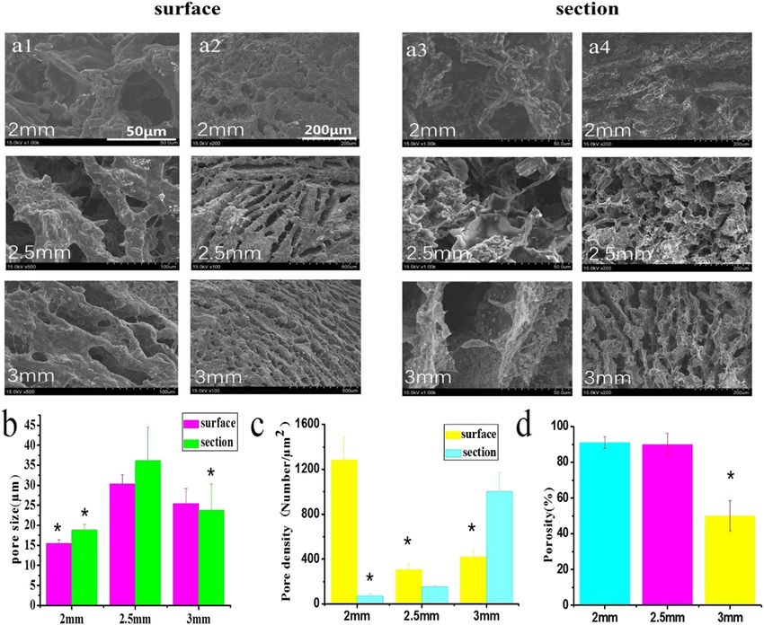
Fabrication of 3D-printed gellan gum/starch composite scaffold 51
significance for promoting nerve regeneration [6], which is difficult to be dissolved in common solvents. For pro-
however has been seldom reported to date. viding an efficient way to prepare natural cellulose hydrogel
Generally, the tissue engineering scaffold prepared under room temperature, a kind of cellulose hydrogel scaf-
by traditional technology, such as freeze-drying and electro- folds was firstly 3D-printed and then cross-linked using
spinning, possesses various geometries with different por- 0.8% N,N′-methylenebisacrylamide (MBA) solution. The
osity, pore size, mechanical properties, etc. However, it is mechanical properties were proven to be largely enhanced
difficult to obtain scaffolds with specially requested or uni- after cross-linking by MBA [15]. Nevertheless, for tissue engi-
form geometries due to the uncontrolled fabrication pro- neering application, the cross-linking reagent may bring
cess. Three-dimensional (3D) printing technology can be toxic substance to the scaffolds and be harmful for cell
used to prepare 3D biomaterials scaffolds with specific growth. Therefore, the development of new bio-ink with
porosity and pore size for better simulating the micro- good mechanical properties and biocompatibility is neces-
environment of cell ingrowth [7]. A composite hydrogel sary [16]. Gellan gum (GG) is an anionic polysaccharide pro-
of alginate, fibrin, and hyaluronic acid/RGD peptide was duced by the Sphingomonas paucimobilis, which has been
prepared using 3D printing technology. Schwann cells widely used in food as a gelling/stabilizing agent and also in
were wrapped in the composite hydrogel to evaluate bacterial culture systems [17,18]. Low concentrationed GG
the effect of the scaffold on the cells. The results showed hydrogels using 3D extrusion techniques had been success-
that the arrangement of cells in the scaffold could be well- fully developed for cartilage regeneration [19–21]. Nanoma-
regulated, indicating a huge application potential in the terials with large specific surface area good characteristics of
field of neural tissue engineering [8]. Ye et al. [9] used biocompatibility and biodegradability have achieved great
gelatin methacrylate (GelMA) hydrogel to print a multi- success in the field of tissue engineering. Nanoparticles fab-
channel hydrogel catheter with a length of 5 mm, and ricated from nanomaterials or composed of general materials
outer diameter of 6 mm, which was used to repair large are commonly used as the reinforcement factors for various
gap nerve injury. The alginate hydrogels with low or high polymer matrix. Thus, nanocomposites have brought huge
concentration of alginate were also used to prepare scaf- potential in the significant enhancement of performance for
folds with shells and cores structure through 3D printing biomedical applications [22]. Starch is a polysaccharide
technology. This unique combination method enables soft composed of glucose units and exists in plant tissues in
hydrogels to obtain 3D scaffolds with stable mechanical the form of amylose and amylopectin. The nano-sized starch
properties [10]. The interconnected pore structure in the has good fluidity when heated in an aqueous solution and
3D printing scaffolds could also provide sufficient space can form firm gels when condensed [23,24]. Huang et al.
for cell connection, cell recognition and interaction, and used a gel-like corn starch network to improve the visco-
nutrient transport [11]. Therefore, 3D printing technology elasticity and strength of the dough [25]. Mehboob et al. [26]
can be used to well control the geometric structure of the fabricated starch with high elasticity and flexibility, which
scaffold for better cell growth and tissue regeneration. showed excellent mechanical properties and low water per-
Bio-ink is one of the key factors that affects the pro- meability. In addition, the biocomposites derived from a
perties of the printed scaffolds. The biomaterials that are starch–glycerol biodegradable matrix reinforced with jute
used as bio-ink should be biocompatible, nontoxic, and fibers aiming for replacing the expensive, hazardous, or
degradable [12]. For example, the enzyme-cross-linked depleting materials were fabricated by Verma et al. [27].
silk fibroin (SF) was used as bio-ink to print a crescent- The addition of starch was shown to increase the viscosity
shaped structural scaffold. The scaffold showed good of the biocomposites and endow a high swelling power and
mechanical properties and to be conducive to the adhe- a low gelatinization temperature. Recently, a quickly formed
sion of living cells. Moreover, the degradation of the scaf- 3D tissue structure-starch foam was prepared by Wen et al.
folds could be well-tailored for cell growth [13]. Besides, [28], which proved that starch foam could encapsulate
the SF/polyethylene glycol (PEG) was also developed as a RSC96 cells and dorsal root ganglion (DRG), proving to be
bio-ink for printing a rectangular parallel-piped scaffold. promising in nerve tissue engineering.
Mixing PEG with SF could result in the formation of SF In this study, a GG/ST hydrogel scaffold was pre-
β-sheet structure, which could lead to gelation and water pared using sol–gel technology and 3D printing tech-
insolubility through physical cross-linking [14]. How- nology. The hydrogel scaffold properties were evaluated
ever, these materials usually need further cross-linking by morphology, swelling test, degradation rate, and pore
to maintain their structural integrity after printing. For analysis. In addition, the toxicity of the scaffolds was
example, cellulose has good biocompatibility and biode- tested using fibroblasts. The proliferation and cell growth
gradation while it is difficult to process, because cellulose morphology of the Schwann cell on the scaffold were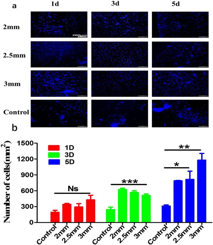
52 Liling Zhang et al.
evaluated to penetrate the potential application prospect temperature is 17°C, the needle gauge is 19, and the thick-
of the scaffold in peripheral nerve regeneration. The ness of the printing layer is 1 mm, The filling gap is 2, 2.5,
study was anticipated to provide an experimental basis and 3 mm, and the air pressure is 0.11–0.3 MPa. Then after
for the development of new artificial nerve grafts for 3D printing, the printed stent needs to be stored at −20°C for
neural tissue engineering. 48 h to fix its shape. Finally, before using the stent, freeze-
dry the stent in a freeze dryer at −50°C for 36 h before use.
2 Materials and methods
2.3 Analysis and test
2.1 Materials
2.3.1 Atomic force microscope (AFM) and rheological
The L929 fibroblast cell line was purchased from the evaluations
ATCC cell bank in Shanghai, China, and the RSC96 rat
Schwann cell line was purchased from the cell bank of The size of starch was detected using AFM (FM-
the Chinese Academy of Sciences. GG was purchased from Nanoview6800). Briefly, the starch was blown to a clean
Yinuo Biotechnology Co., Ltd., China. Starch (ST) and par- coverslip surface to form a single layer, then a tap-mode of
aformaldehyde (PA) come from National Pharmaceutical AFM was used to observe the morphology of starch parti-
Group Chemical Reagent Co., Ltd., China. Tyr-Ile-Gly-Ser- cles. The size of starch particles was then statistically ana-
Arg (YIGSR) peptides were bought from Shanghai Jier lyzed. The rheological properties of the hydrogel at room
Biochemical Co., Ltd., China. Toluidine blue (TBO), temperature were detected using the rheological test with a
Hoechst 33342, Cell Counting Kit-8 kit, penicillin–strepto- rotary rheometer (HAAKE RS6000, Thermo Fisher Science,
mycin (PS), and trypsin (Try) were all from Sigma-Aldrich, USA). The GG/ST hydrogels for rheometer testing were pre-
USA. MTT cell proliferation and toxicity kit and BCA protein pared in advance according to the above method. The gap
concentration determination kit were from Biyuntian Co., between torque and test platform was 1.2 mm, the mode
Ltd., China. Fetal bovine serum (FBS) and DMEM high-sugar was amplitude test, and the stress was determined to be
basal medium come from Gibco, USA. Phosphate buffer 0.1%. Then, the prepared GG/ST hydrogel was added to the
(PBS, pH7) was purchased from HyClone Co., Ltd. test platform with the dosage of 2 mL for measurement. The
shear thinning characteristics of bio-ink were studied in
dynamic mode, and the oscillatory strain sweep test was
performed with 0.1% strain. The values of G′ and G″ under
2.2 Design and fabrication of GG/ST the strain condition of 0.1% were obtained, and finally the
scaffolds loss factor is obtained.
The model printed in this experiment is mainly a cuboid
structure. Firstly, a model diagram of a cuboid structure
was drawn in the software of solid works (2016 version), 2.3.2 Morphological observation
as shown in Figure 1. Then the model diagram was
imported into the computer of the 3D printer for program- After 3D printing was completed, a digital camera (Huawei
ming. Finally, the GG/ST scaffold was prepared by a 3D Nova2s, CN) was first used to observe the general appear-
printer (Shining 3D, CN). The bio-ink was prepared as ance of the scaffolds. An optical microscope was then used
follows: 16 g GG powder with 4, 5.4, 8, and 16 g starch to record the morphology of the scaffold, and the surface
powder separately were dissolved in pure water at a tem- and cross-sectional micro-morphology of the hydrogel
perature of 80°C thoroughly. After stirring and mixing were analyzed by a scanning electron microscope (SEM).
evenly, the solution was left to cool at room temperature Secondly, the scaffold was frozen overnight at −20°C and
and then stored at 4°C overnight to obtain a printable then lyophilized in a freeze dryer (Shanghai De Xiang Co.,
bio-ink. Then, the mixed solution was transferred into Ltd.) for 48 h. The freeze-dried hydrogel samples were cut
the 3D printer, and GG/ST scaffolds were printed according into two halves and divided into surface group and cross-
to the designed model. First follow the steps to turn on the section group. In order to observe the microstructure, two
3D printer, and set the 3D printer to print at a speed of groups of scaffolds were fixed on the aluminum platform
15–20 mm/s, the nozzle temperature is 40°C, the platform and coated with a layer of gold layer. The scaffolds were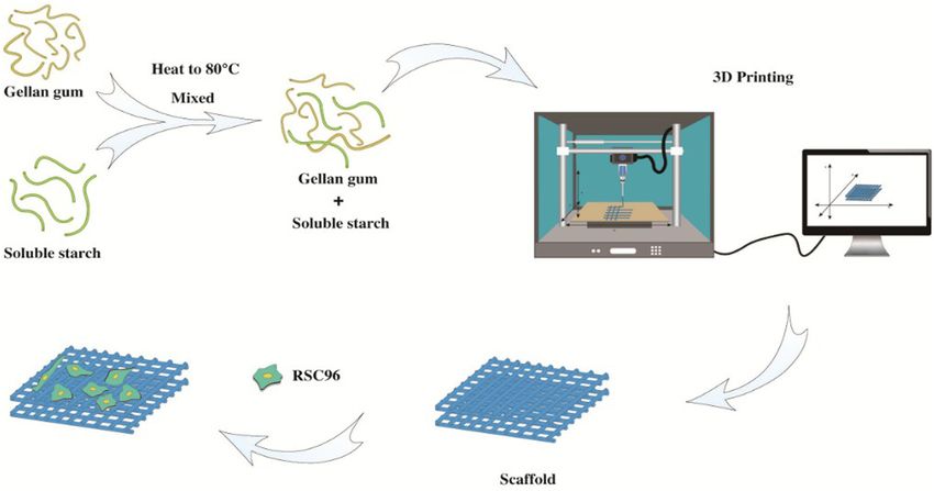
Fabrication of 3D-printed gellan gum/starch composite scaffold 53
Figure 1: The preparation of the GG/ST hydrogel.
then observed using SEM, and the whole process was per- the remaining absolute ethanol was recorded as V3. Three
formed under vacuum condition of 1.4 × 10⁻⁴ bar. parallel samples are tested for each scaffold. The calcula-
tion formula of porosity in this experiment is as follows:
Porosity (%) = (V1 − V3)/(V2 − V3) × 100%
2.3.3 Water uptake
Firstly, the scaffolds with the size of 0.5 × 0.5 × 3 mm were
freeze-dried and weighed; the initial mass was recorded 2.3.5 Degradation test
as W0. Then the freeze-dried scaffolds were immersed in
phosphate buffered saline (PBS, pH = 7.2). Finally, 10, 20, The initial mass of the lyophilized 3D scaffold with a size
30, 60, 90, 120 min and 3 days were chosen as the time of 2 × 2 × 3 mm was recorded as W0, and then the lyophi-
interval for calculating the amount of water uptake. The lized scaffolds were immersed in PBS for 1, 2, 3, 5, 7, and
sample was dried with filter paper at each time point and 10 days, and the scaffolds were lyophilized again and
the mass was recorded as W1. Three parallel samples were weighed as Wnd. Three parallel samples were tested for
tested for each sample. The swelling rate was calculated each sample. The degradation rate Wd of the scaffold
using the following formula: in vitro was calculated by the following formula:
SR = (W1 − W0)/ W0 × 100% Wd (%) = (W0 − Wnd)/ W0 × 100%
2.3.4 Porosity analysis 2.4 Cell culture
The porosity of the GG/ST scaffold is determined by the 2.4.1 Cytotoxicity
gas displacement method. In general, the lyophilized
scaffolds with different printing gaps are immersed in a The three scaffolds were irradiated with ultraviolet light
cylinder filled with absolute ethanol (V1) for 10 min. (365 nm, 10 mW/cm2, ENF-260C/FA, Spectroline, USA)
Then, the total volume of the scaffold immersed in abso- for 30 min, and then soaked in 75% ethanol for 30 min
lute ethanol was recorded as V2. Finally, the scaffold was and washed with PBS (pH = 7.2) 3 times, each time for
taken out of the measuring cylinder, and the volume of 10 min. The scaffold was immersed in 10 mL DMEM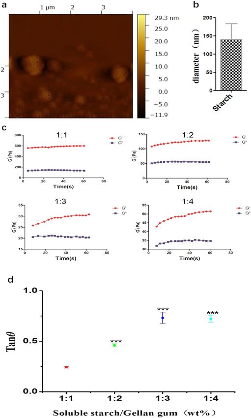
54 Liling Zhang et al.
medium and incubated for 24 h. Then, the medium was expressed as mean ± standard deviation (SD). p values
collected in a 50 mL centrifuge tube and used for L929 of < 0.05 (p < 0.05) were considered as significant.
culture. The cells density was 5 × 10³ cells/mL. After 24 h
and 48 h of culture, 10 μL of MTT reagent was added to
each well and incubated. After further culture for 4 h, the
reagent was removed and 100 μL of formazan lysis buffer 3 Results
was added. After another 4 h, the supernatant with 150 μL
from each sample was transferred to the 96-well plate in a
3.1 AFM and rheological characteristics
microplate reader for absorbance detection at a wave-
length of 570 nm.
Figure 2(a and b) shows the morphology of starch by
AFM. The starch particles were arbitrarily deposited on
the glass slide (Figure (2a)). Through statistical analysis
2.4.2 CCK-8 test
of the image, the size of the starch nanoparticles was
approximately 140 nm (Figure (2b)). Figure 2(c and d) is
The scaffold material was sterilized by ultraviolet light
the rheological characteristics of GG/ST hydrogel. The
for 30 min and immersed in 75% ethanol for 20 min,
storage modulus (G′) of the four ratios of GG/ST scaffolds
and then washed with PBS three times, each time for
were all greater than the loss modulus (G″), which proved
10 min. Then 10 µL of cell suspension with a density of
that the hydrogel was formed. In Figure (2b), The Loss
5 × 10⁴ cells/mL was added to the scaffolds and incubated
factor, namely loss angle (tan θ), is the ratio of loss mod-
at 37°C in a CO2 incubator for 1, 2, and 3 days, respec-
ulus (G″) to storage modulus (G′), which mainly reflects
tively. After that, the culture medium was discarded, and
the viscoelastic ratio of the material. The greater the loss
500 μL of CCK-8 solution was added to each well and
factor, the greater the viscosity of the material, and the
incubated in an incubator for 4 h. Then, 200 µL of the
smaller the loss factor, the greater the elasticity of the
solution was taken from each well and added to a 96-
material. When tan θ > 1, G″ > G′, the hydrogel behaves
well plate, eight parallel samples were used, and the
as a sol, while when tan θ < 1, G″ < G′, the hydrogel
OD was measured at 450 nm.
behaves in a gel state [29]. During the experiment, the
concentration of GG was kept at 16%, and starch was
added in the ratio of 1:4, 1:3, 1:2, 1:1. The increase in
2.4.3 Schwann cells culture
starch concentration had a great effect on the viscoelas-
ticity of the system. When the ratio of ST and GG was 1:1
The RSC96 Schwann cells were used for evaluating the
and 1:2, the elasticity of the material was high, which was
biocompatibility of the printed scaffolds. Before cell cul-
not conducive to material extrusion. When the ratio is
ture, the scaffolds were pretreated using YIGSR polypep-
1:4, the viscosity was too large and the material was too
tide (50 μg/mL) for 2 h for enhancing cell adhesion. Then,
easy to extrude, which was not conducive to maintain the
RSC96 cells were seeded on GG/ST scaffold at a cell den-
scaffold structure; therefore, the concentration ratio of all
sity of 1 × 10⁵ cells/mL and incubated for 1, 3, and 5 days,
samples in the later period remained 1:3. These results
respectively. At each time point, cells were fixed with
indicated that the GG/ST scaffolds had the capability for
4% PA for more than 4 h and washed 3 times with phy-
3D printing.
siological saline. Thereafter, 50 μL of hoechst33342 diluted
1:20 in PBS was added to the cells and kept in the dark
for 30 min. Finally, the scaffolds were washed with physio-
logical saline for five times. The cell growth status was 3.2 Morphology of the scaffolds
observed under an inverted fluorescence microscope
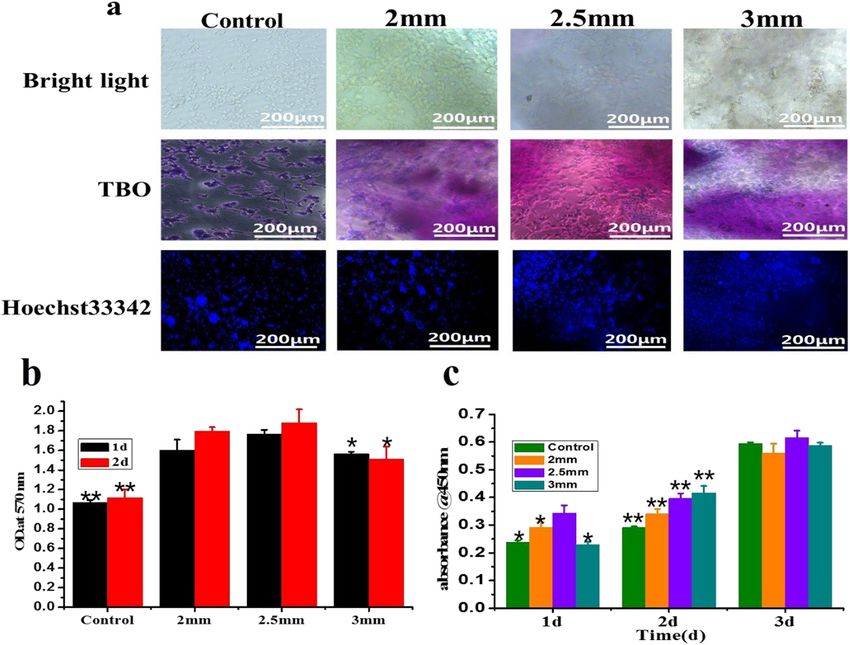
(Leica, Germany). The surface and cross-section of the 3D-printed scaffolds
at different gap were observed by SEM (Figure 3(a1–a4)).
With the increased printing gap, the pore size on the
hydrogel surface increased first and then decreased.
2.5 Statistics The possible reason is that when the length and width
of the printing model are determined, with the increase of
All the experimental results were analyzed by Origin 8.0 the printing gap, the number of turns that the printing
(Origin Lab company) and Image Tool. The results were needle needs to rotate decreases, and the extrusion force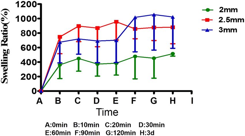
Fabrication of 3D-printed gellan gum/starch composite scaffold 55
and when the printing gap further increased to 3 mm,
the porosity dropped to 45%.
3.3 Swelling behavior
Figure 4 shows the swelling behavior of 3D-printed scaf-
folds with different printing gaps in deionized water.
Each scaffold gradually reached equilibrium swelling
from 0 to 3 days. When the scaffolds’ printing gap was
2 mm, the swelling ratio was between 380 and 440%,
which was lower than that of other groups. When the
printing gap of the scaffold was 2.5 mm, the swelling
rate increased steadily before 1 h, and there was a ten-
dency for the swelling rate to decrease after 1 h. For this
phenomenon, the solvent first infiltrated the surface of
the scaffold, and then slowly dipped into the interior until
equilibrium was reached. Since the pore size on the sur-
face of the 2.5 mm scaffold was smaller than the inner
pore size, the swelling rate increased after 1 h and then
gradually stabilized. When the printing gap of the scaf-
fold was 3 mm, the increased rate of the swelling rate was
less than 2.5 mm before 1 h, but higher than 2.5 mm after
1 h. The reason why the swelling rate of the 3 mm group
was higher than 2.5 mm group at the last time may be that
the cross-section and the number of pores on the surface
of the 2.5 mm group are smaller than those of the 3 mm
group.
Figure 2: AFM of starch granules and rheological properties of
GG/ST scaffolds with different ratios. (a) AFM; (b) starch particle
3.4 Degradation behavior
size; (c) G′ and G″ in amplitude mode; (d) loss angle. ***p < 0.001
by one-way ANOVA test.
The degradation behavior of the prepared three scaffolds
in PBS was studied, and the mass loss from 0 to 10 days
was recorded. As shown in Figure 5, when the printing
will change to a certain extent, so the aperture becomes gap of the scaffolds was between 2.5 and 3 mm, the degra-
larger. At the same time, when the printing distance is dation rate of the scaffolds increased steadily from day
greater than 2.5 mm, the internal extrusion force can 1 to day 2, indicating that the formation of hydrogel did
change in enough time, resulting in the aperture size not reduce its own water absorption performance. The
less than 2.5 mm. Quantitative analysis was shown in degradation rate of the scaffolds dropped sharply from
Figure 3(b); when the gap was 2.5 mm, the surface and 2 to 3 days and showed an upward trend from 3 to 4
internal pore sizes of the hydrogel were larger than other days, and eventually gradually stabilized. The degrada-
hydrogels regardless of the surface or cross-section. As tion rate required multiple freeze-drying and soaking in
shown in Figure 3(c), when the scaffold printing gap PBS. It was noticed that the degradation rate on the third
was 2 mm, the number of pore density reached a maxi- day decreased, which may be caused by the solvent that
mum of 1,220 pores/mm2, and when the gap was 2.5 mm, was not being completely lyophilized. The scaffolds with
the number of pores reached a minimum of about a spacing of 2 mm always maintained a stable degrada-
360 pores/mm2. In Figure 3(d), when the printing gap tion rate, which also was in accordance with the results of
was 2 mm and 2.5 mm, the porosity was around 90%, swelling rate. In contrast, the degradation rates of 2.5 mm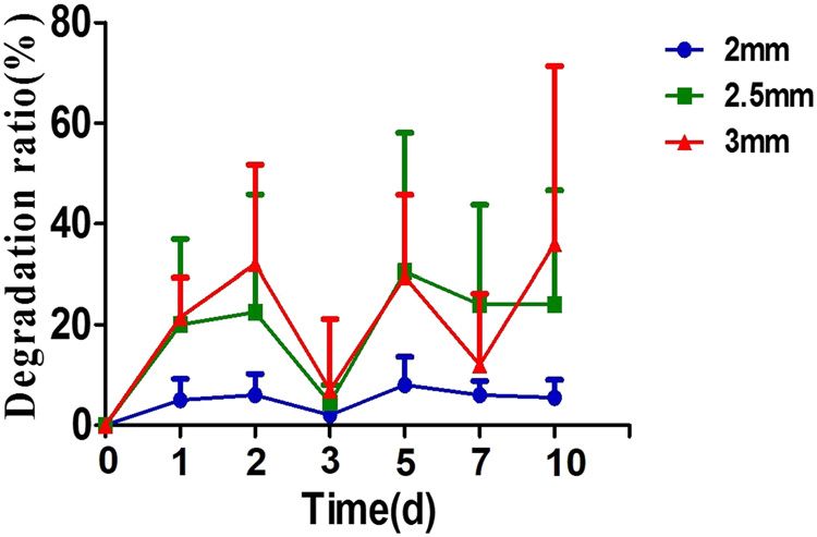
56 Liling Zhang et al. Figure 3: Morphological characterization of GG/ST scaffold. (a) Cross-sectional view of the scaffolds taken by scanning electron micro- scope; (b) quantitative analysis of the cross-sectional pore size; (c) quantitative analysis of the cross-sectional pore density; (d) porosity test. *p < 0.05 by one-way ANOVA test. Figure 4: Swelling ratio test of GG/ST scaffold with various printing gap. *p < 0.05 by one-way ANOVA test. Figure 5: Degradation experiment of GG/ST scaffold.
Fabrication of 3D-printed gellan gum/starch composite scaffold 57
scaffolds and 3 mm scaffolds varied heavily. This result scaffold. The results showed that the prepared composite
indicated that the degradation rate of printed scaffolds hydrogel was suitable for cell adhesion and proliferation
with various printing gap was different. without obvious cytotoxicity. Figure 6(b) shows that the
absorbance of the three scaffolds was significantly higher
than that of the control group, which proved that the
scaffolds were basically nontoxic. The CCK-8 results in
3.5 Cytotoxicity Figure 6(c) show that the absorbance of the three scaf-
folds increased steadily from 1 day to 3 days, which indi-
Figure 6(a) shows the results of L929 fibroblast on printed cated that the cell viability of the cells on the scaffold was
scaffolds with different gaps after 3 days of culture. Cells continuously rising and could grow well on the scaffold.
were distributed on the surface of all scaffolds. However, After 3 days of cell culture, the cell viability on the scaf-
the cells still showed certain aggregation. After 3 days, all fold with 2.5 mm gap was higher than that in the control
the cells on the scaffold were obviously spread out. group. This may be because the pore size of the scaffold
Overall, the cells on the 2 mm scaffold were less than in the 2.5 mm size was larger on the surface and inside
those on 2.5 and 3 mm groups. The cells on the 2.5 and than the other groups, which was good for nutrients to
3 mm groups covered almost the entire surface of all penetrate into the scaffold and cells growth.
Figure 6: Cell morphology and toxicity of GG/ST scaffolds. (a) TBO and immunofluorescence; (b) MTT test cytotoxicity experiment; (c) cell
viability detection. *p < 0.05 and **p < 0.01 by one-way ANOVA test.58 Liling Zhang et al.
sources of autologous transplantation and the loss of
donor site function, autologous nerve transplantation is
usually a clinically conservative treatment [31]. Thus, the
artificial nerve grafts that replace autotransplantation
have been developed. 3D printing technology could con-
struct nerve implants with excellent performance for
nerve regeneration; however, the bio-ink for 3D printing
still needs to be further improved. In this study, a new
type of bio-ink composed of GG and nano-sized starch
was developed. GG/ST scaffolds with different printing
gaps were prepared by 3D printing technology. The
GG/ST scaffold was basically noncytotoxic and the cells
proliferated stably. When the printing distance was
3 mm, the growth state and morphology of Schwann cells
in the scaffold were the best, showing high cell survival
rate and the largest number of cells. Therefore, the GG/ST
scaffold prepared by 3D printing technology may have
potential applications in peripheral nerve regeneration
and provide an important experimental basis for the
development of peripheral nerve grafts.
The maximum concentration of GG for preparing the
GG/ST composite scaffolds was determined to be 16 wt%.
Figure 7: Schwann cells culture. (a) Immunofluorescence images of
At a shear rate of 100 s−1, except for the ratio of GG and
GG/ST scaffold cocultured with RSC96 cells for 1d, 3d, and 5d. (b)
Statistics of cell count results related to immunofluorescence. *p < starch of 1:1, which did not show shear thinning beha-
0.05, **p < 0.01 and ***p < 0.001 by one-way ANOVA test. vior, the other ratios of 1:2, 1:3, and 1:4 all showed this
behavior. As described by Malda et al. [32], the shear
thinning was very important for extrusion-based printers.
Although the three ratios all exhibited shear thinning
3.6 Schwann cells evaluation behavior, the ink with ration of 1:2 could not be printed
depending on the pressure of the 3D printer itself. When
RSC96 cells were cultured in GG/ST scaffold for 1, 3, and the ratio was 1:4, the material was particularly easy to
5 days to evaluate the effect of the scaffold on nerve extrude, but it was not easy to maintain the structural
regeneration. As shown in Figure 7(a), the number of integrity, while easy to collapse. Thus, a ratio of 1:3 was
cells in the scaffold group was significantly more than chosen for the sequential experiment.
that in the control group. Moreover, cells in control group The composite material of GG and starch had a highly
displayed obvious aggregation than the scaffolds group. interconnected pore network and a high total internal
The quantitative analysis of immunofluorescence is shown porosity. All printed scaffolds had a porous structure,
in Figure 7(b). The results show that the number of cells in which was an important indicator of nutrient exchange
the experimental group was higher than that in the control in tissues [33]. In our study, as shown in Figure 3(d), all
group. After 5 days of cultivation, the number of cells in the scaffolds except the 3 mm scaffold exhibited porosity
3 mm group was significantly higher than that in the other higher than 90%, which was of great benefit for material
groups, while the number of cells in the 2 and 2.5 mm exchange and nutrient transport. The reason may be that
groups was basically the same. the density of the surface holes of the 2 mm group was
greater than that of the 3 mm group, and the number of
surface holes was more than that of the 3 mm group.
Compared with the scaffolds of the 2.5 mm group, the
4 Discussion size of the surface and the inner hole was larger than
that of the scaffold of the 3 mm group. The hole diameter
Currently, the clinical application of autologous nerve of the 2 mm and 3 mm scaffolds was smaller than that of
transplantation is still the gold standard for peripheral the 2.5 mm scaffold. The possible reason was that the
nerve regeneration [30]. However, due to the limited printing gap of 2.5 mm was the best setting data for theFabrication of 3D-printed gellan gum/starch composite scaffold 59
3D printer, and the dual network structure of GG/ST nerve cells of the printed GG/ST scaffolds were evaluated.
was obtained. Significant differences in the number of Though GG had been applied for skin tissue regeneration
pores formed and pore size distribution were also in the previous study and cell could proliferate well
observed (Figure 3). It may be speculated that the without cytotoxicity [40], no studies referring to the effect
freeze-dried double-network hydrogel was responsible of GG on nerve cells was reported. Thus, considering the
for these inherent differences. Previous studies reported effect of the scaffolds on cell viability, L929 fibroblast cul-
that in composite scaffolds, the formation of a network ture was first used to evaluate the cytotoxicity of the pre-
structure kept the structure more stable, thereby making pared scaffolds. With the extension of the culture time,
the structure more resistant to shrinkage and expan- the cells proliferated significantly, indicating that all scaf-
sion [34]. folds had no significant toxic effects on the cells. The CCK-
The swelling ability of the hydrogel was further eval- 8 experiment results also confirmed the finding, which
uated. This experiment was achieved by soaking the was in accordance with the previous study. There are
hydrogel in deionized water. Proper swelling of the hydrogel two possible reasons for this. One is that the selected
will help to expand the spacing between the various mole- material, GG, is a food-grade material, an extracellular
cules in the hydrogel and facilitate cell ingrowth and heteropolysaccharide gum produced by aerobic fermenta-
nutrient exchange between cells [35]. The printed scaffold tion of Sphingolipids under neutral conditions. In addi-
structure is easy to diffuse between ions and leads to a soft tion to increasing its mechanical strength, starch itself is
structure, which increases the absorption of water. The pore also a polysaccharide polymerized by glucose. On the
density has a certain relationship with the swelling rate of other hand, since GG/ST mixes to form a loose and porous
the scaffold [36,37]. The larger the pore density, the higher hydrogel, it facilitates nutrient exchange and waste dis-
the swelling rate. It is obvious from Figure 4 that the swelling charge between cells and promotes cell growth. Schwann
rate of 2 mm had always been lower than that of 2.5 and cells are glial cells that play a major supporting role in
3 mm scaffolds, but the swelling rate of 2.5 mm was initially peripheral nerve regeneration [41]. Thus, RSC96 Schwann
higher than 3 mm, and the final expansion rate is lower than cell was cultured on the scaffold to evaluate the effect on
3 mm. This result was the same as that of the 3 mm group peripheral nerve regeneration. As shown in Figure 7(a),
with a higher pore density than the other two groups. The the cells in the experimental group did not have obvious
stability of the hydrogel could provide a stable platform and aggregation when they grew, but the control group could
microenvironment for cell growth and tissue regeneration find obvious aggregation of the cells. Moreover, the
[9]. Three groups of scaffolds were immersed in PBS to eval- number of cells in the experimental group was signifi-
uate the degradation behavior. The 2 mm scaffolds structure cantly more than that in the control group. As shown in
had always maintained a stable degradation rate of around Figure 7(b), RSC96 cells and scaffolds were cultured for 1,
5%, while the degradation rates of the 2.5 and 3 mm scaf- 3, and 5 days later; the number of cells in the experi-
folds were basically maintained at 35%, which was con- mental group was higher than that in the control group.
sistent to the porosity results. Compared with the 2 mm The possible reason is that the printing gap of the 3 mm
scaffold, the scaffold of 2.5 and 3 mm had larger surface group scaffold is larger, the cross-sectional hole density
and internal holes, and the density of the internal pores of is higher than the other 2 groups, and the internal pore
the scaffold was also larger. This facilitated the separation size is larger and connected, which is conducive to mate-
of solvents into the scaffold and the solvent molecules rial exchange and nutrient transportation. Generally,
gradually diffused into the scaffold. Inside the scaffold, cells will have a higher survival rate in a porous network
the volume of the polymer material was continuously with cell binding domains on the scaffold [42]. Lozano
increasing, and the movement of the macromolecular seg- et al. [43] also observed that the large pore density of the
ment was enhanced, and then the movement of the entire scaffold surface and cross-section could form a porous
macromolecular chain was achieved through the coordi- network structure, which was more conducive to the
nated movement of the segment, and the macromolecule exchange of nutrients. The cell survival rate and cell via-
gradually entered the solution to form a thermodynami- bility were better. After 5 days of cell culture, the swelling
cally stable homogeneous system, thus the scaffold with rate of the 3 mm group was the highest among the three
different print gaps showed different degradation ratio. groups of scaffolds and reached a balanced state. The
Our results indicated that these three scaffolds possessed swelling rate of hydrogel is conducive to the exchange
a certain degradation rate, while the degradability of of nutrients in cells [44]. As Akkineni et al. [45] reported,
the scaffold provided a possibility for future clinical appli- the swelling rate of the scaffold was of great significance
cation [38,39]. Finally, the cytotoxicity and effect on for in vitro cell culture experiments and implantation. The60 Liling Zhang et al.
16.7% sodium alginate and 3% GG scaffolds have the Ministry of Education (2020JC08), Qinglan Project of
highest expansion rate in PBS, and human bone marrow Jiangsu Province (2018).
mesenchymal stem cells (HMSC) have the highest cell
number after 7 days of culture on the scaffold. As the Author contributions: All authors have accepted respon-
printing gap increases, the degradation rate of the scaf- sibility for the entire content of this manuscript and
folds increases and the degradation time shortens. approved its submission.
The 2 mm group scaffold has always maintained a low
degradation rate, which was not conducive to peripheral Conflict of interest: The authors state no conflict of
nerve regeneration. Although the degradation rate of the interest.
2.5 mm group has increased, the degradation rate may be
not still insufficient, which needs further investigation.
Shi et al. [46] reported that the SF/gelatin scaffold with a References
degradation rate of about 37% was better for cell growth
and spreading. In this experiment, the degradation rate [1] Grinsell D, Keating CP. Peripheral nerve reconstruction after
injury: a review of clinical and experimental therapies. Biomed
of the 3 mm scaffold group was about 37%, and the
Res Int. 2014;2014:698256.
number of cells was higher than that of other groups. [2] Ebrahimi MH, Samadian H, Davani ST, Kolarijani NR,
Peng et al. [47] and other experiments also proved that Mogharabian N, Salami MS, et al. Peripheral nerve regenera-
the degradation rate can regulate the cell behavior of tion in rats by chitosan/alginate hydrogel composited with
stem cells. It is reasonable to speculate that the degrada- Berberine and Naringin nanoparticles: in vitro and in vivo
study. J Mol Liq. 2020;318:114226.
tion rate may have a certain influence on cell behavior.
[3] Matyga AW, Veen SC, Gasiorowski JZ. Natural and synthetic
extracellular matrix biomaterials for peripheral nerve regen-
eration. In Vitro Cell Dev Biol Animal. 2018;54:27–32.
[4] Jia C, Luo BW, Wang HY, Bian YQ, Li XY, Li SH, et al. Precise and
5 Conclusion arbitrary deposition of biomolecules onto biomimetic fibrous
matrices for spatially controlled cell distribution and func-
tions. Adv Mater. 2017;29(35):282–6.
In this study, GG/ST scaffolds with different printing gaps
[5] Cai SX, Wu CX, Yang WG, Liang WF, Yu HB, Liu LQ. Recent
were prepared by sol–gel technology and 3D printing tech- advance in surface modification for regulating cell adhesion
nology. The scaffolds with cuboid structure possessed and behaviors. Nanotechnol Rev. 2020;9(1):971–89.
suitable physiochemical properties, including porosity, [6] Wu J, Xie LL, Lin WZY, Chen QS. Biomimetic nanofibrous scaf-
swelling, degradation, etc., which may be better for tissue folds for neural tissue engineering and drug development.
Drug Discov Today. 2017;22(9):1375–84.
engineering implantation. Both L929 fibroblasts and RSC96
[7] Hofmann M. 3D Printing gets a boost and opportunities with
Schwann cells showed that the scaffolds had good biocom-
polymer materials. Acs Macro Lett. 2014;3(4):382–6.
patibility without cytotoxicity. Moreover, the proliferation [8] Ning LQ, Sun HY, Lelong T, Guilloteau R, Zhu N, Schreyer DJ,
and cell growth morphology of Schwann cells on the scaf- et al. 3D bioprinting of scaffolds with living Schwann cells for
fold with larger printing gaps were better than that with potential nerve tissue engineering applications.
smaller printing gap. In summary, the GG/ST scaffold Biofabrication. 2018;10(3):234–45.
[9] Ye W, Li H, Yu K, Xie C, Wang P, Zheng Y, et al. 3D printing of
developed by 3D printing technology may have potential
gelatin methacrylate-based nerve guidance conduits with
applications in peripheral nerve regeneration. The study multiple channels. Mater Des. 2020;192:108757.
may provide an important experimental basis for the [10] Akkineni AR, Ahlfeld T, Lode A, Gelinsky M. A versatile method
design and development of artificial implants for treating for combining different biopolymers in a core/shell fashion by
peripheral injuries. 3D plotting to achieve mechanically robust constructs.
Biofabrication. 2016;8:4.
[11] Ma HS, Luo J, Sun Z, Xia LG, Shi MC, Liu MY, et al. 3D printing of
Acknowledgments: The authors gratefully acknowledge
biomaterials with mussel-inspired nanostructures for tumor
the assistance from Yi Zhu. therapy and tissue regeneration. Biomaterials.
2016;111:138–48.
Funding information: The authors gratefully acknowl- [12] Donderwinkel I, van Hest JCM, Cameron NR. Bio-inks for 3D
edge the financial support of the National Natural Science bioprinting: recent advances and future prospects. Polym
Chem UK. 2017;8(31):4451–71.
Foundation of China (31830028, 31771054), Natural
[13] Lode A, Meissner K, Luo YX, Sonntag F, Glorius S, Nies B, et al.
Key Science Research Program of Jiangsu Education Fabrication of porous scaffolds by three-dimensional plotting
Department (19KJA320006), the Open Project of Key of a pasty calcium phosphate bone cement under mild condi-
Laboratory of Organ Regeneration and Transplantation, tions. J Tissue Eng Regen Med. 2014;8(9):682–93.Fabrication of 3D-printed gellan gum/starch composite scaffold 61
[14] Zheng ZZ, Wu JB, Liu M, Wang H, Li CM, Rodriguez MJ, et al. 3D grafts in craniomaxillofacial surgery. Biomed Res Int.
Bioprinting of self-standing silk-based bio-ink. Adv Healthc 2016;2016:3856262.
Mater. 2018;7:6. [31] Jiang CQ, Hu J, Xiang JP, Zhu JK, Liu XL, Luo P. Tissue-engi-
[15] Hu XZ, Yang ZJ, Kang SX, Jiang M, Zhou ZW, Gou JH, et al. neered rhesus monkey nerve grafts for the repair of long ulnar
Cellulose hydrogel skeleton by extrusion 3D printing of solu- nerve defects: similar outcomes to autologous nerve grafts.
tion. Nanotechnol Rev. 2020;9(1):345–53. Neural Regen Res. 2016;11(11):1845–50.
[16] Ferris CJ, Gilmore KG, Wallace GG, Panhuis MIH. [32] Malda J, Visser J, Melchels FP, Jungst T, Hennink WE, Dhert WJ,
Biofabrication: an overview of the approaches used for et al. 25th anniversary article: engineering hydrogels for
printing of living cells. Appl Microbiol Biot. biofabrication. Adv Mater. 2013;25(36):5011–28.
2013;97(10):4243–58. [33] Annabi N, Nichol JW, Zhong X, Ji CD, Koshy S,
[17] Fan YT, Yi J, Hua X, Zhang YZ, Yang RJ. Preparation and char- Khademhosseini A, et al. Controlling the porosity and micro-
acterization of gellan gum microspheres containing a cold- architecture of hydrogels for tissue engineering. Tissue Eng
adapted beta-galactosidase from Rahnella sp R3. Carbohyd Part B Rev. 2010;16(4):371–83.
Polym. 2017;162:10–5. [34] Haque MA, Kurokawa T, Gong JP. Super tough double network
[18] Oliveira IM, Goncalves C, Shin ME, Lee S, Reis RL, Khang G, hydrogels and their application as biomaterials. Polymer.
et al. Anti-inflammatory properties of injectable betametha- 2012;53(9):1805–22.
sone-loaded tyramine-modified gellan gum/silk fibroin [35] Drury JL, Mooney DJ. Hydrogels for tissue engineering: scaffold
hydrogels. Biomolecules. 2020;10:10. design variables and applications. Biomaterials.
[19] Silva-Correia J, Oliveira JM, Caridade SG, Oliveira JT, Sousa RA, 2003;24(24):4337–51.
Mano JF, et al. Gellan gum-based hydrogels for intervertebral [36] Smith AM, Shelton RM, Perrie Y, Harris JJ. An initial evaluation
disc tissue-engineering applications. J Tissue Eng Regen Med. of gellan gum as a material for tissue engineering applica-
2011;5(6):97–107. tions. J Biomater Appl. 2007;22(3):241–54.
[20] Melchels FPW, Dhert WJA, Hutmacher DW, Malda J. [37] Wu XF, Li W, Chen K, Zhang DK, Xu LM, Yang XH. A tough PVA/
Development and characterisation of a new bioink for additive HA/COL composite hydrogel with simple process and excellent
tissue manufacturing. J Mater Chem B. 2014;2(16):2282–9. mechanical properties. Mater Today Commun. 2019;21:100702.
[21] Kesti M, Muller M, Becher J, Schnabelrauch M, D’Este M, [38] Fukushima K. Functionalization and precise synthesis of bio-
Eglin D, et al. A versatile bioink for three-dimensional printing degradable polymers towards biomedical applications. Sen-I
of cellular scaffolds based on thermally and photo-triggered Gakkaishi. 2016;72(12):P571–3.
tandem gelation. Acta Biomater. 2015;11:162–72. [39] Ding HX, Cheng YZ, Niu XL, Hu YC. Application of electrospun
[22] Jian W, Hui D, Lau D. Nanoengineering in biomedicine: current nanofibers in bone, cartilage and osteochondral tissue engi-
development and future perspectives. Nanotechnol Rev. neering. J Biomat Sci Polym E. 2020;110:1–4.
2020;9(1):700–15. [40] Ismail NA, Amin KAM, Razali MH. Novel gellan gum incorpo-
[23] Wang GX, Li S, Feng YL, Hu YX, Zhao GY, Jiang W. Effectively rated TiO2 nanotubes film for skin tissue engineering. Mater
toughening polypropylene with in situ formation of core-shell Lett. 2018;228:116–20.
starch-based particles. Carbohyd Polym. 2020;249:116795. [41] George N, Geller HM. Extracellular matrix and traumatic brain
[24] Shan JQ, Liu DG, Su F, Li MY, Tian HF, Guo MN, et al. injury. J Neurosci Res. 2018;96(4):573–88.
Anisotropic structure and properties of chitin and chitosan [42] Malda J. Engineering of hydrogel-based bioinks for the fabri-
nanofibril-supported starch foams. ACS Sustain Chem Eng. cation of cell-laden 3D constructs. Hum Gene Ther.
2020;8(47):17387–96. 2013;24(12):A27.
[25] Huang Z, Wang JJ, Chen Y, Wei N, Hou Y, Bai WD, et al. Effect of [43] Lozano R, Stevens L, Thompson BC, Gilmore KJ, Gorkin R,
water-soluble dietary fiber resistant dextrin on flour and bread Stewart EM, et al. 3D printing of layered brain-like structures
qualities. Food Chem. 2020;317:126452. using peptide modified gellan gum substrates. Biomaterials.
[26] Mehboob S, Ali TM, Sheikh M, Hasnain A. Effects of cross 2015;67:264–73.
linking and/or acetylation on sorghum starch and film char- [44] Park H, Guo X, Temenoff JS, Tabata Y, Caplan AI, Kasper FK,
acteristics. Int J Biol Macromol. 2020;155:786–94. et al. effect of swelling ratio of injectable hydrogel composites
[27] Verma A, Joshi K, Gaur A, Singh VK. Starch-jute fiber hybrid on chondrogenic differentiation of encapsulated rabbit
biocomposite modified with an epoxy resin coating: fabrica- marrow mesenchymal stem cells in vitro. Biomacromolecules.
tion and experimental characterization. J Mech Behav Mater. 2009;10(3):541–6.
2018;27:5–6. [45] Akkineni AR, Ahlfeld T, Funk A, Waske A, Lode A, Gelinsky M.
[28] Wen XX, Shen MJ, Bai YJ, Xu CL, Han XL, Yang HL, et al. Highly concentrated alginate-gellan gum composites for 3d
Biodegradable cell-laden starch foams for the rapid fabrica- plotting of complex tissue engineering scaffolds. Polymers-
tion of 3D tissue constructs and the application in neural Basel. 2016;8:5.
tissue engineering. J Biomed Mater Res B. [46] Shi WL, Sun MY, Hu XQ, Ren B, Cheng J, Li CX, et al. Structurally
2020;108(1):104–16. and functionally optimized silk-fibroin-gelatin scaffold using
[29] Yoshida S, Kondo K. Fiber volume control of composite mate- 3d printing to repair cartilage injury in vitro and in vivo. Adv
rials by loss angle. Polym Compos. 2016;37(5):1307–11. Mater. 2017;29:29.
[30] Gaudin R, Knipfer C, Henningsen A, Smeets R, Heiland M, [47] Peng YM, Liu QJ, He TL, Ye K, Yao X, Ding JD. Degradation rate
Hadlock T. Approaches to peripheral nerve repair: generations affords a dynamic cue to regulate stem cells beyond varied
of biomaterial conduits yielding to replacing autologous nerve matrix stiffness. Biomaterials. 2018;178:467–80.You can also read